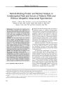
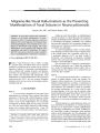
| Title |
Isolated Cortical Visual Loss with Subtle Brain MRI Abnormalities in a case of Hypoxic-ischmic Encephalopathy |
| Creator |
Margolin, E; Gujar, SK; Trobe, JD |
| Affiliation |
Department of Ophthalmology and Visual Sciences (Kellogg Eye Center), University of Michigan, Ann Arbor, Michigan 48109, USA. |
| Abstract |
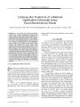
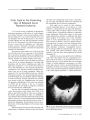
A 16-year-old boy who was briefly asystolic and hypotensive after a motor vehicle accident complained of abnormal vision after recovering consciousness. Visual acuity was normal, but visual fields were severely constricted without clear hemianopic features. The ophthalmic examination was otherwise normal. Brain MRI performed 11 days after the accident showed no pertinent abnormalities. At 6 months after the event, brain MRI demonstrated brain volume loss in the primary visual cortex and no other abnormalities. One year later, visual fields remained severely constricted; neurologic examination, including formal neuropsychometric testing, was normal. This case emphasizes the fact that hypoxic-ischemic encephalopathy (HIE) may cause enduring damage limited to primary visual cortex and that the MRI abnormalities may be subtle. These phenomena should be recognized in the management of patients with HIE. |
| Subject |
Accidents, Traffic; Adolescent; Humans; Hypoxia-Ischemia, Brain, complications; Hypoxia-Ischemia, Brain, pathology; Magnetic Resonance Imaging, methods; Male; Vision Disorders, etiology; Visual Cortex, pathology; Visual Fields, physiology |
| Format |
application/pdf |
| Publication Type |
Journal Article |
| Collection |
Neuro-Ophthalmology Virtual Education Library: Journal of Neuro-Ophthalmology Archives: https://novel.utah.edu/jno/ |
| Publisher |
Lippincott, Williams & Wilkins |
| Holding Institution |
Spencer S. Eccles Health Sciences Library, University of Utah |
| Rights Management |
© North American Neuro-Ophthalmology Society |
| Setname |
ehsl_novel_jno |
| ID |
225590 |
| Reference URL |
https://collections.lib.utah.edu/ark:/87278/s6cr90fr/225590 |