OCR Text |
Show Journal of Neuro- Ophthalmology 21( 3): 214- 216, 2001. • 2001 Lippincott Williams & Wilkins, Inc., Philadelphia Original Contribution 
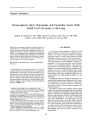
Innervation of the Sternocleidomastoid and Trapezius Muscles by the Accessory Nucleus John C. DeToledo, MD, and Noble J. David, MD Images moving over the retina at velocities as low as a few degrees per second, in movements as head turning, can degrade visual acuity. Visual acuity requires that even minute motion of the head be compensated for, primarily via optokinetic and vestibular reflexes. Whereas we have a good understanding of some the neuronal networks involved in these reflexes, other components of this network, such as the innervation of the paired muscles that turn and tilt the head, are not as well understood. The involvement of the sternomastoid, cleidomas-toid, or trapezius muscles with lesions of the cervicomeduilary junction is often not in conformity with the prevailing neu-roanatomic descriptions of their innervation by the accessory nuclei. We discuss evidence that: 1) the XI nucleus has a rostral and a caudal portion; 2) analogous to the VII nerve, the rostral portion receives projections from both cerebral hemispheres, whereas the caudal portion is innervated preferentially by the contralateral hemisphere; 3) the caudal XI nucleus innervates the ipsilateral cleidomastoid and trapezius with a predominantly crossed corticonuclear innervation; and 4) The rostral XI nucleus innervates both sternomastoids. Each rostral portion receives projections from both cerebral hemispheres. These anatomic features explain the seemingly discrepant findings in patients with cervicomeduilary lesions. Key Words: Sternocleidomastoid- Trapezius- Spinal accessory nerve- Anatomy- Head turning- Head and eye movements. The human sternocleidomastoid is classically described as a single muscle with two attachments of origin- one sternal and one clavicular. From the embryology and comparative anatomy standpoints, however, there is little doubt that the sternocleidomastoid is formed by two separate muscles, the sternomastoid and Manuscript received April 3, 2001; accepted June 25, 2001. From the Department of Neurology ( JCD), University of Miami, Miami, Florida; and the Department of Neuro- Ophthalmology ( NJD), Bascom Palmer Eye Institute, Miami, Florida. Address correspondence and reprint requests to John C. DeToledo, MD, Chief- Neurophysiology Laboratory, Department of Neurology, University of Miami, 1150 NW 14th St., Suite 410, Miami, FL 33136; e- mail: jdetoled@ mednet. med. miami. edu. the cleidomastoid ( 1- 3). In some species, including primates, the sternomastoid and the cleidomastoid blend in a common muscle body near their skull insertion. In other species, however, the two muscles remain separate ( i. e., the cleidocephalicus and sternocephalicus muscles in long- neck ungulates, camel, giraffe, and llama), demonstrating their distinct ontogeny ( 1,2). The XI nucleus also shows variability between species, and its presence appears to be ultimately determined by posture and mode of ambulation ( 4). In the absence of a forelimb, such as in apodial species ( i. e., serpents), the nucleus is absent. CORTICAL REPRESENTATION OF THE STERNOMASTOID, CLEIDOMASTOID, AND TRAPEZIUS Hemispheric strokes are frequently associated with weakness of the contralateral trapezius, weakness of head tilting toward the side of hemiplegia, and weakness of the contralateral trapezius. Frontal lobe seizures may initiate as an isolated head turning, via the sternomastoid, or as an isolated head tilting, via the cleidomastoid ( 5), suggesting that the sternomastoid and cleidomastoid have distinct cortical representations. Seizures that begin with head tilting usually present with concomitant elevation of the ipsilateral shoulder. The cleidomastoid and trapezius muscles appear to have only minor representation in the motor strip ( 6). Stimulation to the supplementary motor cortex in the interhemispheric fissure more often recruits the cleidomastoid and trapezius, resulting in contralateral head tilting and elevation of the shoulder rather than classic ver-sive head movements ( 5,6). Contralateral head tilting or shoulder elevation with stimulation of the frontal convexity had not been observed during intraoperative stimulation or with the use of chronic subdural grids ( 6). CORTICONUCLEAR PATHWAYS TO THE CLEIDOMASTOID AND TRAPEZIUS The corticonuclear fibers to the cleidomastoid and trapezius seem to follow the pyramidal pathway through the 214 INNERVATION OF THE STERNOCLEIDOMASTOID AND TRAPEZIUS MUSCLES 215 internal capsule, ipsilateral peduncle, and basis- pontis, as cleidomastoid weakness often accompanies the more conspicuous trapezius deficits in patients with hemispheric lesions ( 5). The deficits of trapezius and cleidomastoid function seen in patients with focal brain stem lesions indicate that corticonuclear fibers cross the midline rostral to the XI nuclei in the medulla and synapse in the caudal region of the contralateral XI nucleus ( 7,8). TOPOGRAPHY OF LOWER MOTOR NEURONS WITHIN THE ACCESSORY NUCLEUS The organization of the cervical muscles and their innervation by the XI nucleus is determined by posture and mode of ambulation ( Fig. 1) ( 4). The presence of the XI nucleus in a given species seems to be dependent on whether the presence of a forelimb for the nucleus is absent in the apodial species ( i. e., serpents) ( 9). The implications of the anatomic configuration of the human neck and shoulder is readily apparent during movements such as sideward head turning, which in quadrupeds, corresponds to lateral head tilting in humans; flexion/ extension movements of the head in the upright position synergistically use muscles that are reciprocally innervated as agonist- antagonists for horizontal rotation. The sternomastoid, cleidomastoid, and trapezius muscles are innervated by the accessory nuclei. Similar to the facial nucleus, the accessory nucleus is formed by sparse aggregates of neurons, which can be histologically identified as a rostral portion and a caudal portion ( 3). The XI nucleus gives origin to the XI nerve by fusing the exiting root funiculi into a common stem. Although it has not been conclusively demonstrated in humans, the caudal portion of the XI nucleus innervates the cleidomastoid and trapezius muscles in most species, including lower primates ( 1). Regarding the sternomastoid, the most convincing indication that its innervation originates in the rostral XI nucleus derives from observations in comparative anatomy. Willemse ( 2) demonstrated that the muscle analogous to the sternomastoid in ungulates is innervated by the rostrally situated ramus ventralis of the XI nerve, which also innervates the palatopharyngeal muscle ( Hal-let M, Management of myoclonus, Paper presented at the From ipsilnreral cerebral hemisphere ( doubly crossed) To sremomastoid To cleidomnstoid/ IrapeyjiiH Crossed fibers from contraLue hemisphere Rostral XI nucleus Caudal XI nucleus FIG. 1. Cortical inputs to the XI nucleus. The rostral portion receives input from both cerebral hemispheres, largely from bilaterally crossed fibers. Innervation to the sternomastoid originates in this portion of the nucleus. The caudal portion of the nucleus receives input primarily from the contralateral hemisphere from fibers that follow a classic corticonuclear pathway. Innervation to the cleidomastoid and trapezius muscles originates in this portion of the nucleus. Proceedings for Parkinson's Disease and Other Movement Disorders, Vail, 1989). The concomitant involvement of the palatopharyngeal and sternomastoid muscles ( Hallet M, Management of myoclonus, Paper presented at the Proceedings for Parkinson's Disease and Other Movement Disorders, Vail, 1989), but not of the cleidomastoid muscle, in cases of palatal myoclonus provides additional evidence that innervation to the sternomastoid originates in the rostral XI nucleus. Sparing of the sternomastoid in cases where there is loss of cleidomastoid and trapezius because of cervical lesions ( 9) lends further support to the notion that the innervation to the sternomastoid originates more rostrally within the XI nucleus. The analogy between the facial nucleus and the XI nucleus, therefore, goes beyond the rostral and caudal division. The rostral part of the XI innervates the sternomastoid muscles ( which receive input from both cerebral hemispheres), whereas the caudal portion of the XI nucleus innervates the cleidomastoid and trapezius muscles ( which are innervated primarily by the contralateral hemisphere). CONCLUSION The existing neuroanatomic and neurophysiologic observations provide compelling evidence that 1) the sternomastoid and cleidomastoid are distinct muscles with distinct actions and distinct cortical representations; 2) the XI cranial nerve nucleus has a rostral portion and a caudal portion, and similar to the VII cranial nerve, the rostral portion receives projections from both cerebral hemispheres, whereas the caudal portion is preferentially innervated by the contralateral hemisphere; 3) the caudal portion of the XI nucleus supplies the ipsilateral cleidomastoid and trapezius muscles and receives a predominantly crossed innervation; 4) the tracts to the caudal XI nucleus originate in an area of the cortex that is distinct, located more anterior and mesial to that which innervates the sternomastoid; and 5) the rostral portion of each XI nucleus innervates both sternomastoids. Each rostral portion receives projections from both cerebral hemispheres mainly from the prerolandic cortices located anterior to the thumb and face areas. This cortical area is responsible for the volitional rotation of the head toward both sides but with greater strength contralater-ally, and when activating the sternomastoids simultaneously with equal strength, produces anterior flexion of the neck. Acknowledgments: The authors thank Robert S. Dow for his critique and suggestions of an earlier version of this article. REFERENCES 1. Watson DMS. The evolution of the tetrapod shoulder girdle and fore- limb. J Anat 1918; 52: 1- 63. 2. Willemse JJ. The innervation of the muscles of the trapezius J Neuro- Ophthalmol, Vol. 21, No. 3, 2001 216 J. C. DETOLEDO AND N. J. DAVID complex in giraffe, okapi, camel and llama. Arch Neerlandaises Zool 1958; 2: 532- 6. 3. Mckenzie J. Contribution to Embryology, No. 258. Washington: Carnegie Institution, 1966. 4. Jones FW. Attainment of the upright posture of man. Nature 1940; 46: 26- 7. 5. DeToledo JC, Dow R. Sternomastoid function during hemispheric suppression by amytal: insights into the inputs to the spinal accessory nerve nucleus. Mov Disord 1998; 13: 809- 12. 6. Penfield W, Jasper H. Epilepsy and the Functional Anatomy of the Human Brain. Boston: Little, Brown and Co., 1954. 7. Gandevia SC, Applegate C. Activation of neck muscles from the human motor cortex. Brain 1988; 11: 801- 13. 8. Manon- Espaillat R, Ruff RL. Dissociated weakness of the sternocleidomastoid and trapezius with lesions of the central nervous system. Neurology 1988; 38: 796- 7. 9. Strauss WL, Brazier M, Howell A. The spinal accessory nerve and its musculature. Quart Rev Biol 1936; 11: 387^ 05. / Neuro- Ophthalmol, Vol. 21, No. 3, 2001 |