| OCR Text |
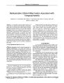
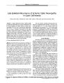
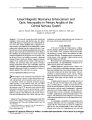
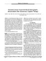
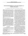
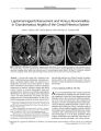
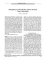
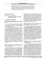
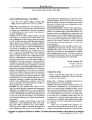
Show STATE OF THE ART Pathogenesis of Nonarteritic Anterior Ischemic Optic Neuropathy Anthony C. Arnold, MD Abstract: Based on histopathology, electron microscopic corrosion cast studies, optic nerve blood flow studies, and clinical data, the pathogenesis of idiopathic nonarteritic ischemic optic neuropathy includes the following features: ( 1) structurally crowded optic discs are predisposed; ( 2) laminar and retrolaminar regions are the most common locations for infarction; ( 3) there is flow impairment in the prelaminar optic disc during the acute phase; ( 4) lack of consistent choroidal flow impairment and the retrolaminar location of infarcts suggest vasculopathy within or distal to the paraoptic branches of the posterior choroidal arteries; ( 5) diabetes is the most consistently identified vas-culopathic risk factor; ( 6) impaired autoregulation of the disc circulation by atherosclerosis, with a possible contribution from serotonin and endothelin- mediated vasospasm, may play a role; and ( 7) progression may be caused by secondary cell death after the initial ischemic insult or compression from cavernous degeneration and mechanical ax-onal distortion. ( JNeuro- Ophthalmol 2003; 23: 157- 163) Nonarteritic anterior ischemic optic neuropathy ( NAION) is presumed to result from circulatory insufficiency within the optic nerve head, but the specific mechanism and location of the vasculopathy remain unproven. In the arteritic form of AION ( AAION), by contrast, histopathologic evidence confirms both inflammatory occlusion of short posterior ciliary arteries ( SPCAs) and infarction within the optic nerve head. Much of the research regarding the etiology of NAION has centered on the SPCA and choroidal circulations and factors that might compromise them. Studies have included anatomic and physiologic ( blood flow) analysis of the optic discs in NAION, along with attempts to link various vascular risk factors to affected subject populations. Several authorities have recently speculated as to the relative contributions of these interrelated Jules Stein Eye Institute, Department of Ophthalmology, University of California, Los Angeles, California. Address correspondence to Anthony C. Arnold, MD, Jules Stein Eye Institute, 100 Stein Plaza, UCLA, Los Angeles, CA 90095- 7005, USA; E- mail: arnolda@ ucla. edu elements ( 1- 5). This review summarizes evidence for the roles of the following factors in the pathogenesis of idiopathic NAION, the form that is not associated with specific precipitating factors such as acute systemic hypotension or anemia: ( 1) Optic disc vasculopathy and infarction: histopathology and electron microscopic corrosion casting studies of optic disc circulation; ( 2) optic disc blood flow impairment: studies of optic nerve head and peripapillary choroidal blood flow, including fluorescein angiography, indocyanine green angiography ( ICG), color Doppler flow ( CDF) studies, and laser Doppler flow ( LDF) studies; ( 3) risk factors for vascular occlusion: prevalence studies of vasculopathic and prothrombotic risk factors; and ( 4) other contributing factors: optic disc structure " crowding," systemic nocturnal hypotension, vasospasm, impaired autoregulation, and secondary neuronal degeneration mechanisms. HISTOPATHOLOGY What is the histopathologic evidence that there is occlusive vasculopathy within the optic disc microcirculation in NAION and that the optic nerve damage is truly ischemic? In AAION, there is extensive histopathologic documentation of infarction in the paralaminar regions of the optic nerve head and inflammation, thrombosis, and occlusion within the SPCAs ( 6- 9). While these findings confirm that SPCA occlusion can and does produce optic disc infarction, the corresponding evidence that this takes place in NAION is lacking. Six cases of nonarteritic optic nerve head infarction have been reported, three of which were atypical cases, including internal carotid occlusion, multiple embolic lesions, and severe acute blood loss, rather than classic idiopathic NAION ( 10- 14). The SPCAs were described only in a case in which emboli within these vessels produced optic disc infarction, and in which the central retinal and pial arteries were also filled with emboli. In other words, this was not a standard case of NAION. No confirmation of lipohyalinosis or other occlusive process within the disc vascular supply has been documented in these or other cases. In a series of 193 eyes collected over 47 years with a histopathologic diagnosis of ischemic optic neuropathy, Knox et al ( 15) documented infarction, but Copyright © Lippincott Williams & Wilkins. Unauthorized reproduction of this article is prohibited. J Neuro- Ophthalmol, Vol. 23, No. 2, 2003 157 JNeuro- Ophthalmol, Vol. 23, No. 2, 2003 Arnold FIG. 1. Photomicrograph of optic nerve with ischemic optic neuropathy and cavernous degeneration. The large cavern is located predominantly in the retrolaminar optic nerve and produces severe leftward displacement of the adjacent intact axons. ( Alcian blue, original magnification X30). Reprinted with permission ( 15). clinical data are largely unreported, and the cases with classic idiopathic NAION are difficult to distinguish. In this series, as in the other cases, the location of vascular compromise is undocumented. Two features are evident from histopathologic studies of both AAION and NAION ( Fig. 1): ( 1) Infarction is primarily located in the retrolaminar region of the optic nerve head, with occasional extension to the laminar and prelaminar layers. This pattern speaks against a primary role for the choroidal circulation in pathogenesis. Although the proportion of the optic disc vasculature supplied by the peripapillary choroid has been controversial, researchers agree that the choroidal contribution is primarily to the prelaminar, not laminar or postlaminar layers. Infarction originating more posteriorly implicates the SPCA branches directly supplying the optic disc. ( 2) Many cases of NAION ( 36% in Knox's series) have shown evidence of cavernous degeneration, with displacement and distortion of adjacent axons ( in some cases dramatically) by an expanded mucopolysaccharide deposit. This finding raises the question of focal secondary compression as a contributing feature in the progressive form of NAION. Unfortunately, correlation of this finding with clinical evidence of progression is lacking. ELECTRON MICROSCOPIC CORROSION CAST STUDIES What is the electron microscopic evidence that there is occlusive vasculopathy within the optic disc microcirculation? Olver et al ( 16) and Onda et al ( 17) have demonstrated in autopsy eyes that the optic disc is supplied by a partial or complete vascular circle ( corresponding to prior descriptions of the circle of Zinn- Haller) derived from the SPCAs, and that this vascular circle may have distinct upper and lower halves consistent with the altitudinal damage of the nerve head commonly seen in NAION ( Fig. 2). These studies also demonstrate the separation of the paraoptic branches of the SPCAs ( which supply the optic nerve head) from the choroidal branches. Unfortunately, no such studies at this level of the circulation in cases of NAION have been performed. OPTIC NERVE HEAD BLOOD FLOW STUDIES What is the evidence that there is decreased blood flow within the optic nerve head in NAION? Fluorescein Angiography Fluorescein angiographic studies in AAION consistently show severely impaired filling of the optic disc and the choroid, in the peripapillary region and diffusely ( 18- 21). In NAION, studies show similarly delayed filling of the prelaminar optic disc ( deeper layers are not well visualized) in the edematous phase, prior to the development of the impaired filling that eventually comes with any form of atrophy ( due to loss of supporting vasculature). This is the most compelling in vivo evidence of optic disc circulatory impairment in NAION ( 18- 20). In studies by Arnold et al ( 22,23), delayed prelaminar optic disc filling (> 5 seconds FIG. 2. Scanning electron photomicrograph of the vasculature of the posterior globe. Superior { long solid arrow) and inferior ( long empty arrow) anastomoses from the medial ( short solid arrow) and lateral ( short empty arrow) short posterior ciliary arteries ( SPCAs) suggest a possible anatomic correlation for the altitudinal pattern of optic nerve damage often seen in NAION. Reprinted with permission ( 16). Copyright © Lippincott Williams & Wilkins. Unauthorized reproduction of this article is prohibited. 158 © 2003 Lippincott Williams & Wilkins NONARTERITIC ANTERIOR ISCHEMIC OPTIC NEUROPATHY JNeuro- Ophthalmol, Vol. 23, No. 2, 2003 Whether the optic disc in NAION lies within a watershed zone between territories supplied by the PC A branches is a controversial issue ( 4,5,22,29,30). Hayreh ( 29,30) has suggested that the " watershed concept" is a major factor in the development of optic nerve ischemia, noting its presence in a substantial number of NAION patients studied with fluorescein angiography and indicating that impaired perfusion pressure within the distribution of a PC A predisposes the optic disc to infarction. In the study of Arnold and Hepler ( 22), however, significantly delayed filling (> 5 seconds) of a vertical watershed zone encompassing at least a quadrant of the optic disc was recorded more often in 43 normal subjects ( 42%) than among 41 ( 27%) patients with NAION ( p = 0.15). Filling of the disc located within these zones, either in normal subjects or in NAION, did not correlate with adjacent choroidal filling, again consistent with a separate source of flow to the optic disc ( the paraoptic branches of the SPCAs). The lack of correlation of disc and choroidal filling would militate against watershed ischemia as a cause of NAION, as disc and parapapillary choroidal flow would be expected to slow together. Moreover, the choriocapillaris, which is the layer visualized on fluorescein angiography and whose absence is interpreted as a watershed zone, does not materially contribute to the laminar region optic disc vascular supply. While the location of the optic disc at the limit of the distribution of a PCA may predispose it to ischemic damage if there is significantly decreased PCA flow, as evidenced by the delayed flow in the PCA and choroid seen in some cases, this is not consistently documented. Fluorescein angiographic findings are more consistent with impaired flow in the direct paraoptic branches to the disc rather than a watershed phenomenon. FIG. 3. A: Fluorescein angiography in NAION. Filling of the optic disc is delayed, but the peripapillary choroid fills normally, suggesting impaired perfusion within the paraoptic branches of the short posterior ciliary arteries ( SPCAs) supplying the optic disc distal to the branching of the choroidal vessels from the SPCAs. B: Fluorescein angiography in nonischemic optic disc edema ( papillitis) shows normal filling of the disc, without filling delay, followed by late hyperfluorescence. A: reprinted with permission ( 22); B: reprinted with permission ( 23). Copyright © Lippincott Williams & Wilkins. Unauthorized reproduction of this article is prohibited. 159 later than choroid and retinal vasculature) was noted in 76% of subjects with acute NAION, compared with no delay in normal controls or in subjects with nonischemic optic disc edema ( Fig. 3). This suggests that the delayed filling is a primary process rather than one secondary to disc edema. In these studies, ( 22,23) the overlying disc surface vasculature, derived from the retinal arterial circulation, showed variable filling patterns: in some cases substantially impaired and in other cases prominently dilated with early filling that precluded evaluation of the filling pattern of the underlying prelaminar layer. It has been postulated that this vascular dilation, most often sectoral, may be analogous to the " luxury perfusion" seen at the junction of perfused and nonperfused regions in cerebral infarctions, perhaps a manifestation of shunting to relatively spared regions of the disc in NAION ( 1,22,24). Arnold and Hepler ( 22) and Siatkowski et al ( 21) have found that segmental parapapillary choroidal filling delay (> 5 seconds) is not a consistent feature in NAION ( it occurred in only 46%> of NAION cases, and similar segmental delays were commonly seen in normal subjects [ 58%]). Furthermore, segments of optic disc and adjacent choroidal filling were poorly correlated. Optic disc filling delay- segmental or complete- was seen adjacent to normally filling choroidal segments, and vice versa ( Fig. 3 A). Similar findings have been reported with indocyanine green ( ICG) studies of the choroidal circulation, which show substantial slowing in AAION but not in NAION ( 25- 28). These data suggest that in NAION, the level of vascular occlusion lies within the distribution of the paraoptic branches of the SPCAs, after their take- off from the choroidal branches. JNeuro- Ophthalmol, Vol. 23, No. 2, 2003 Arnold Color Doppler Flow Studies With current technology, CDF studies have not conclusively localized impaired blood flow in NAION. Fla-harty et al ( 31) measured vascular flow velocities and calculated resistance of the ophthalmic arteries ( OA), central retinal arteries ( CRA), and SPCA in 25 patients with progressive NAION undergoing optic nerve sheath decompression surgery; fellow eyes were used as controls. Preop-eratively, both CRA and SPCA velocities were lower in NAION than in control eyes; postoperatively, an increase in velocity was noted in OA and CRA, and a decrease in calculated resistance was found in the SPCA. While this was interpreted as showing initial flow impairment in NAION, which improved after surgery, the study was widely criticized ( 32). CDF studies of the SPCA parameters have been controversial because: 1. The technique measures flow velocities rather than volume; increase in velocity may actually indicate a decrease in blood flow due to stenosis; flow volume itself cannot be measured accurately because the diameter of the specific vessels measured is not known; 2. Calculations of vascular resistance may be inaccurate if assumptions regarding the autoregulatory characteristics of the vascular bed tested are invalid; 3. Measuring the OA and SPCA flow parameters in the orbit may not represent flow parameters within the paraoptic branches, which supply optic disc; and 4. Measuring the CRA flow parameters is unrelated to the vascular supply of laminar regions of the optic disc. Laser Doppler Flow Studies Laser Doppler flow studies have also not conclusively documented or localized relevant circulatory impairment in NAION. Stationary ( laser Doppler flowmetry, or LDF) or scanning ( Heidelberg Retinal Flowmeter, or HRF) laser imaging methods measure surface blood flow derived from the retinal arterial circulation, rather than flow derived from the SPC As, which supply the deeper layers of the optic disc involved in NAION. Measurements by HRF are limited to 300- to 400- Lim depth. Newer LDF techniques may reach depths of up to 1000 urn, which might include a component of SPCA- derived circulation, but specific contributions from various circulatory components is unproven. In evaluating LDF measurements of optic disc blood flow in rhesus monkey eyes after manipulation of ciliary and retinal circulations, Petrig et al ( 33) found that flow measurements were decreased with occlusion of the CRA, but not the PCAs. These techniques have been used extensively in the study of glaucoma, but they have not been widely applied in NAION. Such measurements are confounded by the disc edema and congestion found in this disorder, in many cases resulting in increased surface vascularity, occasionally profoundly so ( luxury disc perfusion) ( 22,24). However, Leiba et al ( 34) reported that optic nerve head blood flow, as measured by HRF, was diminished in NAION compared with fellow eyes ( n = 14), and in affected and fellow eyes compared with age- matched controls ( n = 7). This study was limited by small numbers of subjects, time delay after onset to study ( up to 6 months), and lack of control for optic disc structure ( cup- disc ratio), which could have a substantial effect on measured surface vasculature, in addition to the issues of the depth of the circulatory bed measured by the technique. Carotid Stenosis: Duplex Studies Carotid stenosis has not been shown to be associated with NAION. Fry et al ( 35) performed carotid Duplex scans in 15 patients with NAION, in 11 patients with transient monocular blindness ( TMB), and in 30 age- matched control subjects. Mean stenosis was not significantly worse in NAION patients ( 19%) than in controls ( 9%), but much worse in those with TMB ( 77%). Two of 15 patients with NAION had stenosis greater than 30%, compared with 5 of 30 controls and 10 of 11 patients with TMB. Muller et al ( 36) did not find hemodynamically significant stenosis in any of 17 subjects with NAION. RISK FACTORS FOR OCCLUSIVE VASCULOPATHY What is the evidence that risk factors for vascular occlusion are associated with NAION? Several cross- sectional case series have estimated the prevalence of systemic diseases that might predispose to vasculopathy in patients with NAION ( 37- 41). Systemic hypertension has been documented in 34 to 47% of patients. However, in those studies that compared these figures to matched population data from the National Health Survey, statistical significance was reached only in the younger age group, those aged between 45 and 64 years. In contrast, Hayreh et al ( 40), found a significantly increased prevalence of vascular risk factors in all age groups. Diabetes has been reported in 10 to 24% of NAION cases, with significantly increased prevalence at all ages in all but one study. Diabetes has also been associated with the development of NAION at a younger age in most series. In these studies, the association of NAION with other cardiovascular events such as stroke and myocardial infarction has been inconsistent. In a case- control study of NAION, Jacobson et al ( 42) addressed these and the additional risk factors of smoking and hypercholesterolemia in 51 patients compared with two separate control groups. While hypertension was found in 57% of patients, it was not found to be significantly more Copyright © Lippincott Williams & Wilkins. Unauthorized reproduction of this article is prohibited. 160 © 2003 Lippincott Williams & Wilkins NONARTERITIC ANTERIOR ISCHEMIC OPTIC NEUROPATHY JNeuro- Ophthalmol, Vol. 23, No. 2, 2003 prevalent than among controls in any age group. Diabetes, found in 34%, was a significant risk factor in all age groups. Neither hypercholesterolemia nor smoking demonstrated significant risk. The 61- patient case control study of Salomon et al ( 43) also confirmed diabetes but not hypertension as a risk factor. Additionally, hypercholesterolemia was found to be a significant risk factor, while smoking was not. In two other case- control studies, ( 44,45) hyperlipid-emia and smoking were both identified as risk factors. A large ( 137 cases) but uncontrolled study by Chung et al ( 46) concluded that smoking was a significant risk factor on the basis that smokers developed NAION at a significantly younger age than nonsmokers. The relation between NAION and elevated plasma homocystine levels remains uncertain. Kawasaki et al ( 47) reported 2/ 17 NAION cases younger than age 50 with elevated homocystine, while Biousse et al ( 48) reported normal values in 14/ 14 NAION cases with a mean age of 43 years. Pianka et al ( 49) reported elevated levels in 45% of 40 NAION patients ( mean age 66 years) but in only 9.8% of control subjects. Weger et al ( 50) also reported significant elevation in 59 NAION patients compared with control subjects. Isolated reports ( 51,52) have documented prothrom-botic risk factors in patients with NAION. But a large study by Salomon et al ( 43) to evaluate lupus anticoagulant, anti-cardiolipin antibodies, prothrombotic polymorphisms, and deficiencies of protein C, S, and antithrombin III in a series of 61patients with NAION and 90 control subjects failed to find any association of these factors with NAION. OTHER FACTORS What is the evidence for other pathogenetic factors in NAION? Mechanical Factors Optic disc structural features play an unknown role in AION. The discs in AAION are normal in diameter and cup size ( 53). Those in NAION are most often small in diameter, with small or absent cups, suggesting to many investigators that " crowding" plays a role in pathogenesis, although exactly how it might do so is unclear ( 54- 5 8). Possibilities include: 1. Crowding produces intracellular axonal swelling secondary to mechanical obstruction to axoplasmic flow, particularly at the most crowded region, the cribriform plate; 2. Subclinical ischemia due to lipohyalinosis and/ or other factors produces additional axoplasmic stasis, with swelling causing compression and further compromise of the microcirculation in the crowded laminar region; 3. Crowding of the disc is associated with an abnormally stiff ( less compliant) cribriform plate region, exaggerating factors 1 and 2; and 4. After acute ischemia, secondary obstruction to axoplasmic flow occurs, with decreased return of neurotrophins, and resultant additional ganglion cell death. Studies of the microstructure andultrastructure of the optic disc in glaucoma suggest that structural aspects of the laminar region may contribute to pathogenesis. Such factors as poor support of axons, or increased shear forces applied to them by the cribriform plate in response to mechanical deformation from elevated intraocular pressure ( due to pore size, collagen content, or abnormal compliance) have been implicated. Similar studies have not been performed in eyes with NAION. Nocturnal Hypotension Hayreh et al ( 59) has proposed that nocturnal systemic hypotension may play a role in the development of NAION, stating that the relative hypotension that normally occurs with sleep may chronically compromise optic disc circulation, particularly in those patients with an exaggerated nocturnal " dip" in blood pressure or in patients, such as those with systemic hypertension, where optic disc circulation autoregulatory mechanisms are impaired ( 59). This effect might be worsened with aggressive antihypertensive therapy, particularly if administered at night, by further exacerbating the nocturnal dip. Hayreh et al ( 59) performed 24- hour ambulatory blood pressure monitoring in 52 cases of NAION compared with 19 cases of primary open angle glaucoma ( POAG) and 65 cases of normal tension glaucoma ( NTG). Mean decrease in systolic and diastolic blood pressure of 25.3% and 31.2%, respectively, were noted in NAION. In general, no significant differences in blood pressure decrease were observed between NAION, NTG, and POAG; however, the 31.2% diastolic decrease in NAION was significantly less ( p = 0.004) than the 36.0% figure for NTG. No control subjects were monitored, but the figures for an age- matched normal population range from 7 to 21%. In patients with NAION and systemic hypertension on medication, nighttime blood pressure figures were significantly lower in those with visual field deterioration. From these data, the authors implied that nocturnal systemic hypotension plays a significant role in the development of NAION in certain susceptible subjects. They subsequently ( 60) reported similar conclusions based on data from 114 NAION, 131 NTG, and 30 POAG subjects, including some patients from the original study ( 59). Landau et al ( 61) performed 24- hour ambulatory blood pressure monitoring in 24 subjects with NAION that was controlled with 24 age- matched, other disease-matched, and medication- matched subjects. Mean decreases of 11% systolic and 18% diastolic were measured Copyright © Lippincott Williams & Wilkins. Unauthorized reproduction of this article is prohibited. 161 JNeuro- Ophthalmol, Vol. 23, No. 2, 2003 Arnold in NAION, compared with 13% and 18% respectively in controls, showing no significant difference. They did, however, see mildly lower mean daytime blood pressures in NAION than in controls, averaging 5 to 7 mm Hg. The largest difference was noted in the morning, indicating a slower morning rise in pressure in NAION subjects when compared with normal control subjects. The contradictory results regarding level of nocturnal " dip" in NAION and whether chronic or intermittent low systemic blood pressure is a factor in etiology remain unresolved. A lively exchange between the authors of the two studies was published in 1997 ( 62,63). Cellular Mechanisms Experimental models of optic nerve ischemia induced by endothelins have been shown to be associated with elevated levels of glutamate in the overlying vitreous ( 68). Levin et al ( 69) have shown evidence of apoptosis in the ganglion cells in a case of NAION. Thus, ischemia-induced cell death may result in release of glutamate, with further cell damage and death by excitotoxic induction of apoptosis. The issue of secondary neuronal damage after ischemic injury by this and other cellular mechanisms, including free radical production and lipid peroxidation, is currently being studied as a potentially treatable component in the progressive process of NAION ( 70). Vasospasm and Impaired Autoregulation Whatever the cause for impaired blood flow in the optic nerve vasculature, persistent hypoperfusion may require impairment in the normal autoregulatory mechanisms of the optic nerve head. Flow is normally maintained constant with variations in perfusion pressure, intraocular pressure, and metabolic conditions ( including tissue oxygen and C02 levels) by factors that vary resistance to flow. Autonomic input to vessels and vasoactive substances ( including vasoconstrictor endothelins and vasodilator nitric oxide) released in response to metabolic influences or mechanical deformation of vascular smooth muscle contribute to the regulation of blood flow in response to these external influences. These autoregulatory mechanisms may be reduced by arteriosclerosis, vasospasm, or antihypertensive medications, including beta- blockers. Hayreh ( 64,65) has postulated that endogenous serotonin, released during platelet aggregation within atherosclerotic plaques, may play a role in the development of ischemic optic nerve damage via its role in vasoconstriction of arterioles and resultant impaired autoregulation, possibly mediated by endothelial- derived vasoactive agents such as endothelins. He has reported serotonin- induced vasoconstriction in CRA and PC As in atherosclerotic monkeys that is reversed by discontinuing an atherogenic diet. The role of endothelins in optic disc ischemia is under intensive study, particularly as endothelin- derived vasospasm may be mediated by intracellular calcium ion influx, which in turn may be counteracted by calcium channel-blocking agents. Intravenous and intravitreal infusions of endothelin- 1 in rabbits have resulted in decreased optic nerve head blood flow. Strenn et al ( 66) showed a decrease in blood flow measured by laser Doppler flowmetry ( LDF) after endothelin- 1 administration that was reversed by administration of the calcium channel blocker nifedipine. Oku et al ( 67) produced chronic optic disc ischemia in rabbits by repeated intravitreal injections of endothelin- 1; capillary blood flow was diminished and a progressive axonal loss and excavation of the nerve heads was noted. REFERENCES 1. Burde RM. Optic disk risk factors for nonarteritic anterior ischemic optic neuropathy. Am J Ophthalmol 1993; 116: 759- 64. 2. Lessell S. Nonarteritic anterior ischemic optic neuropathy: enigma variations. Arch Ophthalmol 1999; 117: 386- 8. 3. Feldon SE. Anterior ischemic optic neuropathy: trouble waiting to happen. Ophthalmology 1999; 106: 651- 2. 4. Hayreh SS. Anterior ischemic optic neuropathy: trouble waiting to happen [ Letter]. Ophthalmology 2000; 107: 407- 9. 5. Feldon SE. Anterior ischemic optic neuropathy: trouble waiting to happen [ Author's reply]. Ophthalmology 2000; 409- 10. 6. Spencer WH, Hoyt WF. A fatal case of giant- cell arteritis ( temporal or cranial arteritis) with ocular involvement. Arch Ophthalmol 1960; 64: 862- 7. 7. MacFaul PA. Ciliary artery involvement in giant cell arteritis. Br J Ophthalmol 1967; 51: 505- 12. 8. Henkind P, Charles NC, Pearson J. Histopathology of ischemic optic neuropathy. Am J Ophthalmol 1970; 69: 78- 90. 9. Mac Michael IM, Cullen JF. Pathology of ischaemic optic neuropathy. In: Cant JS, ed. The Optic Nerve, Proceedings of the Second William MacKenzie Memorial Symposium. London: Henry Kimpton; 1972: 108- 16. 10. KnoxDL, Duke JR. Slowly progressive ischemic optic neuropathy. Trans Am Acad Ophthalmol Otolaryngol 1971; 75: 1065- 8. 11. Cogan DG. Neurology of the Visual System. Springfield, Illinois: Charles C. Thomas; 1966, 186. 12. Lieberman MF, Shahi A, Green WR. Embolic ischemic optic neuropathy. Am J Ophthalmol 1978; 86: 206- 10. 13. Rootman J, Butler D. Ischaemic optic neuropathy- a combined mechanism. Br J Ophthalmol 1980; 64: 826- 31. 14. Johnson MW, Kincaid MC, Trobe JD. Bilateral retrobulbar optic nerve infarctions after blood loss and hypotension. A clinicopatho-logic case study. Ophthalmology 1987; 94: 1577- 84. 15. Knox DL, Kerrison JB, Green WR. Histopathologic studies of ischemic optic neuropathy. Tr Am Ophth Soc 2000; 98: 203- 22. 16. Olver JM, Spalton DJ, McCartney AC. Microvascular study of the retrolaminar optic nerve in man: the possible significance in anterior ischemic optic neuropathy. Eye 1990; 4: 7- 24. 17. Onda E, Cioffi GA, Bacon DR, et al. Microvasculature of the human optic nerve. Am J Ophthalmol 1995; 120: 92- 102. 18. Eagling EM, Sanders MD, Miller SJ: Ischaemic papillopathy. Clinical and fluorescein angiographic review of forty cases. Br J Ophthalmol 1974; 58: 990- 1008. 19. Hayreh SS: Anterior ischaemic optic neuropathy. Differentiation of arteritic from non- arteritic type and its management. Eye 1990; 4: 25- 41. 20. Mack HG, O'Day J, Currie JN: Delayed choroidal perfusion in giant cell arteritis. J Clin Neuro- Ophthalmol 1991; 11: 221- 7. 21. Siatkowski RM, Gass JD, Glaser JS, Smith, JL, Schatz NJ, Schiff-man J: Fluorescein angiography in the diagnosis of giant cell arteritis. Am J Ophthalmol 1993; 115: 57- 63. Copyright © Lippincott Williams & Wilkins. Unauthorized reproduction of this article is prohibited. 162 © 2003 Lippincott Williams & Wilkins NONARTERITIC ANTERIOR ISCHEMIC OPTIC NEUROPATHY JNeuro- Ophthalmol, Vol. 23, No. 2, 2003 22. Arnold AC, Hepler RS: Fluorescein angiography in acute nonarter-itic anterior ischemic optic neuropathy. Am J Ophthalmol 1994; 117: 222- 30. 23. Arnold AC, Badr MA, Hepler RS. Fluorescein angiography in nonischemic optic disc edema. Arch Ophthalmol 1996; 114: 293- 8. 24. Friedland S, Winterkorn JM, Burde RM. Luxury perfusion following anterior ischemic optic neuropathy. J Neuro- Ophthalmol 1996; 16: 163- 71. 25. Meadows M, Slavin M, Beer PM, et al. Indocyanine green angiography of anterior ischemic optic neuropathy and giant cell arteritis. NANOS Annual Meeting, 1998. 26. Sadun F, Pece A, Brancato R. Fluorescein and indocyanine green angiography in arteritic anterior ischaemic optic neuropathy. Br J Ophthalmol 1998; 82: 1344- 5. 27. ValmaggiaC, SpeiserP, Bischoff P, etal. Indocyanine green versus fluorescein angiography in the differential diagnosis of arteritic and nonarteritic anterior ischemic optic neuropathy. Retina 1999; 19: 131- 4. 28. Oto S, Yilmaz G, Cakmakci S, Aydin P. Indocyanine green and fluorescein angiography in nonarteritic anterior ischemic optic neuropathy. Retina 2002; 22: 187- 91. 29. Hayreh SS. The blood supply of the optic nerve head and the evaluation of it- myth and reality. Progr Retin Eye Res 2001; 20: 563- 93. 30. Hayreh SS. Blood flow in the optic nerve head and factors that may influence it. Progr Retin EyeRes 2001; 20: 595- 624. 31. Flaharty PM, Sergott RC, Lieb W, Bosley TM, Savino PJ. Optic nerve sheath decompression may improve blood flow in anterior ischemic optic neuropathy. Ophthalmology 1993; 100: 297- 302. 32. Hayreh SS, Beach KW. [ Discussion of: Flaharty PM, Sergott RC, Lieb W, Bosley TM, Savino PJ. Optic nerve sheath decompression may improve blood flow in anterior ischemic optic neuropathy]. Ophthalmology 1993; 100: 303- 5. 33. Petrig BL, Riva CE, Hayreh SS. Laser doppler flowmetry and optic nerve head blood flow. Am J Ophthalmol 1999; 127: 413- 25. 34. Leiba H, Rachmiel R, Harris A, et al. Optic nerve head blood flow measurements in non- arteritic anterior ischemic optic neuropathy. Eye 2000; 14: 828- 33. 35. Fry CL, Carter JE, Kanter MC, Tegeler CH, Tuley MR. Anterior ischemic optic neuropathy is not associated with carotid artery atherosclerosis. Stroke 1993; 24: 539- 42. 36. Muller M, Kessler C, Wessel K, et al. Low- tension glaucoma: a comparative study with retinal ischemic syndromes and anterior ischemic optic neuropathy. Ophthalmic Surg 1993; 24: 835- 8. 37. Boghen DR, Glaser JS. Ischaemic optic neuropathy. The clinical profile and history. Brain 1975; 98: 689- 708. 38. Repka MX, Savino PJ, Schatz NJ, et al. Clinical profile and long-term implications of anterior ischemic optic neuropathy. Am J Ophthalmol 1983; 96: 478- 83. 39. Guyer DR, Miller NR, Auer CL, et al. The risk of cerebrovascular and cardiovascular disease in patients with anterior ischemic optic neuropathy. Arch Ophthalmol 1985; 103: 1136- 42. 40. Hayreh SS, Joos KM, Podhajsky PA, et al. Systemic diseases associated with nonarteritic anterior ischemic optic neuropathy. Am J Ophthalmol 1994; 118: 766- 80. 41. Ischemic Optic Neuropathy Decompression Trial Study Group. Characteristics of patients with nonarteritic anterior ischemic optic neuropathy eligible for the Ischemic Optic Neuropathy Decompression Trial. Arch Ophthalmol 1996; 114: 1366- 74. 42. Jacobson DM, Vierkant RA, Belongia EA. Nonarteritic anterior ischemic optic neuropathy. A case- control study of potential risk factors. Arch Ophthalmol 1997; 115: 1403- 7. 43. Salomon O, Huna- Baron R, Kurtz S, et al. Analysis of profhrom-botic and vascular risk factors in patients with nonarteritic anterior ischemic optic neuropathy. Ophthalmology 1999; 106: 739^ 12. 44. Giuffre G. Hematological risk factors for anterior ischemic optic neuropathy. Neuro- ophthalmology 1990; 10: 197- 203. 45. Talks SJ, Chong NH, Gibson JM, et al. Fibrinogen, cholesterol, and smoking as risk factors for non- arteritic anterior ischaemic optic neuropathy. Eye 1995; 9: 85- 8. 46. Chung SM, Gay CA, McCrary JA. Nonarteritic ischemic optic neuropathy. The impact of tobacco use. Ophthalmology 1994; 101: 779- 82. 47. Kawasaki A, Purvin VA, Burgett RA. Hyperhomocysteinaemia in young patients with non- arteritic anterior ischaemic optic neuropathy. Br J Ophthalmol 1999; 83: 1287- 90. 48. Biousse V, Kerrison JB, Newman NJ. Is non- arteritic anterior ischaemic optic neuropathy related to homocysteine? Br J Ophthalmol 2000; 84: 555. 49. Pianka P, Almog Y, Man O, et al. Hyperhomocystinemia in patients with nonarteritic anterior ischemic optic neuropathy, central retinal artery occlusion, and central retinal vein occlusion. Ophthalmology 2000; 107: 1588- 92. 50. Weger M, Stanger O, Deutschmann H, et al. Hyperhomocysteinaemia, but not MTHFR C677T mutation, as a risk factor for nonarteritic ischemic optic neuropathy. Br J Ophthalmol 2001; 85: 803- 6. 51. Acheson JF, Sanders MD. Coagulation abnormalities in ischaemic optic neuropathy. Eye 1994; 8: 89- 92 52. Worrall BB, Moazami G, Odel JG, et al. Anterior ischemic optic neuropathy and activated protein C resistance. J Neuro- Oph-thalmology 1997; 17: 162- 5. 53. Jonas JB, Gusek GC, Naumann GOH. Anterior ischemic optic neuropathy: nonarteritic form in small and giant cell arteritis in normal sized optic discs. Int Ophthalmol 1988; 12: 119- 25. 54. Beck RW, Savino PJ, Repka MX, Schatz NJ, Sergott RC. Optic disc structure in anterior ischemic optic neuropathy. Ophthalmology 1984; 91: 1334- 7. 55. Feit RH, Tomsak RL, Ellenberger C Jr.. Structural factors in the pathogenesis of ischemic optic neuropathy. Am J Ophthalmol 1984: 98: 105- 8. 56. Doro S, Lessell S. Cup- disc ratio and ischemic optic neuropathy. Arch Ophthalmol 1985; 103: 1143- 4. 57. Beck RW, Servais GE, Hayreh SS. Anterior ischemic optic neuropathy. IX. Cup- to- disc ratio and its role in pathogenesis. Ophthalmology 1987; 94: 1503- 8. 58. Mansour AM, Shoch D, Logani S. Optic disk size in ischemic optic neuropathy. Am J Ophthalmol 1988; 106: 587- 9. 59. Hayreh SS, Zimmerman MB, Podhajsky P, et al. Nocturnal arterial hypotension and its role in optic nerve head and ocular ischemic disorders. Am J Ophthalmol 1994; 117: 603- 24. 60. Hayreh SS, Podhajsky P, Zimmerman MB. Role of nocturnal arterial hypotension in optic nerve head ischemic disorders. Ophthal-mologica 1999; 213: 76- 96. 61. Landau K, Winterkorn JM, Mailloux LU, et al. 24- hour blood pressure monitoring in patients with anterior ischemic optic neuropathy. Arch Ophthalmol 1996; 114: 570- 5. 62. Hayreh SS, Zimmerman MB, Podhajsky P, et al. Nonarteritic anterior ischemic optic neuropathy: role of nocturnal arterial hypotension ( letter). Arch Ophthalmol 1997; 115: 942- 3. 63. Landau K, Winterkorn JMS, Napolitano B. Nonarteritic anterior ischemic optic neuropathy: role of nocturnal arterial hypotension ( reply to letter). Arch Ophthalmol 1997; 115: 943- 5. 64. Hayreh SS, Piegors DJ, Heisted DD. Serotonin- induced constriction of ocular arteries in atherosclerotic monkeys. Arch Ophthalmol 1997; 115: 220- 8. 65. Hayreh SS. Retinal and optic nerve head ischemic disorders and atherosclerosis: role of serotonin. Progr Retin Eye Res 1999; 18: 191- 221. 66. Strenn K, Manilla B, Wolzt M, et al. Reversal of endothelin- 1- induced ocular hemodynamic effects by low- dose nifedipine in humans. Clin Pharmacol Ther 1998; 63: 54- 63. 67. Oku H, Sugiyama T, Kojima S, et al. Experimental optic cup enlargement caused by endothelin- 1 - induced chronic optic nerve head ischemia. Surv Ophthalmol 1999; 44 ( suppl l): S74- 84. 68. Kim TW, Kang KB, Choung H- K, et al. Elevated glutamate levels in the vitreous body of an in vivo model of optic nerve ischemia. Arch Ophthalmol 2000; 118: 533- 6. 69. Levin LA, Louhab A. Apoptosis of retinal ganglion cells in anterior ischemic optic neuropathy. Arch Ophthalmol 1996; 114: 488- 91. 70. Potarazu SV. Ischemic optic neuropathy: models for mechanism of disease. Clin Neurosci 1997; 4: 264- 9. Copyright © Lippincott Williams & Wilkins. Unauthorized reproduction of this article is prohibited. 163 |