| OCR Text |
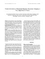
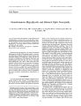
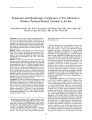
Show Journal of'A] euro- Ophthalmology 21( 1): 8- 11, 2001. © 2001 Lippincott Williams & Wilkins, Inc., Philadelphia Visual Activation in Functional Magnetic Resonance Imaging at Very High Field ( 4 Tesla) Atsushi Miki, MD, PhD, Grant T. Liu, MD, Jonathan Raz, PhD, Sarah A. Englander, PhD, Gabrielle R. Bonhomme, MD, David O. Aleman, BS, Edward J. Modestino, MLA, Chia- Shang J. Liu, BA, and John C. Haselgrove, PhD Objectives: Functional magnetic resonance imaging ( fMRI) at very high field strengths provides functional brain mapping with the enhanced signal to noise ratio and the larger blood oxygenation level- dependent ( BOLD) effect. We report activated areas in the standard space detected by fMRI at 4 Tesla ( T) during simple visual stimulation. Materials and Methods: Twelve healthy young subjects were scanned using a 4 T scanner during binocular flashing visual stimulation. Functional images were realigned to the first scan and then spatially normalized. Individual and group data analyses were performed to identify areas of visual activation. Results: Activation of the bilateral primary visual cortex ( V1/ V2) was observed along the entire calcarine fissure in all subjects. The activated area extended to the extrastriate cortex in all subjects. Activation of the bilateral lateral geniculate nucleus ( LGN) was detected in all subjects. The group data showed activation of the bilateral primary visual cortex and the bilateral lateral geniculate nucleus. Conclusions: Robust activation of the vision- related areas was successfully obtained in all subjects using a 4 T magnetic resonance scanner. These results suggest that fMRI at very high field strengths may be effective in showing visual system physiology, and that it can be a promising method to assess visual function of human subjects. Key Words: Functional magnetic resonance imaging- Visual activation- 4 Tesla- Primary visual cortex- Lateral geniculate nucleus. Manuscript received April 7, 2000; accepted December 5, 2000. Supported in part by Prevent Blindness America, Postdoctoral Research Fellowship, PD98017 ( AM), and Uehara Memorial Foundation ( AM). From the Division of Neuro- ophthalmology, Departments of Neurology and Ophthalmology ( AM, GTL, EJM), and Department of Radiology ( SAE), Hospital of the University of Pennsylvania; Division of Biostatistics, University of Pennsylvania ( JR); University of Pennsylvania School of Medicine ( GRB, CSJL), Philadelphia, Pennsylvania; Department of Pediatrics ( DOA), and Department of Radiology ( CSJL, JCH), the Children's Hospital of Philadelphia, Philadelphia, Pennsylvania. Address correspondence and reprint requests to Atsushi Miki, MD, PhD, Division of Neuro- ophthalmology, Department of Neurology, Hospital of the University of Pennsylvania, 3400 Spruce Street, Philadelphia, PA 19104, USA; e- mail: amiki@ mail. med. upenn. edu. Functional magnetic resonance imaging ( fMRI) of the brain with very high field strengths (> 3.0 Tesla [ T]) has been performed during visual, motor, and cognitive tasks ( 1- 11). The signal to noise ratio is greater at very high field strengths than at field strengths of most conventional scanners ( e. g., 1.5 T scanners) ( 12), and the contribution of the capillaries to the signal change relative to that of large vessels can be increased by the use of magnetic resonance ( MR) scanners with higher field strengths ( 13,14). Although signal changes in conventional MR scanners are relatively small ( usually 1- 3% for 1.5 T), previous studies at very high field strengths have shown greater signal changes of brain regions ( 1 - 3,12). Signal intensity changes can be more than 20% in the visual cortex ( 3). However, to our knowledge, activation pattern of fMRI at very high field during simple visual stimulation has not been described after image transformation to the standard brain, and variability in activation among subjects has not been well established. The aim of this study was to describe the vision-related areas detected in 12 subjects by fMRI at 4 T. We used spatial normalization of the functional images to the standard space to report the location of activated areas and to perform group data analysis. METHODS Subjects and data acquisition Twelve healthy volunteers ( 8 men and 4 women), ranging in age from 23 to 32 years, gave informed consent and participated in this study. Approval of the consent and protocol for this study was given by the Committee on Studies Involving Human Beings of the University of Pennsylvania. No subjects had a history of neurologic or ophthalmologic disease. All studies were performed with a 4 T Signa scanner ( General Electric Medical Systems, Milwaukee, WI) with a quadrature head coil. Three- dimensional axial images were acquired covering the whole brain for anatomic images ( 28 slices; slice thickness = 5 mm). Subsequently, 8 VISUAL ACTIVATION IN FUNCTIONAL MRI 9 we selected a volume that covered the occipital lobe for functional image acquisition. Functional images were obtained using a gradient- echo echo- planar image ( EPI) sequence ( time to repeat [ TR] = 2000 ms; echo time [ TE] = 28 ms; matrix size = 40 x 64; field of view = 150 x 240 mm2; 21 slices; slice thickness = 5 mm) after data for distortion correction were collected. The first 20 seconds of EPI data ( ten scans) were discarded to remove the magnetic saturation effects. Binocular full- field visual stimulation was provided by the light- emitting diode goggles ( S10VSB; Grass Instruments, Quincy, MA). Subjects were wearing flashing goggles and were instructed to keep their eyes open. During the " rest" condition, the goggles were turned off for 20 seconds ( ten scans). During the " task" condition, the goggles flashed at a rate of 8 Hz for 20 seconds ( ten scans). The task condition alternated with the rest condition; the cycle was repeated six times, resulting in the acquisition of 120 scans during 4 minutes. Data analysis Data analysis was performed on UNIX workstations. IDL ( Interactive Data Language, Boulder, CO) and SPM96 ( Wellcome Department of Cognitive Neurology, London, UK) were used for data analysis. The first ten scans were discarded before transferring the data from the scanner to the workstations. Distortion correction files were used to correct geometric distortion in EPI images caused by residual magnetic field inhomogeneity, and this information was applied for the two EPI data sets of each subject. The anterior commissure ( AC) was determined, and the origin for the EPI images was set on the superior edge of the AC. The EPI scans were realigned to the first scan. The realigned EPI images were spatially normalized to the standard space described by the Talairach and Tournoux atlas (" Talairach space") ( 15) using the parameter derived from the 8- parameter affine transformation between the anatomic images and Tl template. The data were smoothed using an 8 x 8 x 10 mm full width at half maximum Gaussian kernel. A boxcar delayed by 6 seconds was used as a reference function to account for the delay in hemodynamic response. The t values for each subject were calculated for each voxel and converted to Z scores. Peak Z values within the visual cortex and lateral geniculate nucleus ( LGN) of the thalamus were recorded. In addition, the mean Z value within the anatomically defined primary visual cortex was measured in each subject. The region of interest for the primary visual cortex was determined on the SPM Tl template. The group SPMs were then constructed using the random effects kit ( ftp:// ftp. fil. ion. ucl. ac. uk/ spm/ spm96_ RFX. tar. gz). Each subject's EPI scans were collapsed into one image per condition ( i. e., two images for each subject). Finally, the SPM was created from twelve subjects' images using the positron- emission tomography ( PET) statistics routine. The activation maps were overlaid on the corresponding Tl- weighted images of each subject or the Tl template of SPM96. RESULTS Single subject analysis Robust activation was observed in the bilateral visual cortex in all subjects ( Fig. 1 and Table 1). The area of activation with the highest Z scores extended along the entire calcarine fissure. The mean Z score within the anatomically defined primary visual cortex was 6.66. Although women ( mean Z = 6.95, n = 4) had slightly higher Z values than men ( mean Z = 6.54, n = 8), there was no statistically significant difference between sexes in the magnitude of primary visual cortex activation ( standard normal test, p = 0.216, a two- sided test). In most subjects, distinct areas with high Z values comparable to the primary visual cortex were present more laterally and bilaterally ( Fig. 1). The Talairach coordinates of these are consistent with those of MT/ V5 ( 16). sagittal coronal ROIL J FIG. 1. Visual activation of fMRI at 4 T of two subjects ( A and B). Suprathreshold ( p < 0.001) areas are displayed on the transverse anatomic images of each subject. Robust activation of occipital cortex, including area MT/ V5, can be seen ( right: Talairach coordinates 52,- 56,0, Z = 7.45, left: Talairach coordinates - 41,- 64,- 5, Z = 7.46, for subject A). In addition, bilateral LGN activation is clearly visible. J Neuro- Ophthalmol, Vol. 21, No. 1, 2001 10 A. MIKIETAL. TABLE 1. Peak activation of visual cortex in Talairach coordinates Left hemisphere location - 11,- 64,- 5 - 4,- 90,15 - 8,- 86,40 - 8,- 86,10 - 8,- 71,- 10 - 8,- 86,20 - 11,- 75,- 15 - 15,- 79,- 5 - 8,- 82,0 - 8,- 90,- 10 - 8,- 79,5 - 4,- 90,25 - 4,- 90,25 Z score 7.95 7.61 7.27 7.55 7.66 7.47 7.55 6.88 7.70 8.25 7.99 7.63 6.80 Right hemisphere location 8,- 68,0 26,- 79,5 15,- 94,30 15,- 79,15 19,- 90,10 8,- 75,20 8,- 86,0 11,- 98,0 22,- 98,0 8,- 86,- 10 4,- 86,20 19,- 86,25 26,- 75,30 Z score 7.76 7.89 7.18 7.43 8.45 7.62 7.88 8.12 7.90 8.49 8.09 8.14 5.37 * The peak activation of group data was determined on the activation map constructed from 12 subjects' data using random- effect analysis. All subjects showed activation in the bilateral LGN ( 17,18) ( Fig. 1). Talairach coordinates of the maxima in the LGN activation are shown in Table 2. Group analysis The group analysis showed activation of occipital areas spanning along the calcarine fissure, and the maximal activation was found in this cluster ( Fig. 2). The area extended to the parieto- occipital sulcus, where the most peripheral visual field is represented ( 19). The activated area possibly contained the area V2 as well as VI. The bilateral LGN activation was distinctly observed in the group data analysis ( corrected/? < 0.05), suggesting consistency in the location of the small area of LGN activation among subjects ( Fig. 2). DISCUSSION In this study, we performed fMRI at very high field and postprocessed the functional images using spatial TABLE 2. Peak activation of lateral geniculate nucleus in Talairach coordinates Left LGN location - 26,- 19,- 5 - 22,- 26,5 - 26,- 26,- 5 - 26,- 38,5 - 19,- 34,5 - 26,- 30,- 5 - 30,- 26,- 5 - 26,- 22,0 - 22,- 26,0 - 22,- 26,- 5 - 22,- 30,0 - 22,- 30,0 - 22,- 30,0 Z score 6.94 3.99 4.19 3.94 4.34 5.47 4.78 6.43 4.23 5.57 7.01 3.80 4.50 Right LGN location 22,- 22,- 5 26,- 19,0 26,- 22,- 5 22,- 22,5 22,- 34,5 22,- 30,5 22,- 30,0 22,- 26,0 22,- 30,0 30,- 22,- 5 26,- 26,0 22,- 22,- 5 22,- 26,0 Z score 6.74 4.31 4.96 3.41 4.41 5.05 6.48 5.67 3.99 5.46 6.56 5.14 4.53 * The peak activation of group data was determined on the activation map constructed from 12 subjects' data using random- effect analysis. LGN, lateral geniculate nucleus. ja^ v. a coronal FIG. 2. Activation of visual cortex and LGN in the group analysis ( n = 12). Suprathreshold ( p < 0.01) areas are displayed on the T1 template of SPM96. normalization, which transforms a single subject's images into a standard space. Spatial normalization enables us to perform a group data analysis, which is more appropriate in making inferences at the population level than an individual data analysis ( 20). The activated areas along the calcarine fissure were identified in individual and group data analyses, suggesting that this technique is capable of demonstrating activity of the primary visual cortex accurately. Also, the activated area included ex-trastriate cortex, although the extent of the area is variable among subjects. Such variability may result from the difference in subjects' attention levels ( 21). The activation of MT/ V5 in most subjects can be explained by the fact that this area is well known to respond to flickering stimuli ( 22,23). In addition to the visual cortex activation, bilateral activation of LGN was reliably obtained in all of the volunteers. The sensitivity of this measure was proven in the group data analysis as well as the standard individual data analysis. Chen and associates ( 8,9) used a similar visual stimulus and detected activation of the pulvinar nucleus of the thalamus as well as LGN activation. However, we did not find reliable activation of the pulvinar nucleus in this study. The discrepancy between their findings and ours may be explained by the difference in the statistical procedures, especially in the filter size in the spatial smoothing. Because the pulvinar nucleus is spatially close to LGN, it remains to be studied whether fMRI with higher spatial resolution reveals activation of the pulvinar nucleus using otherwise the same procedure as ours. A robust response is the most important factor in clinical applications of fMRI and enhances the success rates of fMRI examinations ( 24). In this context, fMRI at very / Neuro- Ophthalmol, Vol. 21, No. 1, 2001 VISUAL ACTIVATION IN FUNCTIONAL MRI 11 high field is a powerful tool for mapping cortical function. Further studies may include the use of more specific visual stimuli ( e. g., motion, color, or form) to identify higher visual cortex. REFERENCES 1. Ogawa S, Tank DW, Menon R, et al. Intrinsic signal changes accompanying sensory stimulation: functional brain mapping with magnetic resonance imaging. Proc Natl Acad Sci USA 1992; 89: 5951- 5. 2. Kim S- G, Ashe J, Georgopoulos AP, et al. Functional imaging of human motor cortex at high magnetic field. J Neurophysiol 1993; 69: 297- 302. 3. Turner R, Jezzard P, Wen H, et al. Functional mapping of the human visual cortex at 4 and 1.5 Tesla using deoxygenation contrast EPI. Magn Reson Med 1993; 29: 277- 9. 4. Ugurbil K, Garwood M, Ellermann J, et al. Imaging at high magnetic fields: initial experiences at 4 T. Magn Reson Q 1993; 9: 259- 77. 5. Ellerman JM, Flament D, Kim S- G, et al. Spatial patterns of functional activation of the cerebellum investigated using high field ( 4 T) MRI. NMR Biomed 1994; 7: 63- 8. 6. Thulborn KR, Chang SY, Shen GX, et al. High resolution echo-planar fMRI of human visual cortex at 3.0 Tesla. NMR Biomed 1997; 10: 183- 90. 7. Menon RS, Gati JS, Goodyear BG, et al. Spatial and temporal resolution of functional magnetic resonance imaging. Biochem Cell Biol 1998; 76: 560- 71. 8. Chen W, Kato T, Zhu X- H, et al. Mapping of LGN activation during visual stimulation in human brain using fMRI. Magn Reson Med 1998; 39: 89- 96. 9. Chen W, Kato T, Zhu X- H, et al. Human primary visual cortex and lateral geniculate nucleus activation during visual imagery. Neu-roreport 1998; 9: 3669- 74. 10. Thulborn KR. Clinical rationale for very- high- field ( 3.0 tesla) functional magnetic resonance imaging. Top Magn Reson Imaging 1999; 10: 37- 50. 11. Ugurbil K, Hu X, Chen W, et al. Functional mapping in the human brain using high magnetic fields. Philos Trans R Soc Land B Biol Sci 1999; 354: 1195- 213. 12. Gati JS, Menon RS, Ugurbil K, et al. Experimental determination of the BOLD field strength dependence in vessels and tissue. Magn Reson Med 1997; 38: 296- 302. 13. Ogawa S, Menon RS, Tank DW, et al. Functional brain mapping by blood oxygenation level- dependent contrast magnetic resonance imaging. Biophys J 1993; 64: 803- 12. 14. Menon RS, Ogawa S, Hu X, et al. BOLD based fMRI at 4 Tesla includes a capillary bed contribution: echo- planar imaging correlates with previous optical imaging using intrinsic signals. Magn Reson Med 1995; 33: 453- 9. 15. Talairach J, Tournoux P. Co- planar Stereotaxic Atlas of the Human Brain. Stuttgart: Thieme, 1988. 16. Buchel C, Friston KJ. Modulation of connectivity in visual pathways by attention: cortical interactions evaluated with structural equation modelling and fMRI. Cereb Cortex 1997; 7: 768- 78. 17. Horton JC, Landau K, Maeder P, et al. Magnetic resonance imaging of the human lateral geniculate body. Arch Neurol 1990; 47: 1201- 6. 18. Buchel C, Turner R, Friston K. Lateral geniculate activations can be detected using intersubject averaging and fMRI. Magn Reson Med 1997; 38: 691- 4. 19. Horton JC, Hoyt WF. The representation of the visual field in human striate cortex: a revision of the classic Holmes map. Arch Ophthalmol 1991; 109: 816- 24. 20. Friston KJ, Holmes AP, Worsley KJ. How many subjects constitute a study? Neuroimage 1999; 10: 1- 5. 21. Buchel C, Josephs O, Rees G, et al. The functional anatomy of attention to visual motion. A functional MRI study. Brain 1998; 121: 1281- 94. 22. Tootell RBH, Reppas JB, Kwong KK, et al. Functional analysis of human MT and related visual cortical areas using magnetic resonance imaging. J Neurosci 1995; 15: 3215- 30. 23. Sunaert S, Van Hecke P, Marchal G, et al. Motion- responsive regions of the human brain. Exp Brain Res 1999; 127: 355- 70. 24. Thulborn KR. Why neuroradiologists should consider very- high-field magnets for clinical applications of functional magnetic resonance imaging. Top Magn Reson Imaging 1999; 10: 1- 2. J Neuro- Ophthalmol, Vol. 21, No. 1, 2001 |