| OCR Text |
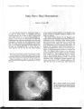
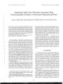
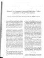
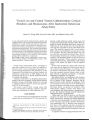
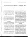
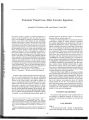
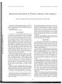
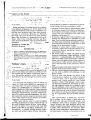
Show Journal of Neuro- Ophthalmology 20( 1): 59- 60, 2000. © 2000 Lippincott Williams & Wilkins, Inc., Philadelphia Cysticercosis of the Optic Nerve Vimala Menon, MS, Radhika Tandon, MD, Sangeeta Khanna, MD, Pradeep Sharma, MD, Sudarshan Khokhar, MD, Sushma Vashisht, MD, and Indu Garg, MD Cysticercosis of the optic nerve has been reported only twice in the literature. A case of optic nerve cysticercosis in a 50- year-old woman with atypical optic neuritis is reported. Computerized tomography showed a thickened left optic nerve with a ring- enhancing lesion containing an eccentric nodule. An enzyme- linked immunosorbent assay test for cysticercosis further established the diagnosis. The patient was treated with oral prednisolone and albendazole, with no improvement in vision. Key Words: Albendazole- Computed tomography- Cysticercosis- Optic nerve. Cysticercosis is the most common parasite affecting the human nervous system ( 1). It is also the most common ocular platyhelminthic infection ( 2). Despite a high incidence of brain, orbital, and intraocular involvement, cysticercosis in the optic nerve is uncommon and previously has been reported only twice ( 3- 5). We report a case of optic nerve cysticercosis that presented as atypical optic neuritis, with a lesion demonstrated on high-resolution computerized tomography. CASE REPORT A 50- year- old woman experienced decreasing vision OS over 4 months, associated with left- sided headache and pain on movement OS. She had already been treated with a course of oral prednisolone 40 mg/ day for 4 days, followed by a taper over 2 weeks, without any improvement. Visual acuity was 6/ 6 OD, and there was no light perception OS. The visual field OD was normal. A left afferent pupillary defect was present. Hertel exophthal-mometry measured 1- mm proptosis OS. The anterior segment was normal OU. Fundus examination OS revealed disc edema with pallor and peripapillary exudates and hemorrhages ( Fig. 1). Enlargement of the left retrobulbar optic nerve was detected on B- scan ultrasonography with an 8- MHz probe, but no focal lesion was detected. Computerized Manuscript received January 14, 1999; accepted December 1, 1999. From the R. P. Centre for Ophthalmic Sciences, New Delhi, India. Address correspondence and reprint requests to Dr. Menon, R. P. Centre for Ophthalmic Sciences, A. I. I. M. S., Ansari Nagar, New Delhi, 110029 India. tomography with contrast of the head and orbit had been obtained earlier by a local practitioner. When reviewed, it showed enlargement of the anterior part of the left optic nerve with a ring- enhancing lesion. Gadolinium-enhanced magnetic resonance imaging of the orbits suggested the presence in the left optic nerve of an abscess cavity with an enhancing rim ( Fig. 2). Based on these imaging findings, a possibility of inflammatory granuloma within the left optic nerve was considered. High- resolution computed tomography ( 1- mm sections) demonstrated an eccentric nodule within the ring-enhancing lesion ( Fig. 3), confirming the diagnosis of cysticercosis. There was no evidence of cysticercosis infection elsewhere in the body. Results of an enzyme-linked immunosorbent assay for cysticercosis were positive. The patient was treated with oral prednisolone 1 mg/ kg/ day for 1 week, followed by albendozole 15 mg/ kg/ day, but she did not show any improvement in her vision. DISCUSSION The purpose of this report is to document a case of optic nerve cysticercosis that presented as atypical optic neuritis and to describe the role of imaging studies in its diagnosis. Cysticercosis is a serious public health problem in the developing countries. The central nervous system, skeletal muscles, and the eye are common sites of larval invasion, probably because of their high glycogen content ( 5). Cysticercosis of the optic nerve is rare, and only two previous reports have been published ( 3,4). Both of those cases were initially believed to be optic nerve tumors, and they were diagnosed retrospectively on histopathology. The computed tomography images of our patient were similar to those in the histopathologi-cally proven case reported previously ( 3). The optic nerve appeared thickened and contained an area of low attenuation. In our patient, further imaging by high-resolution computed tomography with 1- mm sections revealed a tiny area of increased attenuation within the lesion, pathognomonic of the scolex. Our patient presented to us quite late in her course, when she already had no light perception and optic atrophy OS. She was treated initially with steroids because of lack of conclusive evidence of the etiology, and only later after the diagnosis of cysticercosis was she given oral albendazole. Albendazole is currently the preferred 59 60 V. MENON ETAL. FIG. 1. Fundus ( OS) showing disc edema with pallor and peripapillary exudates and hemorrhages. therapy for neurocysticercosis ( 6,7) and has been reported to produce cyst regression in neurocysticercosis, as well in orbital cysticercosis ( 6- 9). However, our patient did not recover vision. Because she was reluctant to FIG. 2. T2- weighted, gadolinium- enhanced, axial magnetic resonance imaging of the left orbit shows a linear hypointense structure adjacent to an oval hypenntense lesion in the anterior part of the optic nerve, suggesting an abscess within the optic nerve. FIG. 3. High- resolution computed tomography with 1- mm sections demonstrates an eccentric nodule within the intraneural ring- enhancing lesion. undergo surgery and was already blind in that eye with an atrophic disc, we did not feel surgical intervention was clinically justified. Whether earlier medical therapy of optic nerve cysticercosis could be successful in restoring optic nerve function remains to be evaluated. Acknowledgment: The editor thanks Dr. J. Winlerkohn for editing this paper for publication. REFERENCES 1. Barry M, Kaldiian LC. Neurocysticercosis. Semin Neurol 1993; 13: 131- 43. 2. Duke- Elder S, Perkin ES. Cysticercosis. In: Duke- Elder S, ed. System of Ophthalmology. London: Henry Kimpton, 1966;: 478- 88. 3. Madan VS, Dhamija RM, Gill HS, et al. Optic nerve cysticercosis: a case report. J Neurol Neurosurg Psychiatry 1991; 54: 470- 1. 4. Bousquet CF, Dufour TF, Derome PC. Retrobulbar optic nerve cysticercosis. J Neurosurg 1996; 84: 293- 6. 5. Diseases caused by helminths. In: Miller NR, ed. Walsh and Hoyt's clinical neurophthalmology. Philadelphia: William and Wilkins, 1995: 3318^ 3. 6. Escobedo F, Penagos P, Rodriguez S, Sotelo J. Albendazole therapy for neurocysticercosis. Arch Intern Med ) 987; 147: 738^ 41. 7. Takayanagui OM, Jardim E. Therapy for neurocysticercosis: comparison between albendazole and praziquantel. Arch Neurol 1992; 49: 290- 4. 8. Menon V, Kumar G, Prakash P. Cysticercosis of extraocular muscle. J Pediatr Ophthalmol Strabismus 1994; 31: 126- 8. 9. Sihota R, Honavar SG. Oral albendazole in the management of extraocular cysticercosis. Br J Ophthalmol 1994; 78: 621- 3. J Neuro- Ophthalmol, Vol. 20, No. I, 2000 |