| OCR Text |
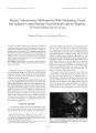
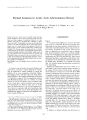
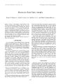
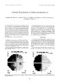
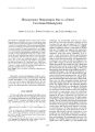
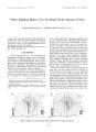
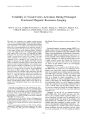
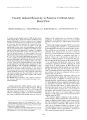
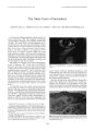
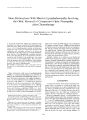
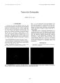
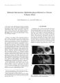
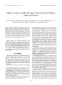
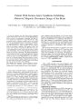
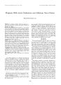
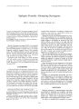
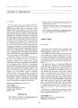
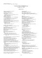
Show Journal of Neum- Ophthalmology 18( 4): 246- 249, 1998. © 1998 Lippincott Williams & Wilkins, Philadelphia Unusual Presentations of Sellar Arachnoid Cyst Benjamin B. Chun, M. D., Andrew G. Lee, M. D., William F. Coughlin, M. D., David T. Floyd, M. D., and Eugene F. May, M. D. This report describes two unusual cases of parasellar arachnoid cyst with different neuro- ophthalmologic manifestations and clinical courses: a 33- year- old woman with parasellar arachnoid cyst, manifested by incongruous homonymous hemiano-pia, and a 64- ycar- old man with a presumed parasellar arachnoid cyst and bitemporal hemianopia that subsequently decompressed spontaneously. Parasellar arachnoid cyst is uncommon, and the clinical course has been incompletely described in the literature. Optimal treatment of patients with these cysts necessitates better understanding of their signs, symptoms, and clinical course. Key Words: Sellar- Arachnoid- Suprasellar. Manuscript received February 10, 1998; accepted April 2, 1998. From the Departments of Ophthalmology ( B. B. C., E. F. M.), Radiology ( W. F. C.), and Neurosurgery ( D. T. F.), Madigan Army Medical Center, Taconia, Washington; and the Departments of Ophthalmology, Neurology, and Neurosurgery, Baylor College of Medicine, and the Division of Neurosurgery, M. D. Anderson Cancer Center, University of Texas ( A. G. L.), Houston, Texas, U. S. A. Address correspondence and reprint requests to Benjamin Chun, M. D., Ophthalmology Department, Madigan Army Medical Center, Tacoma, WA 98431, U. S. A. Parasellar arachnoid cyst is rare, and its origin is not very well understood. It may also be confused with other pituitary cyst. This suprasellar cyst is infrequently encountered in the literature ( 1,2), and thus the clinical course has not been well defined. The patient's typical initial symptoms are a bitemporal hemianopic visual field defect and pituitary abnormalities ( 3- 5). We present two unusual cases of parasellar arachnoid cyst with neuro- ophthalmologic manifestations. CASE ONE A 33- year- old woman had painless, progressive loss of vision and diplopia in 1987. Neuroimaging demonstrated a suprasellar arachnoid cyst. The cyst was decompressed, and the patient's vision improved. No other information is available from the surgical or postoperative period. Between 1989 and 1995, her vision was subjectively stable. In 1996, she experienced pain when moving her eyes, diplopia, and worsening vision. Best corrected visual 23 DEC 96 GOT: OUTSIDE NORHflL LIMITS FL 3/ 20 15.73 DB GOT: OUTSIDE NORMAL LIMITS • • • • • • • • • • • • • • • • • • • • • FL 8/ 16 If) • 13.33 D6 FIG. 1. Humphrey visual field test ( 30- 2) in December 1996 demonstrates an incongruous, left homonymous hemianopic field defect that is denser superiorly. 246 SELLAR ARACHNOID CYST 247 FIG. 2. Coronal T1 - weighted ( repetition, 417 msec; echo time 15 msec; excitations, 1) magnetic resonance image shows upward displacement of posterior chiasm and optic tracts by cerebrospinal fluid isointense mass ( arrow). acuity was 20/ 20 in the right eye ( RE) and 20/ 60 in the left eye ( LE). She had a relative afferent pupillary defect LE. Visual field testing by Humphrey automated perimetry ( 30- 2) demonstrated an incongruous, left homonymous hemianopic field defect that was denser superiorly ( Fig. I). Ophthalmoscopy showed diffuse optic atrophy in both eyes. Magnetic resonance ( MR) imaging of the sella demonstrated a suprasellar arachnoid cyst compressing the optic chiasm and right optic tract ( Fig. 2). The patient underwent frontal craniotomy with fenestration of the cyst and placement of an Ommaya shunt in the cyst. After surgery, visual acuity and visual field testing showed improvement in both eyes, compared with the visual fields taken before surgery. CASE TWO A 64- year- old man underwent screening evaluation for glaucoma because of a positive family history. The patient reported having headaches but had no visual or other neurologic symptoms. Visual acuity was 20/ 25 in both eyes. Intraocular pressures were 30 mmHg OD and 24 mmHg OS. There was a right relative afferent pupillary defect. Visual fields tested by Humphrey automated perimetry ( 30- 2) revealed a bitemporal hemianopic visual field defect ( Fig. 3). Ophthalmoscopy showed a normal optic nerve in each eye. Magnetic resonance scan of the head demonstrated a cyst within the sella turcica that extended superiorly, with upward displacement of the optic chiasm consistent with arachnoid cyst. ( Figs. 4 and 5). A transsphenoidal decompression of the cyst was recommended. Before surgery, another visual field test by Humphrey perimetry demonstrated resolution of the visual field defect ( Fig. 6). Repeat MR scan of the head showed that the suprasellar component of the cyst had spontaneously decompressed, with resolution of the mass effect on the optic chiasm ( Figs. 7 and 8). DISCUSSION Suprasellar arachnoid cyst may cause visual dysfunction usually manifested as a bitemporal visual field defect. The homonymous hemianopia observed in case l is Fixation Losses 0/ 17 DATE 03- 13- 96 Fixation Losses 1/ 18 DATE 03- 13- 96 . « *$ MD - 1.42 DB MD - 2.66DB FIG. 3. Humphrey visual field test ( 30- 2) in March 1996 demonstrates a bitemporal superior hemianopia. ./ Ni'uw- Ophtlwlmol, Vol. 18, No. 4. 1998 248 B. B. CHUN ET AL. J: J* ,0- A^ ll- 1SS 5 llBBlf • » r l S* hnr W hft^ H^ . Em > A us: as LC * rt , -- fe^ i tOfJHti:-; HI. i J 1 V ^ H* dHF « i r i jAOHElOW 1HEAC H- + : T H • at o FOV IB 0* 1 2 - 6 » H » FIG. 4. Contrast- enhanced T1- weighted ( repetition time, 425 msec; echo time, 15 msec; excitations, 1) sagittal magnetic resonance image shows a cerebrospinal fluid isointense suprasellar cyst ( arrow) with both intrasellar and suprasellar components. The infundibulum is displaced anterosuperiorly. exceptional and is probably explained by anatomic variation in the relation between the dorsum sellae and the chiasm, which may result in a variety of visual deficits ( 6). If the chiasm is more anterior to the sella, a suprasellar cyst may encroach on the posterior aspect of the chiasm or optic tract and thus may result in a posterior chiasmal macular involving a visual field defect, such as a contralateral homonymous hemianopia. We suspect i ; * B a Atfh- lSSf H1WB l i « vmt o r I . ID * t£* k 1 it i ^ RTBT sactf » Lkd. lg- m Army K* rt> r> HIU3HI1TUU H- 3F + u ti i v"' kjTi ^ WSL Hi IMfcCI U* 2 3 . r A iff * 1* 1 Fa • ai FIG. 5. Contrast- enhanced T1- weighted coronal magnetic resonance image shows the cystic mass displacing the optic chiasm ( arrow) in a slightly cephalad direction. that this was the cause of our first patient's homonymous hemianopia. The differential diagnosis of a suprasellar cyst includes cystic craniopharyngioma, cystic pituitary adenoma, epidermoid tumor, and epithelial or Rathke's cleft cyst. Although histologic confirmation was made from pathologic examination in our first patient, the diagnosis in our second patient was based on MR imaging characteristics. Unless histologic examination is made, diagnosis cannot be certain. However, the differential diagnosis Fixation Losses 0/ 14 DATE 05- 15- 96 3T ., j| ifl ;;;;;;; i;; iffc ) | • | '*.^ AMUJJ v 1 t I Fixation Losses 1/ 15 DATE 05- 15- 96 **?::; MD + 0.74DB MD - 2.31 DB FIG. 6. Humphrey visual field test ( 30- 2) in May 1996 demonstrates resolution of the previous bitemporal hemianopia. J Neuro- Ophlhulmol, Vol. IS, No. 4. 199$ SELLAR ARACHNOID CYST 249 FIG. 7. T1- weighted sagittal magnetic resonance image shows decrease in size of suprasellar component of cystic mass. Intra-sellar appearance is unchanged ( arrow). can be reasonably narrowed, using the specific MR characteristics of each cyst ( 7,8). Although other suprasellar cysts, such as Rathke's cleft cyst and craniopharyngioma, may manifest characteristic signals or even calcification on MR imaging, our patient demonstrated cerebrospinal isointense fluid within the cyst on all imaging sequences. This appearance is consistent with arachnoid cyst ( 7). W\^ ihJtl*^ Bl^ 2* to .3 J 30 : M* CX 91 IT 1 . 31 n iti. fe [ fl 49*. » HE LS. ft/ l Cfc H i ll V^ 1 • * * dt • ^ ^ ' i 11 CC raBneVIS* H- flP VJUi 4 : T * M W H f Wl • E . .^ HL H; IS " S C: 4 FIG. 8. T1- weighted coronal image shows resolution of the mass-effect against optic chiasm ( arrow). To the best of our knowledge, case 2 is the first suprasellar arachnoid cyst reported to undergo spontaneous asymptomatic decompression. Steinberg et al. ( 9) reported a case of a 47- year- old man with a Rathke's cleft cyst, which repeatedly decompressed spontaneously but which caused recurrent aseptic meningitis. Although there are several theories in the literature concerning the genesis of suprasellar arachnoid cyst, a one- way valve theory appears most plausible ( 2,6,10- 12). Hornig proposed that during a Valsalva maneuver, cerebrospinal fluid ( CSF) could enter the sella turcica through a slit defect in the diaphragm sellae with a oneway valve effect allowing entry of CSF into the cyst and trapping the CSF within ( 13). We hypothesize that our patient in case 2 had a slit defect in the diaphragm sella, which consequently allowed fluid to enter with Valsalva, but without any avenue of egress. The cyst may have enlarged and stretched to the point that mechanical forces deformed or opened the valve, causing CSF to leak, thus decompressing the cyst. The treatment options for suprasellar arachnoid cyst include observation, open surgical or stereotactic aspiration of cyst contents, or total surgical resection. Patients who are asymptomatic may be observed and, as our patient 2 demonstrated, may experience spontaneous decompression without intervention. Symptomatic patients, especially those with visual loss, may benefit from decompression. REFERENCES 1. Harter LP, Silverbcrg GD, Branl- Zawad/ ki M. Intrasellar arachnoid cyst: case report. Neurosurgery 1980; 7: 387- 90. 2. Ring BA, Waddington M. Primary arachnoid cysts of Ihc sella turcica. A. JR Am J Roentgenol 1966; 98: 611- 5. 3. Meyer FB, Carpenter SM, Laws ER Jr. Intrasellar arachnoid cysts. Surg Neurol 1987; 28: 105- 10. 4. Baskin DS, Wilson CB. Transsphenoidal treatment of nonneoplastic intrasellar cysts. ./ Neurosurg 1984; 60: 8- 13. 5. Weber EL, Vogel FS, Odom GL. Cysts of Ihc sella turcica. ./ Neurosurg 1970; 33: 48- 53. 6. Bergland RM, Ray BS, Torack RM. Anatomical variations in the pituitary gland and adjacent structures in 225 human autopsy cases. J Neurosurg 1968; 28: 93- 9. 7. Ross DA, Norman D, Wilson CB. Radiologic characteristics and results of surgical management of Rathke's cysts in 43 patients. Neurosurgery 1992; 30: 2: 173- 8. 8. Osborne AG. Brain tumors and tumorlike masses: classification and differential diagnosis. Diagnostic Neurology. New York: Mosby, 1994: 401- 528. 9. Steinberg FK, Koenig GH, Golden JB. Symptomatic Rathke's cleft cysts. J Neurosurg 1982; 5: 290- 5. 10. Obrador S. The Empty sella and some related syndromes. ./ Neurosurg 1972; 36: 162- 8. 11. Lee WM, Adams JE. The empty sella syndrome. ./ Neurosurg 1968; 28: 351- 6. 12. Rengachary SS, Watanabe 1, Brackett CE. Pathogenesis of intracranial arachnoid cysts. Surg Neurol 1978: 9: 139- 44. 13. Hornig FW, Zervas NT. Slit defect of the diaphragm sellae with valve effect: Observation of a " slit valve." Neurosurgery 1992; 30: 265- 7. ./ Neiim- Ophllmhiiol. Vol. 18. No. 4, 1998 |