| OCR Text |
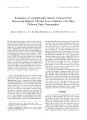
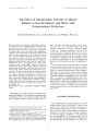
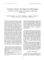
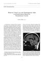
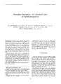
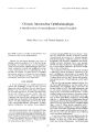
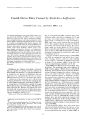
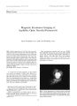
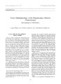
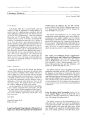
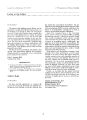
Show Journal of Neuro- Ophthalmology 17( 1): 39^ 3, 1997. © 1997 Lippincott- Ravcn Publishers, Philadelphia Nemaline Myopathy: An Unusual Cause of Ophthalmoparesis R. Alan Wright, M. B., ch. B., M. D., F. R. A. C. P., Gordon T. Plant, M. D., F. R. C. P., David N. Landon, M. B., B. S., B. S. C, L. R. C. P., M. R. C. S., and John A. Morgan- Hughes, M. D., F. R. C. P. Ophthalmoparesis and ptosis are extremely rare in nemaline myopathy. A 45- year- old man with a long history of bilateral ptosis and a 1- year history of diplopia is reported. Leg and arm weakness and wasting had been present since childhood, with a very slow deterioration over time. On examination, there was nonfatigueable bilateral ptosis that was more marked on the right. There was diplopia on left gaze. Extraocular movements showed limitation of elevation and adduction of the right eye. There was bilateral facial weakness, as well as proximal and distal wasting and weakness in the arms and legs. Electromyography ( EMG) showed a combination of myopathic and neurogenic changes. Triceps muscle biopsy showed small multiple collections of rod- like structures in > 50% of fibers. This patient presented with a clinical picture that did not primarily suggest nemaline myopathy. This case illustrates the heterogeneity of this disorder and the need for muscle biopsy to make an accurate diagnosis in patients with ptosis and progressive external ophthalmoparesis. Key Words: Nemaline myopathy- Ophthalmoparesis- Ptosis- Muscle biopsy. Manuscript received August 29, 1995. From the Neurology Department, The National Hospital for Neurology and Neurosurgery, and the Neuro- ophthalmology Department of Moorfields Eye Hospital, London, England. Address correspondence and reprint requests to Dr. Gordon Plant, Neurology Department, The National Hospital for Neurology and Neurosurgery, Queen Square, London WC1N 3BG, England. Presented in part at the Association of British Neurologists meeting June 1995, Berlin, Germany. Ophthalmoparesis may be seen in a wide variety of myopathies. However, ptosis and ophthalmoparesis has only rarely been reported in nemaline myopathy ( 1,2). Nemaline myopathy was first described in 1963 by Shy et al. ( 3) and Conen et al. ( 4). The term nemaline was coined by Shy et al. ( 3) because of the thread- like ( Greek = nema) appearance of the abnormal structures. CASE REPORT A 45- year- old man was seen in 1994 for evaluation of bilateral ptosis, diplopia, and generalised muscle weakness. The patient ( and his mother) reported that he had had bilateral ptosis dating back to childhood. There had been a very slow deterioration in his ptosis over time. There was no significant fluctuation in the degree of ptosis and no fa-tigueability. The patient had not had diplopia until 1 year prior to review, since which time he had had diplopia on looking to the left. Gait difficulties had been present since childhood with bilateral leg weakness most marked distally. At age 9 years, the patient had undergone orthopaedic procedures to both feet ( the details of which are unknown). He reported that his distal legs " had always been thin" and he thought his gait had only deteriorated very slowly over the last 20 years. The patient walked unaided. Similarly, his weakness 39 40 R. A. WR and wasting in both arms was of longstanding duration with only very slow deterioration. There were no speech or swallowing problems, and the patient did not think his facial appearance had changed. There was no family history of neurological disease. His mother, aged 70 years, was well; his father had died at age 67 years from a myocardial infarction, and a 40- year- old brother was normal. His parents were unrelated. He was not taking any medications. General examination showed prognathism and slightly irregular teeth, but no other dysmorphic features. Higher functions were normal. Corrected visual acuities were 20/ 20 ( 6/ 6) and N4.5 on the right and 20/ 15 ( 6/ 5) and N4.5 on the left. Ishihara testing was normal. There was bilateral ptosis with frontalis overactivity. Palpebral apertures measured 7 mm on the right and 9 mm on the left; levator function was 6 mm bilaterally ( Fig. 1A, B). There was no increase in ptosis with prolonged upward gaze or repeated eye opening and closing. There was no Cogan's lid twitch sign. There was horizontal diplopia on left gaze with, at times, some additional vertical separation of the images. Extraocular movements showed a limitation of the images. Extraocular movements showed a limitation of upgazc and adduction in the right eye ( Fig. 2) that was not improved by oculocephalic testing. There was no FIG. 1. A: Patient in 1984 ( aged 35 years) showing bi Patient in 1994 ( aged 45 years) showing increased bilal TETAL. nystagmus. Fundi were normal with no evidence of pigmentary retinopathy. There was bilateral mild facial weakness as well as mild weakness of neck flexion and extension. The tongue was normal, as was speech. The patient was generally thin, with wasting both proximally and distally in the upper and lower limbs. Tone was normal. There was no scapular winging. There was mild symmetric proximal and distal weakness in the arms. In the legs, there was mild weakness of hip flexion with bilateral foot drop and severe weakness of toe dorsiflexion. The feet were high arched. Reflexes required reinforcement to be elicited. Plantar responses were flexor. Cerebellar function and sensation were normal. He walked with a high stepping gait without ataxia. Creatine kinase was normal as were blood screen, biochemistry, thyroid function tests, and acetylcholine receptor antibodies. Electrocardiogram was normal. Nerve conduction studies were normal. Electromyography ( EMG) showed mainly myopathic changes with small motor units. No myotonia was seen. Some muscles also showed large polyphasic units, raising the possibility of both neurogenic and myopathic pathologies. Computed tomography ( CT) scan of the orbits showed a normal size and configuration of the extraocular muscles. Muscle biopsy of the left triceps showed small al ptosis, slight prognathism and irregular teeth. B: ptosis with frontalis overactivity. J Neuro- Ophthalmol, Vol. 17, No. 1, 1997 NEM ALINE MYOPATHY AND OPHTHALMOPARESIS 41 FIG. 2. Patient's extraocular movement's showing a limitation of upgaze and adduction in the right eye. multiple collections of rod- like structures in > 50% of fibers ( Fig. 3A- D). The rods were usually in the central regions of the fibers, but occasionally they accumulated beneath the sarcolemma. With oxidative enzyme stains, these fibers had a mottled appearance due to patchy disorganisation of the inter-myofibrillar network. There were no ragged red fibers, but very occasional fibers showed reduced or absent cytochrome oxidase staining. The majority of fibers ranged from 40- 70 ^ m. Occasional atrophic fibers were seen. Relative proportions of types 1, 2A, and 2B fibers was normal. Findings were consistent with nemaline myopathy. Electron microscopy confirmed the presence of collections of rods in many fibers ( Fig. 4A, B). These were usually superficial in location and were often associated with myonuclei. There was some evidence of generalised fiber atrophy, with occasional small angular fibers, but no other significant abnormalities; mitochondria were normal in number and morphology, and there was no increase in glycogen content. The basal laminae of the majority of the endomysial capillaries were substantially thickened. DISCUSSION Three broad categories of nemaline myopathy are recognised, although the boundaries between the various subtypes are not always clearcut [ see review by Fardcau and Tome ( 5) 1. Firstly, a severe neonatal myopathy, from which most patients succumb, during the first few weeks or months of life has been reported. Secondly, a more mild congenital myopathy, which may be nonprogressive or slowly progressive, is also recognised. Thirdly, adult onset cases, which include a wide spectrum of manifestations and severity, are also seen. In the vast majority of reported cases of nemaline myopathy, extraocular muscles and levator palpe-brae superioris arc unaffected. The 4th edition of Walsh and Hoyt's textbook of neuro- ophthalmol-ogy ( 6) states that " . . . no patient with nemaline myopathy has been reported to have ocular findings. . ." Rare case reports have documented extraocular movement abnormalities. Hopkins ct al. ( 1) described a 39- year- old woman who had ". . . mild ptosis. The external ocular movements were full except for questionable weakness of the left inferior rectus muscle with inconstant diplopia in extreme downward gaze." This patient's 63- year- old mother had " bilateral ptosis but external ocular movements were full and conjugate and nystagmus was not present." Muscle biopsy findings in these patients showed nemaline myopathy. Fukunaga ct al. ( 2) described a 35- year- old man born of consanguineous parents whose clinical findings included " ptosis of the left eyelid. There was limitation of adduction, upward and downward movement of the left eye." The authors also commented that the patient's elder sister, who had a similar clinical course, had died at age 40 years, although no further clinical details of her extraocular movements were provided. Muscle biopsy in this patient showed nemaline myopathy. These authors also reported finding abnormal mitochondria with crystal- like inclusions in muscle that they believed were nonspecific. The longstanding slowly progressive arm and leg weakness, wasting associated with facial weakness, and dysmorphic features seen in this patient would be consistent with nemaline myopathy. The predominantly myopathic EMG findings ( but with some muscles also showing coexisting neurogenic features) is also seen in nemaline myopathy Lsee review by Fardeau and Tome ( 5)]. No abnormalities suggestive of mitochondrial dysfunction were seen in this patient's muscle biopsy. The finding of occasional cytochrome oxidase negative fibers has previously been reported in patients with a variety of neuromuscular diseases, including 3 of 19 patients with nemaline myopathy ( 7). Yamamoto et al. ( 7) found no relationship between cytochrome oxi- J Neuro- Ophthalmol, Vol. 17, No. I, 1997 42 R. A. WRIGHT ET AL. FiG. 3. A: Left triceps muscle biopsy showing small multiple collections of rod- like structures ( see arrows). No ragged red fibers are seen. Modified Gomori trichrome stain. Scale = 100 | xm. B: Rod- like structures in central and peripheral locations within fibers ( see arrows). Modified Gomori trichrome stain. Scale = 50 fim. C: Cytochrome oxidase staining showing patchy staining in some type 1 fibers. Scale = 100 ( xm. D: ATPase staining at pH 4.6 showing a normal distribution of type 1 ( dark), type 2A ( light), and type 2B ( intermediate) fibers. Scale = 100 ( xm. dase- negalive fibers and fibers with rods, and concluded that the cytochrome oxidase negativity was probably a secondary phenomenon. No other causes to account for our patient's extraocular muscle weakness were found, and it seems most likely that his nemaline myopathy is responsible. This report highlights an uncommon feature of nemaline myopathy and illustrates an unusual cause of ptosis and progressive external oph-thalmoparesis. M( j. 4. A: A cluster of rod bodies adjacent to a nucleus at the periphery of a fiber in longitudinal section. Scale 2 p. m. B: A group of rod bodies in transverse section at the periphery of a fiber. Scale = 1 | xm. J Neuro- Ophthalmol, Vol. 17, No. I, 1997 NEM ALINE MYOPATHY AND OPHTHALMOPARESIS 43 Acknowledgment: We gratefully acknowledge the help of the patient in allowing us to publish his photograph, Linda Peachey for preparation of the light microscopy, and Kerrie Riley for preparation of the electron microscopy. REFERENCES 1. Hopkins IJ, Lindsey JR, Ford FR. Nemaline myopathy. A long- term clinicopathologic study of affected mother and daughter. Brain 1966; 89: 299- 310. 2. Fukunaga H, Osame M, Igata A. A case of nemaline myopathy with ophthalmoplegia and mitochondrial abnormalities. J Neurol Sci 1980; 46: 169- 77. 3. Shy GM, Engel WK, Somers JE, Wanko T. Nemaline myopathy. A new congenital myopathy. Brain 1963; 86: 793- 810. 4. Conen PE, Murphy EG, Donohue WL. Light and electron microscopic studies of " myogranules" in a child with hypotonia and muscle weakness. Can Med Assoc J 1963; 89: 983- 6. 5. Fardeau M, Tome FMS. Congenital myopathies. In: Engel AG, Franzini- Armstrong C, eds. Myology. 2nd ed. New York: McGraw Hill, 1994: 1487- 532. 6. Miller NR, ed. Walsh and Hoyt's clinical neuro-ophthalmology. 4th ed, vol 2. Baltimore: Williams and Wilkins, 1985: 785- 891. 7. Yamamoto M, Koga Y, Ohtaki E, Nonaka I. Focal cytochrome c oxidase deficiency in various neuromuscular diseases. J Neurol Sci 1989; 91: 207- 13. J Nciiro- Oplilhalmol. Vol. 17. No. 1. 1997 |