| OCR Text |
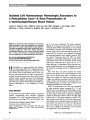
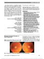
Show Asymmetric Papilledema in Idiopathic Intracranial Hypertension Samuel Bidot, MD, Beau B. Bruce, MD, PhD, Amit M. Saindane, MD, Nancy J. Newman, MD, Valérie Biousse, MD Background: Very asymmetric papilledema in idiopathic intracranial hypertension (IIH) is rare, and few studies have dealt with this atypical presentation of IIH. Our aim was to describe the clinical and radiologic features of patients with IIH and very asymmetric papilledema. Methods: We identified all adult patients from our IIH database with very asymmetric papilledema defined as a $2 modified Frisén grade difference between the 2 eyes. Demo-graphic data and initial symptoms were collected, and all brain imaging studies performed at our institution were reviewed. Results: Of the 559 adult patients with definite IIH, 20 (3.6%; 95% confidence interval [CI], 2.3-5.6) had very asymmetric papilledema at initial evaluation. They were older (39 vs 30 years; P , 0.001), had lower cerebrospinal opening pressure (35.5 vs 36 cmof water; P = 0.03), and were more likely to be asymptomatic compared with patients with symmetric papil-ledema (27% vs 3%; P , 0.001). Visual fields were worse on the side of the highest-grade papilledema (P = 0.02). The bony optic canal was smaller on the side of the lowest-grade edema in all 8 patients (100%) in whom the imaging was sufficient for reliable measurements (P = 0.008). Conclusions: IIH with very asymmetric papilledema is uncommon. Very asymmetric papilledema may result from differences in size of the bony optic canals, supporting the concept of compartmentation of the perioptic subarachnoid spaces. Journal of Neuro-Ophthalmology 2015;35:31-36 doi: 10.1097/WNO.0000000000000205 © 2014 by North American Neuro-Ophthalmology Society Very asymmetric papilledema in idiopathic intracranial hypertension (IIH) is an uncommon finding that can raise concern for alternate diagnoses, such as unilateral optic neuropathy. Few studies have systematically addressed the issue of asymmetric papilledema in IIH, and most are only case reports or small case series (1-11). The largest series of 38 patients focused specifically on visual function outcome (12), and only 1 study has detailed orbital imaging findings (13). Very asymmetric papilledema offers a unique oppor-tunity to study factors proposed in the pathogenesis of papilledema. Although several mechanisms have been sug-gested to explain very asymmetric papilledema, such as optic nerve sheath defects and loss of lamina cribrosa com-pliance (1,6), its mechanism remains unclear. Our aim was to describe the clinical and radiologic features of patients with IIH and very asymmetric papil-ledema and to characterize factors associated with this unusual presentation of IIH. METHODS Clinical Evaluation Our study was approved by the Emory University Institutional Review Board. Using our established database of patients seen in our center between 1989 and 2013, we identified all adult patients (age 18 years or older) with definite IIH according to the modified Dandy criteria (14). Although this study was a retrospective chart review, all patients were evaluated in a standardized fashion, and all had fundus photographs at presentation. Departments of Ophthalmology (SB, BBB, NJN, VB) and Neurology (BBB, NJN, VB), Emory University School of Medicine, Atlanta, Georgia; Department of Epidemiology (BBB), Rollins School of Public Health and Laney Graduate School, Atlanta, Georgia; and Departments of Radiology and Imaging Science (AMS) and Neurological Surgery (NJN), Emory University School of Medicine, Atlanta, Georgia. Supported in part by an unrestricted departmental grant (Department of Ophthalmology) from Research to Prevent Blindness, Inc, New York and by NIH/NEI core grant P30-EY06360 (Department of Ophthalmology). Presented at French Society of Ophthalmology, May 11, 2014, Paris, France. S. Bidot receives research support from Berthe Fouassier Foundation (Paris, France) and Philippe Foundation (New York, NY). B. B. Bruce receives research support from the NIH/NEI (K23-EY019341). N. J. Newman received the Research to Prevent Blindness Lew R. Wasserman Merit Award. The authors report no conflicts of interest. Address correspondence to Valérie Biousse, MD, Neuro-Ophthalmology Unit, Emory Eye Center, The Emory Clinic, 1365-B Clifton Road NE, Atlanta, GA 30322; E-mail: vbiouss@emory.edu Bidot et al: J Neuro-Ophthalmol 2015; 35: 31-36 31 Original Contribution Copyright © North American Neuro-Ophthalmology Society. Unauthorized reproduction of this article is prohibited. We first selected all patients with IIH from our IIH database, who had been recorded as having a $1 Frisén grade difference in papilledema between the 2 eyes at initial pre-sentation. All fundus photographs were then unpaired and graded independently using the modified Frisén scale (15) in random order by 2 neuro-ophthalmologists (S.B. and B.B.B.), who were masked to the patients' clinical data. In case of disagreement, the third neuro-ophthalmologist (V.B.) was asked to grade the papilledema. We defined very asym-metric papilledema as a $2 modified Frisén grade difference between the 2 eyes and selected all patients with IIH with at least 2 grades of difference in their papilledema. Demographic data of all patients with very asymmetric papilledema including age, gender, body mass index (BMI), and race were collected. Initial symptoms (visual loss, transient visual obscurations, diplopia, tinnitus, and headaches), Snellen visual acuity, intraocular pressure (IOP), automated visual fields, and cerebrospinal fluid (CSF) opening pressure (OP) were recorded. Radiologic Evaluation We reviewed all brain imaging and selected those performed at our institution (either magnetic resonance imaging [MRI] or contrast-enhanced computed tomography [CT]). MRI was performed on either 3.0-T (Siemens Trio, Erlangen, Germany) or 1.5-T (Siemens Avanto, Erlangen, Germany or GE Signa, Milwaukie, WI) scanner using a standard head coil. The MRI protocol included a T2 sequence at a slice thickness of 5 or 3 mm. Patients received intravenous gadolinium contrast mate-rial at a standard dose (0.1 mmol/kg Multihance; Bracco Diagnostics Inc, Monroe Township, NJ), followed by post-contrast T1 axial and T1 sagittal volumetric images. The magnetic resonance venography (MRV) protocol included either a noncontrast MRV using an oblique sagittal 2D time-of- flight (TOF) technique or a contrast-enhanced MRV, which included an axial precontrast MRV mask, followed by repetition of the sequence after contrast administration. The precontrast MRV dataset was subtracted from the postcontrast dataset, and multiple oblique maximum intensity projections were generated from this subtracted dataset. The CT exami-nation included orbital cuts and was performed on a 64 detector row scanner (GE Lightspeed VCT, Milwaukee, WI) using 70 mL iodinated contrast agent (Isovue 370; Bracco Imaging) with 5-mm slice reconstructions. All MRI and CT examinations were reviewed by an experienced neuroradiologist (A.M.S.) blinded to the neuro-ophthalmic examination. Previously described orbital imag-ing findings associated with increased intracranial pressure were recorded on all patients with imaging. These findings included protrusion of the optic nerve head, flattening of the posterior sclera, increased perioptic nerve CSF (0: none; 1: mild; 2: moderate; 3: severe [measured at the level of maximal dilation of the orbital optic nerve sheath]), and vertical tortuosity of the intraorbital optic nerve. The presence of meningoceles, as defined by CSF containing structures outside the expected confines of the cranial vault, was recorded. The presence or absence and side of transverse sinus stenosis was evaluated from either the contrast-enhanced MRV, postcontrast T1 (16), 2D TOF MRI, postcontrast CT, or transverse sinus flow voids on T2 images when there was no contrast-enhanced image set. When possible, and before recording the radiologic findings, cross-sectional area of both the right and left optic canals were measured using either precontrast T1 volumetric 1 mm isotropic images, by refor-matting into a coronal oblique plane orthogonal to the axis of each optic canal, or thin-section CT examination that was also reformatted in a similar fashion using images of 0.625 mm to obtain cross-sectional area (Fig. 1). Statistical Analysis All statistical analyses were performed with R: A language and environment for statistical computing (R Foundation for Statistical Computing, http://www.R-project.org). Continu-ous and ordinal variables were compared between groups using the Mann-Whitney U test. Pearson x2 with Yates' continuity correction or Fisher exact test, as appropriate, were used to compare categorical variables. The eyes of patients with very asymmetric papilledema were compared using the Wilcoxon signed rank test for continuous variables and paired proportion test for categorical variables. These tests were 2 tailed, and significance was set at 5%. RESULTS Of the 559 adult patients with definite IIH seen over 24 years, 20 (3.6%; 95% confidence interval [CI], 2.3-5.6) had very asymmetric papilledema defined as a $2 modified Frisén grade difference between 2 the eyes at initial evalua-tion. Patients with very asymmetric papilledema were signif-icantly older (39 vs 30 years; P , 0.001), had significantly lower CSF OP (35.5 vs 36 cm of water; P = 0.03), and were significantly more likely to be asymptomatic compared with patients with more symmetric papilledema (27% vs 3%; P , 0.001) (Table 1). The highest-grade edema was on the right side in 9 of 20 patients (45%). Papilledema was strictly unilateral in 8 of 20 patients (40%) and was located on the right side in 3 of 8 patients (38%). When comparing the eye with the highest-grade edema to the fellow eye, visual fields were significantly worse (mean deviation, 23.0 vs 22.1 dB; P = 0.02), but visual acuities and IOPs were not different (Table 2). Neuro-imaging (11 MRIs and 1 CT) was performed at our institution for 12 of 20 patients. Nine of 11 patients with MRI received intravenous gadolinium contrast mate-rial, and 3 of 11 patients had an MRV (contrast-enhanced MRV: 2/3 patients). The optic canal was smaller on the side of the lowest-grade edema in all 8 patients (100%, 7 MRIs and 1 CT) in whom it could be reliably evaluated (Table 2). The cross-sectional optic canal measurement was 14.9% smaller on average on the side of the lowest-grade edema 32 Bidot et al: J Neuro-Ophthalmol 2015; 35: 31-36 Original Contribution Copyright © North American Neuro-Ophthalmology Society. Unauthorized reproduction of this article is prohibited. FIG. 1. Cross-sectional area measurement technique. Figures on the right and left match with the right and left side, respectively. Figures (A-C) come from the same patient, and measurement was obtained by using precontrast T1 volumetric MRI (B and C). Figure (D) come from another patient to illustrate the technique using thin-section CT scan reconstruction. A. Optic disc appearance. Asymmetric papilledema, grade 2 in the right eye and grade 4 in the left eye. B. Positioning of the guides in the axial plane. Measurement of the cross-sectional area of each optic canal using MRI (C) and CT (D). CT, computed tomography; MRI, magnetic resonance imaging. Bidot et al: J Neuro-Ophthalmol 2015; 35: 31-36 33 Original Contribution Copyright © North American Neuro-Ophthalmology Society. Unauthorized reproduction of this article is prohibited. (range: 2.5%-31.0%; P = 0.008) (Fig. 1). Asymmetric peri-optic nerve CSF was reported in 6 of 12 patients (50%) and was always less prominent on the side of the lowest-grade edema (P = 0.01). Optic nerve protrusion into the globe and scleral flattening, both trended toward being more com-mon on the side of the highest-grade edema (P = 0.07). DISCUSSION Our study confirms that very asymmetric papilledema is rare in IIH, occurring in less than 4% of patients with definite IIH. Interestingly, we showed that the bony optic canal was always smaller on the side of the lowest-grade edema. Few reports (1-13) have dealt with asymmetric papille-dema in IIH. Three studies reported its prevalence in a ter-tiary neuro-ophthalmic setting (1,11,12), and found heterogeneous results. Using the same definition of asym-metric papilledema, Wall and White (12) and the Idio-pathic Intracranial Hypertension Treatment Trial (11) have shown that IIH with very asymmetric papilledema was uncommon (z7.5%), but a smaller series of 6 patients (1) found a higher prevalence (23%). Surprisingly, our study found a much lower prevalence of 3.6%. Several factors may have contributed to these discrepancies. First, we believe that the high prevalence found by Lepore (1) should be interpreted cautiously because the sample was TABLE 2. Comparison of ophthalmic and radiologic features between the eye with the highest-grade papilledema versus the eye with the lowest-grade papilledema in patients with definite idiopathic intracranial hypertension and asymmetric papilledema Eye With Papilledema P value Highest Grade, Median (IQR) or n (%) Lowest Grade, Median (IQR) or n (%) Ophthalmic findings (n = 20) Modified Frisén grade 3 (2 to 4) 1 (0 to 2) NA VA, logMAR* 0.0 (20.12 to 0.7) 0.0 (20.12 to 2.0) 0.39 IOP 15.5 (13 to 17) 15.5 (13 to 18) 0.90 HVF, mean deviation† (n = 19) 23.0 (25.1 to 22.1) 22.1 (22.9 to 20.7) 0.02 Radiologic findings (n = 12) Optic canal measurement, mm2 (n = 8) 18.5 (13.6 to 29.8) 15.8 (12.1 to 27.8) 0.008‡ Prominent peri-ON CSF 6 (50) 0 (0) 0.01 Scleral flattening 8 (67) 3 (25) 0.07 Vertical tortuosity of the ON 1 (8) 1 (8) 1.0 ON head protrusion 9 (75) 3 (25) 0.07 Meningocele 5 (42) 5 (42) 1.0 Transverse sinus stenosis 11 (92) 10 (92) 1.0 *Range provided instead of IQR. †Mean deviation in decibels. ‡All patients (100%) had a smaller optic canal on the side of the lowest-grade papilledema. CSF, cerebrospinal fluid; HVF, automated visual field; IOP, intraocular pressure; IQR, interquartile range; NA, not applicable; ON, optic nerve; VA, visual acuity. TABLE 1. Demographics, initial symptoms, and cerebrospinal fluid opening pressure in patients with idiopathic intracranial hypertension with asymmetric and symmetric papilledema Papilledema Asymmetric, n = 20, n (%) or Median (IQR) Symmetric, n = 539, n (%) or Median (IQR) P value Demographics Age, yr 39 (33-45) 30 (25-36) ,0.001 Gender, men 3 (15) 39 (7) 0.20 Race, white 13 (65) 292 (54) 0.35 BMI, kg/m2 37.8 (32.5-45.1) 38.0 (32.2-44.0) 0.83 Initial symptoms Headache 7 (35) 417 (77) ,0.001 TVOs 7 (35) 80 (15) ,0.001 Decreased vision 2 (9) 127 (24) ,0.001 Diplopia 0 (0) 30 (6) 0.62 Tinnitus 0 (0) 37 (7) 0.64 None 6 (27) 17 (3) ,0.001 CSF OP, cm 35.5 (27.0-37.0) 36.0 (31.0-44.0) 0.03 BMI, body mass index; CSF OP, cerebrospinal fluid opening pressure; IQR, interquartile range; TVO, transient visual obscurations. 34 Bidot et al: J Neuro-Ophthalmol 2015; 35: 31-36 Original Contribution Copyright © North American Neuro-Ophthalmology Society. Unauthorized reproduction of this article is prohibited. small (6/27 patients; 95% CI, 7.1-38.9) and no details regarding the definition of asymmetric papilledema were provided. Second, we had a strict definition of very asym-metric papilledema to emphasize the potential differences between symmetric and asymmetric papilledema in terms of demographics, clinical presentation, and radiologic features; therefore, we might have underestimated its prevalence. Regarding patient demographics, we found that patients with IIH with very asymmetric papilledema were older compared with patients without asymmetric papilledema, which also was reported by Lepore (1). Lepore (1) suggested a possible loss of compliance in the aging lamina cribrosa, buffering the effect of the perioptic CSF pressure. However, he did not address how this would result in asymmetric papilledema. Wall and White (12) found an overrepresenta-tion of men (29%); we also found a higher frequency of men (15%) among patients with very asymmetric papilledema, but this did not reach significance when compared with the group of patients with symmetric papilledema. In our study, race and BMI of patients with very asymmetric papilledema were similar to those with symmetric papilledema. The clinical presentation of patients with IIH with very asymmetric papilledema seems to differ from that of patients with IIH with symmetric papilledema, with a high pro-portion of asymptomatic cases (27%) and a lower proportion of patients with headaches (35%) in the asymmetric papilledema group. In a previous series including patients from the same IIH database (17), we emphasized that men with IIH experience headache less often. Our higher fre-quency of men with very asymmetric papilledema, none of whom had headache, may account partially for this differ-ence. In addition, although no correlation between headaches and CSF OP in IIH has been reported, the significantly lower CSF OP we found among patients with very asymmetric papilledema might have contributed to the difference in head-ache frequency. Regarding visual function, as previously re-ported (12), visual fields were significantly worse on the side of the highest-grade papilledema. The most interesting result from our study is that the bony optic canals were consistently smaller on the side of the lowest-grade papilledema. The pathophysiology of CSF dynamics in IIH is not fully understood (18). The lack of a clear relationship between the degree of papilledema and the CSF OP (14,19) suggests that an underlying mechanism may sometimes prevent the optic disc from swelling in some cases of IIH. The pathogenesis of papilledema depends on the translaminar pressure gradient at the optic nerve head, and therefore on the balance between the CSF pressure in the perioptic subarachnoid spaces and the IOP. Hayreh (20) showed that high CSF pressure in the perioptic subarachnoid spaces or low IOP cause identical microscopic changes and axonal flow stasis. However, our study and others (2,7,13) have shown that asymmetric IOP, although reported in anec-dotal cases (3,5,10), is not the primary mechanism of asym-metric papilledema. We believe that, in the absence of optic atrophy (20), asymmetric papilledema is most likely related to asymmetric transmission of the CSF pressure to the lamina cribrosa (21). Two mechanisms for asymmetric transmission of the CSF pressure along the perioptic subarachnoid spaces previously have been proposed, namely asymmetric structural changes either in the lamina cribrosa (1) or along the optic nerve sheath (6). These mechanisms remain debated. Our study shows compelling data regarding the role of asymmetry of the bony optic canals in the genesis of very asymmetric papilledema. It is well known that the orbital portion of the perioptic subarachnoid spaces shows distension in IIH (22,23). A study of 15 patients with IIH, 10 with IIH, failed to demonstrate asymmetric distension of the perioptic sub-arachnoid spaces in patients with unilateral papilledema, but no details regarding the grading of papilledema were provided (13).We included only very asymmetric papilledema to iden-tify obvious potential differences between the 2 eyes. We showed that asymmetric distension of the perioptic subarach-noid spaces occurred in half of our cases, always less prom-inent on the side of the lowest-grade edema, suggesting that the CSF pressure may be lower in the perioptic subarachnoid spaces on the side of the lowest-grade edema. Possibly, our imaging or grading system might not have been sensitive enough to capture subtle asymmetry in the remaining 50% of cases with symmetric perioptic CSF. The concept of compartmentation of the perioptic sub-arachnoid spaces (4,24,25), in which the perioptic subarach-noid spaces are partially separated from the suprasellar cisternal spaces, seems more likely to explain asymmetric papilledema. Although the orbital portion of the perioptic subarachnoid spaces is able to distend under increased CSF pressure, the intracanalicular portion, the narrowest (4,19), is characterized by tight relationships between the surrounding bone and the optic nerve (24). Because of its unique anatomy, the region of the optic canal plays a crucial role in CSF flow dynamics between the suprasellar cistern and the perioptic subarachnoid spaces. We have demonstrated that the bony optic canal was always smaller on the side of the lowest-grade edema. This anatomic configuration probably allows CSF pressure to be less easily transmitted along the optic nerve on the side of the smaller canal, thereby resulting in lower local intraorbital CSF pressure and less optic disc edema. However, longitudinal data on the optic canal diameter in patients with IIH are needed to better understand whether this asymmetry in size is congenital or results from bony erosion related to longstanding CSF hypertension, as described in other skull base locations with chronic IIH (26,27). The fact that we and others (1) have found that patients with IIH with asymmetric papilledema are older than other patients with IIH may support an acquired etiology. Despite our small sample, there seems to be a definite relationship between the severity of papilledema and the cross-sectional area of the optic canal, suggesting that asymmetric papilledema may result from asymmetries in Bidot et al: J Neuro-Ophthalmol 2015; 35: 31-36 35 Original Contribution Copyright © North American Neuro-Ophthalmology Society. Unauthorized reproduction of this article is prohibited. the bony optic canal. Our study lends further support to the concept of compartmentation of the perioptic subarachnoid spaces developed by Killer and Subramanian (24) and sug-gests that the bony optic canal may be a "bottleneck" inter-fering with the CSF flow between the perioptic subarachnoid spaces and the suprasellar cistern. Whether the asymmetry in size of the optic canal is congenital or acquired requires further study. REFERENCES 1. Lepore FE. Unilateral and highly asymmetric papilledema in pseudotumor cerebri. Neurology. 1992;42:676-678. 2. Brosh K, Strassman I. Unilateral papilledema in pseudotumor cerebri. Semin Ophthalmol. 2013;28:242-243. 3. Abegg M, Fleischhauer J, Landau K. Unilateral papilledema after trabeculectomy in a patient with intracranial hypertension. Klin Monbl Augenheilkd. 2008;225:441-442. 4. Killer HE, Jaggi GP, Flammer J, Miller NR, Huber AR, Mironov A. Cerebrospinal fluid dynamics between the intracranial and the subarachnoid space of the optic nerve. Is it always bidirectional? Brain. 2007;130:514-520. 5. Greenfield DS, Wanichwecharungruang B, Liebmann JM, Ritch R. Pseudotumor cerebri appearing with unilateral papilledema after trabeculectomy. Arch Ophthalmol. 1997;115:423-426. 6. Moster ML, Slavin M, Wall M. Unilateral disk edema in a young woman. Surv Ophthalmol. 1995;39:409-416. 7. Strominger MB, Weiss GB, Mehler MF. Asymptomatic unilateral papilledema in pseudotumor cerebri. J Clin Neuroophthalmol. 1992;12:238-241. 8. To KW, Warren FA. Unilateral papilledema in pseudotumor cerebri. Arch Ophthalmol. 1990;108:644-645. 9. Sher NA, Wirtschafter J, Shapiro SK, See C, Shapiro I. Unilateral papilledema in "benign" intracranial hypertension (pseudotumor cerebri). JAMA. 1983;250:2346-2347. 10. Kawasaki A, Purvin V. Unilateral optic disc edema following trabeculectomy. J Neuroophthalmol. 1998;18:121-123. 11. Wall M, Kupersmith MJ, Kieburtz KD, Corbett JJ, Feldon SE, Friedman DI, Katz DM, Keltner JL, Schron EB, McDermott MP. The idiopathic intracranial hypertension treatment trial: clinical profile at baseline. JAMA Neurol. 2014;71:693-701. 12. Wall M, White WN. Asymmetric papilledema in idiopathic intracranial hypertension: prospective interocular comparison of sensory visual function. Invest Ophthalmol Vis Sci. 1998;39:134-142. 13. Huna-Baron R, Landau K, Rosenberg M, Warren FA, Kupersmith MJ. Unilateral swollen disc due to increased intracranial pressure. Neurology. 2001;56:1588-1590. 14. Friedman DI, Jacobson DM. Diagnostic criteria for idiopathic intracranial hypertension. Neurology. 2002;59:1492-1495. 15. Scott CJ, Kardon RH, Lee AG, Frisén L, Wall M. Diagnosis and grading of papilledema in patients with raised intracranial pressure using optical coherence tomography vs clinical expert assessment using a clinical staging scale. Arch Ophthalmol. 2010;128:705-711. 16. Saindane AM, Mitchell BC, Kang J, Desai NK, Dehkharghani S. Performance of spin-echo and gradient-echo T1-weighted sequences for evaluation of dural venous sinus thrombosis and stenosis. AJR Am J Roentgenol. 2013;201:162-169. 17. Bruce BB, Kedar S, Van Stavern GP, Monaghan D, Acierno MD, Braswell RA, Preechawat P, Corbett JJ, Newman NJ, Biousse V. Idiopathic intracranial hypertension in men. Neurology. 2009;72:304-309. 18. Batra R, Sinclair A. Idiopathic intracranial hypertension; research progress and emerging themes. J Neurol. 2014;261:451-460. 19. Hayreh SS. Pathogenesis of oedema of the optic disc (papilloedema). A preliminary report. Br J Ophthalmol. 1964;48:522-543. 20. Hayreh SS. Optic disc edema in raised intracranial pressure. V. Pathogenesis. Arch Ophthalmol. 1977;95:1553-1565. 21. Tso MO, Hayreh SS. Optic disc edema in raised intracranial pressure. III. A pathologic study of experimental papilledema. Arch Ophthalmol. 1977;95:1448-1457. 22. Bäuerle J, Nedelmann M. Sonographic assessment of the optic nerve sheath in idiopathic intracranial hypertension. J Neurol. 2011;258:2014-2019. 23. Hoffmann J, Schmidt C, Kunte H, Klingebiel R, Harms L, Huppertz HJ, Ludemann L, Wiener E. Volumetric assessment of optic nerve sheath and hypophysis in idiopathic intracranial hypertension. AJNR Am J Neuroradiol. 2014;35:513-518. 24. Killer HE, Subramanian PS. Compartmentalized cerebrospinal fluid. Int Ophthalmol Clin. 2014;54:95-102. 25. Killer HE. The optic nerve: a new window into cerebrospinal fluid composition? Brain. 2006;129:1027-1030. 26. Pérez MA, Bialer OY, Bruce BB, Newman NJ, Biousse V. Primary spontaneous cerebrospinal fluid leaks and idiopathic intracranial hypertension. J Neuroophthalmol. 2013;33:330-337. 27. Bialer OY, Pérez MA, Bruce BB, Newman NJ, Biousse V, Saindane AM. Meningoceles in idiopathic intracranial hypertension. AJR Am J Roentgenol. 2014;202:608-613. STATEMENT OF AUTHORSHIP Category 1: a. Conception and design: S. Bidot, V. Biousse, A. M. Saindane, B. B. Bruce, and N. J. Newman; b. Acquisition of data: S. Bidot, A. M. Saindane, and B. B. Bruce; c. Analysis and interpreta-tion of data: S. Bidot, V. Biousse, A. M. Saindane, B. B. Bruce, and N. J. Newman. Category 2: a. Drafting the article: S. Bidot, A. M. Saindane, and B. B. Bruce; b. Revising it for intellectual content: S. Bidot, V. Biousse, A. M. Saindane, B. B. Bruce, and N. J. Newman. Category 3: a. Final approval of the completed article: S. Bidot, V. Biousse, A.M. Saindane, B. B. Bruce, and N. J. Newman. 36 Bidot et al: J Neuro-Ophthalmol 2015; 35: 31-36 Original Contribution Copyright © North American Neuro-Ophthalmology Society. Unauthorized reproduction of this article is prohibited. |