| OCR Text |
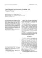
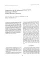
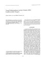
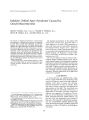
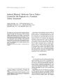
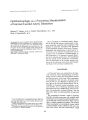
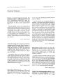
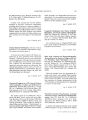
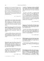
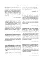
Show Journal of Clinical Neuro- ophthalmology 12( 4): 226- 229, 1992. © 1992 Raven Press, Ltd., New York Papillophlebitis and Arteriolar Occlusion in a Pregnant Woman Michael Humayun, M. D., Jorge Kattah, M. D., Thomas R. Cupps, M. D., Suresh R. Limaye, M. D., and Georgia A. Chrousos, M. D. Among the pathologic findings affecting the eye during pregnancy, microvascular abnormalities affecting both choroidal and retinal circulation have been reported in cases of complicated pregnancy. We report a case of papillophlebitis and arteriolar occlusion in a pregnant woman without any complications throughout her pregnancy. The patient was placed on glucocorticoids and her vision, visual fields and funduscopic appearance improved almost to normal. Despite the improvement with treatment, the contributing role of glucocorticoids in this case could not be fully determined. Papillophlebitis and arteriolar occlusion should be included among gestational- related vasculopathies. Key Words: Retina- Ischemia- PapillophlebitisVasculitis-- Optic nerve- Pregnancy. From the Center for Sight ( M. H., S. R. L., G. A. C), the Departments of Neurology and Medicine O. K.), and Division of Rheumatology ( T. R. C), Georgetown University Medical Center, Washington, D. C, U. S. A. Address correspondence and reprint requests to Dr. Georgia A. Chrousos at Georgetown UniVersIty, 3800 Reservoir Road N. W., Washington, DC 20007, U. S. A. ' 226 Pregnancy has a wide range of systemic effects that have a potential impact on virtually any ocular condition ( 1). Changes in intraocular pressure ( 2,3), corneal sensitivity ( 4), visual field ( 5), and retina ( 1) have been reported. Most of the retinal changes associated with pregnancy are related to toxemia. Characteristically, proteinuria, edema, and arterial hypertension have been associated with deterioration of proliferative retinopathy ( 6), retinopathy of toxemia ( 7), retinal detachment ( 8,9), choroidal detachment ( 10), choroidal vascular insufficiency ( 11), central serous chorioretinopathy ( 12), and ischemic papillophlebitis ( 13). We report a case of papillophlebitis and arteriolar occlusion in a woman with an uncomplicated pregnancy. To our knowledge, this is the first such case displayed in the absence of toxemia. CASE REPORT A 32- year- old woman at 37- week gestation was in good general health and ocular health until she suddenly developed a paracentral scotoma and blurred vision in her left eye. On examination, best corrected visual acuity was 20/ 20 in the right eye and counting fingers at 1 m in the left. An afferent pupillary defect was present on the left. The funduscopic examination was normal in the right eye. In the left she had disk edema, retinal hemorrhages, tortuosity of the vessels, sheathing along the inferior temporal venuole, and retinal edema alo~ g the distribution of the superior temporal artenole above the macula ( Fig. 1A). Automated static perimetry showed central and paracentral defects in the left eye ( Fig. 2A). The remainder of the ocular examination was unremarkable. The patient's laboratory workup was essentially PAPILLOPHLEBITIS AND ARTERIOLAR OCCLUSION 227 FIG. 1. ( A) Fundus of the patient's left eye. Note optic disk edema, hemorrhages, tortuosity of the vessels, sheathing along the inferior temporal venuole and retinal edema along the distribution of the superior temporal arteriole above the macula. ( 8) Same eye 4 months later. unremarkable except for her sedimentation rate, which was 39 mmlh. Complete blood and platelet counts were normal. Complement C3, C4, and CH 50 were within normal limits. Antinuclear antibody ( ANA), anti- deoxyribonucleic acid antibody ( ADNA), antineutrophile cytoplasmic antibody ( ANCA), anticardiolipin antibody ( ACA), and extractable nuclear antigen ( ENA) were also negative. Quantitative immunoglobulins were within normal limits except for IgG, 361 mgldl, and IgA, 64.9 ngldl. Magnetic resonance imaging ( MRI) study of the brain was negative. Three days after the onset of her symptoms, the patient underwent an elective caesarean section and delivered a healthy male infant. She was then subsequently placed on a 3- day course of pulsed intravenous methylprednisolone administered 250 mg every 6 hours. This was followed by oral prednisone at a dose of 80 mg per day. Fluorescein angiography after completion of her intravenous glucocorticoid treatment depicted a delay in filling of the superior temporal arteriole, and peripapillary fluorescein dye leakage that extended along the inferior temporal venuole. Visual acuity gradually improved over the ensuing week to 20/ 30 in the left eye. Funduscopic examination 1 month later revealed less disk edema and vascular tortuosity. Resolving hemorrhages of the eye were still visualized. Visual field testing continued to show similar central and paracentral scotomas and improved paracentral defects of the left eye. Four months later, the patient's visual acuity was 20/ 20 in both eyes. Funduscopic examination showed residual tortuosity of the vessels in the left eye, which was otherwise normal ( Fig. 18). Visual field further improved with only a few central and paracentral defects remaining in the left eye ( Fig. 2B). The remainder of the ocular examination was unremarkable. Administration of oral prednisone was slowly tapered and then discontinued 2 months later. The laboratory workup was repeated a number of times during the year following the onset of her problem, and it was always negative, including normalization of her sedimentation rate. DISCUSSION Retinal changes are frequently seen in pregnancy associated with toxemia. Funduscopic changes were first described by von Graefe in 1855 ( 14). They are believed to occur in 60% to 100% of all women with toxemia ( 7). The pathogenesis of this is not fully understood. Some authors have described toxemic retinopathy as sharing features similar to hypertensive retinopathy ( 6,7). Recently, Brancato et al. ( 6) have proposed low- grade disseminated intravascular coagulation as the main cause of toxemic proliferative retinopathy, implicating microvascular occlusive phenomena. Serous exudative retinal detachments occur in 10% of patients with toxemia ( 1). Although both retina and choroid have been implicated as the possible sources of subretinal fluid, Mabie et al. ( 9) believe that retinal detachment is secondary to an isolated choroidal vasculopathy. Retinal vasculitis has been described in association with toxemic pregnancy J Clin Neurcrophthalmol, Vol. 12, No. 4, 1992 228 M. HUMAYUN ET AL. FIG. 2. ( A) Visual field of the left eye on presentation. Note the presence of central and paracentral field defects. ( B) Follow- up visual fields 4 months later reveal much smaller defects. B by Price et al. ( 13). Their patients had extensive periphlebitis, retinal hemorrhages and edema, optic nerve head swelling, and widespread capillary occlusion. Visual outcome was poor ( personal communication with Dr. Price). Uncomplicated pregnancy, as in our patient, has been rarely associated with retinal changes in general and even less with retinal phlebitis and arterial obstruction. Only a patient with phlebitis of the retina early in pregnancy ( 15) and two pregnant women with retinal obstruction and migraine disorders ( 16) have been reported. Our patient had papillophlebitis with the addition of branch retinal artery occlusion and perivenuolar sheathing. Papillophlebitis is believed to be a type of central retinal vein occlusion in young people, the exact cause of which is not known. Recently, Schatz et al. ( 17) reported papillophlebitis and cilioretinal artery occlusion as a separate entity in young adults. These authors suggest that the cilioretinal occlusion occurred subsequently and probably secondary to papillophiebitis and perfusion pressure changes in the cilioretinal artery. In our case, the occluded artery was not cilioretinal, but a superior temporal branch. Central or branch retinal artery occlusion is rarely seen in young patients. It has been seen in association with systemic vascular diseases such as diabetes, collagen vascular diseases, embolic diseases, optic nerve tumor, and inflammation ( retinitis) ( 17). The patient's workup failed to disclose any systemic disease or optic nerve tumor. The mechanism of the branch retinal artery occlusion may lOin Nfllro- ophthalmol. Vol. 12. No. 4. 1992 have been inflammation similar to the one seen in previous retinal vasculitis cases reported by Price et al. ( 13). The association of perivenuolar sheathing in other areas of the retina, although not pathognomonic, was suggestive of inflammation too. There is, however, the distinct possibility that the branch retinal artery obstruction was secondary to the optic nerve swelling and that the whole retinal picture was due to occlusion ischemia rather than inflammation. In that case, glucocorticoids may have had nothing to do with the clearing of the vision. The fluorescein angiography, which showed a patent superior branch artery despite the ischemic whitening of the retina, was not particularly helpful either in providing us with more information, since it was performed after the completion of the high- dose intravenous methylprednisolone treatment. Papillophlebitis and arteriolar occlusion should be added to the list of ocular manifestations associated with pregnancy, even in the absence of toxemia, with which the ophthalmologist should be familiar. REFERENCES 1. Sunness JS. The pregnant woman's eye. Suru Ophthalmol 1988; 32: 219- 38. 2. Kass MA, Sears ML. Hormonal regulation of intraocular pressure. Suru OphthalmoI1977; 22: 153- 76. 3. Phillips cr, Gore SM. Ocular hypotensive effect of late pregnancy with and without high blood pressure. Br JOphthaimol 1985; 69: 117- 19. 4. Riss B, Riss P. Corneal sensitivity in pregnancy. Ophthalmologica 1981; 183: 57-- 62. PAPILLOPHLEBITIS AND ARTERIOLAR OCCLUSION 229 5. Walsh FB, Hoyt WF. Clinical neuro- ophthalmology 3rd ed. Baltimore: Williams & Wilkins, 1969: 2124- 5. 6. Brancato R, Menchini U, Bandello F. Proliferative retinopathy and toxemia of pregnancy. Ann Ophthalmol 1987; 19: 182- 3. 7. Kline LB. Retinopathy in toxemia of pregnancy. South Med J 1981; 74: 34-<). 8. Oliver M, Uchenik D. Bilateral exudative retinal detachment in eclampsia without hypertensive retinopathy. Am J Ophthalmol 1980; 90: 792-{'. 9. Mabie Wc, Ober RR. Fluorescein angiography in toxaemia of pregnancy. Br J Ophthalmol 1980; 64: 666- 71. 10. Kenny GS, Cerasoli JR. Color fluorescein angiography in toxemia of pregnancy. Arch OphthalmoI1972; 87: 383- 8. 11. Fastenberg OM, Fetkenhour CL, Choromokos E, et aJ. Choroidal vascular changes in toxemia of pregnancy. Am I OphthalmoI1980; 89: 362- 8. 12. Gass JDM. Central serous chorioretinopathy and white subretinal exudation during pregnancy. Arch Ophthalmol 1991; 109: 677- 81. 13. Price J, Marouf L, Heine MW, et aJ. New angiographic findings in toxemia of pregnancy. Ophthalmology 1986; 93 ( suppl): 125. 14. von Graefe A. Ueber eine Krebsablagerung im lnnern des Auges, des surspruenglicher Sitz zwischen Sclera und Chorioidea war. Arch Ophthalmol 1855; 2: 214- 30. 15. Spitzberg DH. Retinal phlebitis associated with pregnancy. Ann OphthalmoI1982; 14: 101- 2. 16. Brown Gc, Magargal LE, Shields JA. Retinal arterial obstruction in children and young adults. Ophthalmology 1981; 88: 18-- 25. 17. Schatz H, Fong ACO, McDonald HR, et aJ. Cilioretinal artery occlusion in young adults with central retinal vein occlusion. Ophthalmology 1991; 98: 594-<) 01. JClin Neuro- ophthalmol. Vol. 12, No. 4, 1992 |