| OCR Text |
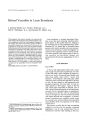
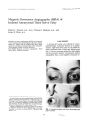
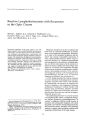
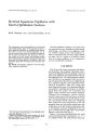
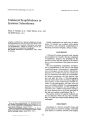
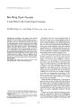
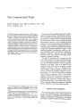
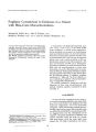
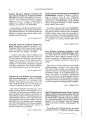
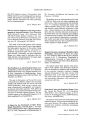
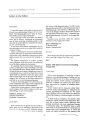
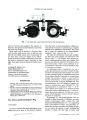
Show Journal of Clinical Neuro- ophthalm% gy 11( 1): 19- 24, 1991. © 1991 Raven Press, ltd., New York Rapid Change in Visual Fields Associated with Suprasellar Lymphocytic Hypophysitis Maryla Stelmach, M. B. B. S., and Justin O'Day, F. R. A. C. O., F. R. A. C. S. A young woman presented in the third trimester of pregnancy with a rapid onset of bitemporal hemianopia and reduced visual acuity caused by an unusual steroid responsive suprasellar tumor. A computerized tomography scan revealed a 2- cm suprasellar mass which was thought to be a tuberculum sellae meningioma. Surgery was delayed because of pregnancy. A short course of high- dose steroids was given to promote fetal lung maturity. This produced an unexpected and dramatic resolution of the field loss. As the steroid dose was reduced, the visual fields deteriorated, necessitating a craniotomy. The histology revealed lymphocytic hypophysitis, a rare but distinct clinicopathological entity affecting the anterior pituitary lobe. The significance of the suprasellar site and a possible role for the use of steroids in the preoperative management of this condition is discussed. Key Words: Autoimmune disease-- Bitemporal hemianopia- Lymphocytic hypophysitis- Pituitary tumorPregnancy- Suprasellar mass. From the Department of Anatomy, University of Melbourne, Parkville ( M. S.), and St. Vincent's Hospital, Melbourne, Victoria, Australia. Address correspondence and reprint requests to Dr. M. Z. Stelmach % Dr. O'Day's Consulting Rooms at 55 Vlctona Parade, Fitzroy, Victoria 3065, Australia. 19 Lymphocytic hypophysitis is a rare disease of probable autoimmune origin which affects the anterior pituitary lobe. It is most commonly seen in women and has a close temporal relationship to pregnancy. In all previously reported cases the masses have been intrasellar, with or without suprasellar extension. This is the first reported case of lymphocytic hypophysitis involving the suprasellar site alone. It is also the first case which documents the rapid deterioration of visual fields associated with this condition as well as the sudden and dramatic improvement of visual fields following a short course of high- dose steroids. CASE REPORT A 21- year- old Caucasian primigravida presented at 28 weeks gestation with a lO- day history of sudden onset of blurred vision in the right eye associated with a history of nocturnal bitemporal headaches. Her pregnancy had been uneventful until these symptoms arose, and there was no relevant past history. Goldmann perimetry demonstrated a bitemporal field loss which was more complete in the right eye than in the left eye ( Fig. 1A). Visual acuity with myopic correction was 20/ 30 in the right eye and 20/ 20 in the left eye. Testing color vision with Ishihara plates demonstrated loss of color vision in both eyes, particularly in the right eye. There was also a right relative afferent pupil defect. Examination of the posterior segments was normal. In particular, the optic discs were pink, and venous pulsation was present. Clinically there was no evidence of hypopituitarism. The computerized tomography ( CT) scan showed a uniformly enhancing 2- cm suprasellar mass lying directly above the diaphragma sellae ( Fig. 2). The mass impinged upon the optic nerves and chiasma and appeared to be attached to the tuberculum sellae. There was no obvious calcifica- 20 M. STELMACH AND ]. O'DAY -' 4e ." ," 19' 4 · 84 0' ' HO_ I4e - - · ' le ." B FIG. 1. A: Visual fields at the time of presentation demonstrate a bitemporal field loss with a complete right temporal hemianopia and a left superior temporal quadrantanopia. B: Visual fields six days after presentation demonstrate a small island of vision remaining in the upper nasal quadrant of the right eye and a progressing left superior quadrantanopia. ' 0;": A A FIG. 2. A: The CT scan demonstrates a large enhancing suprasellar mass. B: The CT scan taken at the level of the sella turcica demonstrates a normal pituitary fossa with no apparent intr2" 81lar mass. :,:: 11( 1- 0 ' . " I. 194/ //--------""" SUPRASELLAR LYMPHOCYTIC HYPOPHYSITIS 21 tion and the pituitary fossa appeared normal. A presumptive diagnosis of tuberculum sellae meningioma was made. The full blood examination was normal except for a neutrophil leucocytosis of 20.0 x 10⁹/ 1 ( normal range: < 11 x 10911) which was consistent with pregnancy. On biochemical evaluation there was an elevated prolactin of 5,196 IU/ ml ( normal range in pregnancy: 50- 800 IU/ ml), with normal serum FSH, LH and thyroid indices. Over the next 6 days from the time of presentation, the visual fields continued to deteriorate rapidly, with the bitemporal field defect becoming more complete ( Fig. IB), but because of the pregnancy, immediate surgery was deferred. The patient was started on a short course of high dose betamethasone ( 5.7 mg i. m., b. i. d. x 6) in order to promote fetal lung maturation and to reduce the potential risks of prematurity should spontaneous labor occur. This was followed by a reducing dose of oral prednisolone over 9 days, commencing with 30 mg orally daily. When the initial dose of steroid was given, Bromocriptine ( 2.5 mg orally b. i. d.) was also commenced in order to reduce the secretion of prolactin. Bromocriptine was continued after the prednisolone was ceased. Three days after commencing the steroid treatment, an unexpected and dramatic improvement in the visual fields was noted, with recovery of vision in the right inferior quadrants ( Fig. 3). At FIG. 3. Visual fields 10 days after presentation and 1 day after completing the 3- day course of intramuscular betamethasone. There is a significant reduction of the right temporal hemianopia. together with resolution of the left superior quadrantanopia. 34. l8 · 4 · 84 OS ----'"' i- e l3O_ l~ --- I1.~ I Clin Neuro- ophthalmol, Vol, 11, No, 1, 1991 M. STELMACH AND J. O'DAY the same time the visual acuity remained unaltered, but the bitemporal headaches ceased. However, once the steroid dose was reduced, contraction of the visual fields developed, prompting surgical intervention. A transfrontal craniotomy was performed, removing a rubbery grey suprasellar mass arising from the pituitary stalk. The tumor was associated with dense arachnoid fibrosis adhering to the optic nerves, chiasma, and superior hypophyseal arteries. Histological examination demonstrated that the mass consisted of loci of normal pituitary tissue densely infiltrated by lymphocytes and plasma cells, with a small number of neutrophil polymorphs and moderate interstitial fibrosis ( Fig. 4). On the basis of the extensive lymphocytic infiltration, a diagnosis of lymphocytic hypophysitis was made. Immunoperoxidase staining with anterior pituitary hormones identified the presence of normal anterior pituitary endocrine cells without apparent loss of any cell line. Both T and Bcells were identified and plasma cells showed polyclonal kappa and lambda light chains. Immunological investigations, including anterior pituitary antibodies, were negative. An HLA typing and complement allotyping were performed as part of a research study into autoimmune disorders ( personal communication from Dr. J. B. Best). Her HLA typing is A2 Aw68 B13 B58/ 4 DR4/ 7 DRw53 DQw2/ 3 and complement allotyping is C4A3 C4Bl BfFl BfS. In the immediate postoperative period, the patient's visual acuity deteriorated, later returning to 20/ 25 in both eyes. The visual fields demonstrated a persistent bitemporal field loss which slowly improved postoperatively with a residual permanent field loss affecting the upper temporal quadrants. A healthy female infant was delivered by cesarean section near term. The patient remained on anterior pituitary hormone replacement therapy. DISCUSSION Lymphocytic hypophysitis is a rare but distinct clinicopathological entity affecting the anterior pituitary lobe. The true incidence of this condition is probably under- reported in the literature. It has a close temporal association with pregnancy- 16 of 21 cases described in the literature were reported in women either during pregnancy or within 14 months postpartum. Only one case has been reported in a man ( 1). The exact pathogenesis of this condition is un-certain. Since lymphocytic hypophysitis was first reported in the literature in 1962, considerable evidence has been gathered to suggest that it has an autoimmune basis ( 2,3). Some patients have been found to have associated autoimmune diseases such as thyroiditis ( 3- 6), pernicious anemia ( 5,7) and adrenalitis ( 8). Future investigations into HLA typing may yield further information. Lymphocytic hypophysitis is a potentially fatal condition. In 8 cases, this was the antecedent _ cause of death, diagnosed only at autopsy. In every case, death had occurred within a year of onset of the symptoms and in 5 cases was due to acute adrenal insufficiency secondary to hypopituitarism ( 4- 6,8,9). Some of the presenting symptoms relate to the pressure effect of a space- occupying mass. Reduced visual acuity and rapidly progressing bitemporal hemianopia, or superior temporal quadrantanopia, have been reported in 6 of the cases ( 2,10- 13). The nonophthalmological symptoms relate to pituitary hypofunction, which may be generalized or limited to one or two hormones. As yet, there is no certain method of establishing the diagnosis by radiological, clinical, or laboratory methods. Diagnosis is made on histology alone. The common denominator in all reported cases has been the profound, diffuse lymphocytic infiltration of the anterior pituitary sparing the stalk and posterior pituitary lobe. Microscopically, this is characterized by a diffuse infiltrate of lymphocytes and plasma cells associated with interstitial fibrosis. A spectrum of changes, including focal dense aggregates of lymphocytes ( 4- 7,11), lymphocytic nodules ( 10,11), and true lymphocytic follicles with germinal centers, have been described ( 1- 3,8, 14,15, 17). At present, surgical excision of the mass with decompression of neighboring structures remains the treatment of choice. The patient in this study exemplifies one of the most common presentations of lymphocytic hypophysitis- that of a previously well, young, pregnant female. However, this case is unique in two respects: ( a) in the fluctuation of the visual field loss associated with steroid treatment; and ( b) in the suprasellar site of the mass, since in all previously reported cases only intrasellar masses, with or without suprasellar extension, have been described. The unexpected and dramatic resolution of the field loss occurred 3 days after commencing the intramuscular high dose betamethasone and oral bromocriptine. This improvement was not sustained, even . in the presence of bromocriptine, once a reducmg dose of oral prednisolone was • I 1" 1" 1/ /------ ""'"" A B SUPRASELLAR LYMPHOCYTIC HYPOPHYSITIS FIG. 4. A: The excised suprasellar mass, demonstrating smail foci of ancen'o pltl. l'itary cells surrounded by extensive lymphocytic infiltrate with some plasma cells and moderate interstitial fibrosis. ( Hematoxylin and eosin; original magnification x 128). B: The excised tissue under higher magnification. ( Hematoxylin and eosin; original magnification x320). 23 I Gill Neuro- ophthalmol, Vol. 11, No. I. 1991 M. STELMACH AND J. O'DAY commenced. It is probable that the marked improvement of visual field loss was primarily due to the eiiect of the high- dose steroids on lymphocytic hypophysitis, either by reducing local swelling or by a direct eiiect on the inflammatory cells within the tissue. Steroids have been used in two other cases with resolution of symptoms ( 12,14), and have been suggested as possible treatment by McGrail et al. ( 13). In the first case oral prednisolone ( 60 mg orally daily) and acetazolamide ( 250 mg b. i. d.) were used in a 23- year- old postpartum woman presenting with headaches and a sixth nerve palsy, who was thought to have pseudotumor cerebri ( 14). Once the treatment was ceased, the symptoms recurred. She was later diagnosed as having lymphocytic hypophysitis. In the second case, dexamethasone ( 4 mg orally q. i. d.), which has the same relative potency as betamethasone, was used in a 24- year- old postpartum woman presenting with severe headaches and a right temporal superior visual field defect ( 12). Although the headaches improved markedly, no mention was made of an improvement in the field loss. However, it is of interest to note that bilateral improvement of visual acuity and partial unilateral resolution of a bitemporal hemianopia have been reported in one patient with elevated serum prolactin levels following the administration of bromocryptine ( 2), as has the spontaneous resolution of a bitemporal hemianopia, although signs of sellar expansion persisted ( 7). The second unique feature of this case was the site of the mass. Lymphocytic hypophysitis classically occupies the sella turcica. Intrasellar masses with suprasellar extension have been described, but no case has been reported of a suprasellar mass alone. The differential diagnosis of a suprasellar mass producing visual symptoms includes meningiomas, craniopharyngiomas, and related tumors ( Rathke's cleft cyst and suprasellar epidermoid cyst), gliomas, germ cell tumors, hemangiomas, and metastatic tumors ( 16). In this case, the CT scan showed a suprasellar mass, hence a pituitary tumor was thought unlikely, and a diagnosis of tuberculum sellae meningioma was made. During craniotomy, the ectopic pituitary tissue was found to be arising from the infundibular stalk and had no extension below the diaphragma sellae. The site of this ectopic pituitary mass could explain the elevated serum prolactin, because compression of the infundibulum would disrupt the normal hypophyseal transport of prolactin inhibitary factor. Although lymphocytic hypophysitis is a rare disorder, it should be considered in pre- or postpartum women who present with a sudden onset of rapidly progressing visual field loss with or without associated symptoms of hypopituitarism. In such instances, a trial of steroids of similar dosage and relative potency as was used in this case may be of therapeutic as well as diagnostic value. Acknowledgment: We extend our thanks to Prof. Ross Anderson and Dr. David Machet from the Pathology Department, St. Vincent's Hospital, Melbourne, Australia, for their generous assistance with and advice on the pathology discussed in this paper. REFERENCES 1. Guay AT, Agnello V, Tronic BC, Gresham DG, Friedberg SR. Lymphocytic hypophysitis in a man. I Clin Endocrinol Metabol 1987; 65: 631-- 4. 2. Asa SL, Bilbao jM, Kovacs K, josse RG, Kreines K. Lym · phocytic hypophysitis of pregnancy resulting in hypopituitarism: a distinct clinicopathologic entity. Ann Int Med 1981; 95: 166- 71. 3. jensen MD, Handwerger BS. Scheithauer BW, Carpenter pc, Mirakian R. Banks PM. Lymphocytic hypophysitis with isolated corticotropin deficiency. Ann Intern Med 1986; 105: 200- 3. 4. Goudie RB. Pinkerton PH. Anterior hypophysitis and Hashimoto's disease in a young woman. I Pathol Bacterial 1962; 83: 5~ 5 5. Hume R. Roberts GH. Hypophysitis and hypopituitarism: a report of a case. Br Med I 1967; 2: 548- 50. 6. Richtsmeier Aj. Henry RA, Bloodworth JMB, Ehrlich EN: LymphOid hypophysitis with selective adrenocorticotropic hormone deficiency. Arch Intern Med 1980; 140: 1243- 5. 7. Mazzone T. Kelly W. Ensick J. Lymphocytic hypophysitis associated with antiparietal antibodies and vitamin B12 deficiency. Arch Intern Med 1983; 143: 1794- 5. 8. Lack fE. Lymphoid hypophysitis with end organ insufficiency. Arch Pathol Lib Med 1975; 99: 215- 9. 9. Gleason TH. Stebbins PL, Shanahan MF. Lymphoid hypophysitis in a patient with hypoglycemic episodes. Arch Pathol Lib Med 1978; 102: 46- 8. 10. Hungerford GO. Biggs P). Levine JH. Shelley BE. Perot PL, Chambers jK.. LymphOid adenohypophysitis with radiologICal and chmcal findings resembling a pituitary tumor. A/ NR 1982; 3:~. 11. Baskin OS. Townsend JJ. Wilson CD. Lymphocytic adenohypophysitis of pregnancy simulating a pituitary adenoma: a distinct pathological entity. I Neurosurg 1982; 56: 148- 53. 12. Meichner RG. Riggio S. Manz H). Earll JM. Lymphocytic adenohypophysitis causing pituitary mass. Neurology 1987; 37: 158-- 61. 13. McGrail KM. Beyerl BD, Black PMcL, Klibanski A. Zervas NT. Lymphocytic adenohypophysitis of pregnancy with complete recovery. Neurosurgery 1987; 20: 791- 3. 14. Mayfield RK. Levine JH, Gordon L, Powers). Galbraith RM. Rawe SE. Lymphoid adenohypophysitis presenting as a pituitary tumor. Am I Med 1980; 69: 619- 23. 15. Portocarrero CJ.. Robinson AG. Taylor AL. Klein I. LymphOid hypophysltis: an unusual cause of hyperprolactinerrua and enlarged sella turcica. lAMA 1981; 246: 1811- 2. 16. Miller NR. Walsh and Hayt's clinical neuro- opthalmology. Vol. 3. 4th ed. Baltimore. MD: Williams & Wilkins, 1988: 1150. 17. Ceblin MS, Velasco ME, De Las Mulas JM, Drnet RL. Galactorrhea associated with lymphocytic adenohypophysitis. Br I Obstet Gynecol 1981; 88: 675- 80. , '.' ~ , '" I 1"" 1 |