| OCR Text |
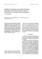
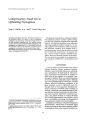
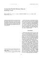
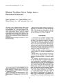
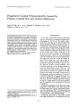
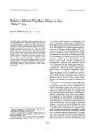
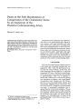
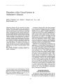
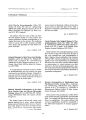
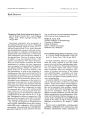
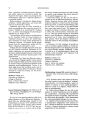
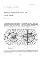
Show ] ou77IJll of Clinical Neuro- ophthalmology 10( 1): 27- 31, 1990, Compensatory Head Tilt in Upbeating Nystagmus Jorge c. Kattah, M. D., and 1. Forcht Dagi, M. D. © 1990 Raven Press, Ltd., New York Upbeating nystagmus has been described in lesions of the posterior fossa. We report a case of upbeating nystagmus accompanying a focal hemorrhagic lesion of the left brachium conjunctivum, the anterior vermis, and the anterior superior left cerebellar hemisphere. The nystagmus was suppressed by a contralateral head tilt. We postulate that in this instance, acquired central nystagmus was inhibited by the otolith- ocular reflex. Key Words: Upbeating nystagmus- Focal hemorrhagic lesion- Compensatory head tilt. From the Department of Neurology, Georgetown University Medical Center a. c. K.), and the Department of Neurosurgery, Walter Reed American Medical Center ( T. F. D.), Washington, D. C., U. s. A. Address correspondence and reprint requests to Dr, J. C. Kattah, 3800 Reservoir Road, N. W., Washington, D. C., 20007, U. S. A. 27 The purpose of this article is to describe a patient who displayed primary position upbeat nystagmus that was completely suppressed by rightward head tilt. All other head positions were associated with large- amplitude upbeat nystagmus. He initially appeared with headaches, ataxia, oscillopsia, and positional vomiting, all of sudden onset. A focal hemorrhagic lesion was identified involving the left brachium conjunctivum, the anterior vermis, and the anterior superior cerebellar hemisphere. Recovery followed resection of the lesion. Our case serves as an example of an acquired central nystagmus inhibited by a compensatory head tilt. CASE REPORT A 14- year- old boy experienced the acute onset of bifrontal headache, nausea, positional vomiting, inability to stand, left hand clumsiness, oscillopsia, and a spontaneous 30° right head tilt. The neurologic examination demonstrated an inability to stand with the feet together or to perform a tandem gait. In addition, there was marked dysmetria of the left arm and a bilaterally positive Babinski sign. Neurophthalmological examination revealed primary positional upbeating nystagmus in all head positions except a rightward head tilt, through which it was suppressed. Standard positional maneuvers provoked either vomiting or retching. Additional neuroophthalmological findings included saccadic horizontal and upward pursuit and occasional hypometric horizontal saccades. The patient showed no vertical deviation that could be demonstrated with red glass, screen cover, and Maddox rod testing. The remainder of the examination results were unremarkable. Multiplanar unenhanced computed tomography demonstrated a round lesion of increased signal density involving the left anterior superior cerebellar hemisphere, part of the superior cerebellar ver- 28 f. C. KATTAH AND T. FORCHT DAGI mis, and the left brachium conjunctivum ( Fig. 1). A discrete mass effect was noted, with moderate displacement of the fourth ventricle and the left lateral midbrain. The mass was not enhanced on i. v. contrast injection. Cerebral angiography confirmed the presence of an avascular mass in the left superior cerebellar hemisphere. Surgical exploration of the left cerebellar hemisphere disclosed a hemorrhagic cavity in this location whose contents on microscopic examination consisted of an organizing clot. Following drainage of the clot, the patient's condition gradually improved, with disappearance of the nystagmus, ataxia, and other abnormalities. The cause of the hemorrhage was not definitively elucidated; we postulate rupture of a small arteriovenous malformation or a cavernous hemangioma in the cerebellar cortex. EYE MOVEMENT RECORDINGS Conventional electrooculogram recording of horizontal and vertical eye movements was carried out shortly after admission. The testing sequence included visual fixation and effects of static head tilt on fixation, horizontal and vertical saccades, horizontal and vertical pursuit, vestibulo- ocular reflexes, and standard positional testing. Primary gaze upbeat nystagmus was maintained as long as the head was straight or tilted to the left ( Fig. 2) but was suppressed by a rightward head tilt ( Table 1). Lateral head tilt angles of 5° or more to the right abolished the nystagmus completely. The nystagmus velocity and frequency were maximal when the patient looked at a near target and the head was straight. Nystagmus was unchanged in the extreme right and left gaze. It decreased in downgaze and increased in amplitude in upgaze and in the supine position. Nystagmus became downbeating in the hanging head ( HH) and the left HH ( hyperextended) positions and was not present in the right HH position. It disappeared with simultaneous trunk and head hyperflexion, regardless of the angle of lateral head rotation. Body rotation with the head held still had no effect FIG. 1. Computed tomography scan demonstrates a round lesion of increased signal density involving the left anterior superior cerebellar hemisphere. part of the superior cerebellar vermis, and the left brachium conjunctivum. COMPENSATORY HEAD TILT IN UPBEATING NYSTAGMUS RIGHT HEAD TilT 29 ,8' ~ , ,' V " P" eo • " be I 8° r~ " L" VV. I ,., b' f ., 1 $- 4 ....... ' " .... .. ."". ,... eo LEFT HEAD TILT FIG. 2. A conventional electro- oculogram recording of horizontal and vertical eye movements shows primary gaze upbeat nystagmus was maintained as long as the head was straight or tilted to the left but was suppressed by a rightward head tilt. te' r UP f Down! l. ± 8e:> r HEAD STRAIGHT SUPINE 1m "" i''' 1 fl" li" l ml fm ''' I ~ l" m•• c on the nystagmus. Horizontal saccades of ± 15° amplitude showed normal velocity and latency, but 20% of them were hypometric. Horizontal pursuit was saccadic with a gain of 0.7. While upward pursuit was saccadic, downward pursuit was normal. Rotationally induced nystagmus, with the patient looking at a fixation target, could not be suppressed with either clockwise or counterclockwise directions. The gain of the optokinetic nystagmus was decreased in both the clockwise ( 0.15) and counterclockwise ( 0.3) directions ( normal value, 0.6). Attempts at caloric testing induced retching and precluded the recording of eye movements. DISCUSSION Head tilt occurs in a variety of clinical conditions and with the exclusion of local musculoskeletal conditions involving the neck may be classified as ocular, labyrinthine, or central in origin. Ocularly induced head tilts are by far the most frequent and typically result from superior oblique muscle paresis. Here, the head is tilted in the direction opposite to the paretic muscle to avoid diplopia ( 1). Compensatory tilts and turns may be induced by other vertical extraocular muscle palsies ( 2) and may also accompany congenital nystagmus and I Cli" Neuro- ophllullmol, Vol. 10, No. 1, 1990 30 1. c. KATTAH AND T. FORCHT DAGI TABLE 1. Nystagmus characteristics spasmus nutans ( 3-- 6). By decreasing the amplitude of the nystagmus, these head attitudes may improve visual acuity. Peripheral labyrinthine lesions may be signalled by head tilt ipsilateral to the affected ear ( 7,8). Both the labyrinth and the brain stem lesions have been imputed in the so- called " ocular tilt reaction" ( OTR), a synkinesis characterized by head tilt and vertical divergence and torsion of the eyes ( 9). Experimental animal data and clinical observations in man suggest that OTR may result from midbrain lesions at the level of the interstitial nucleus of Cajal ( 10,11) that disturb the tonic otolithvestibular regulatory mechanism that controls vertical ocular alignment and head posture. The OTR may be either paroxysmal or sustained, with a head tilt ipsilateral to the lesion ( 9- 12). Likewise, medullary lesions may be associated with OTR ( 12). Finally, cerebellar mass lesions also result in head tilt. The direction is unpredictable, and the mechanism is not defined. The head may tilt toward or away from the lesion ( 8). We concluded that our patient probably had a voluntary compensatory head tilt away from the lesion that curbed the oscillopsia. This head tilt stimulated a normal otolith- ocular reflex with nystagmus suppression. Additional inhibitory signals from the neck could have contributed to nystagmus suppression. The abnormal head postures that develop in torticollis and other dystonic conditions are different. They are distinguished from other attitudinal abnormalities of the head by the rigidity. They are generally held to be primary rather than compensatory. Furthermore, eye movements are generally normal in dystonic conditions with the exception of the oculogyric crises induced by phenothiazines and other neurotropic agents. Primary position upbeating nystagmus has been described with lesions at different levels of the brain stem. The majority of cases studied pathologically involved the pontomedullary junction ( 13) and the perihvpoglossal medullary nuclear " I'ni~ l' ( ll a recently described Head position Right head tilt Head center left head tilt Head center and convergence Bending forward Hanging head Direction o Upbeating Upbeating Upbeating o Downbeating Slow phase velocity ( deg/ s) o 27 17 40 o 10 Frequency ( Hz) o32 4o2 pathway that mediates the vertical vestibulocular reflex, the ventral tegmental pathway, may explain most of the cases with medullary and caudal pontine lesions ( 15). Lesions affecting the brachium conjunctivum, similar to that seen in our patient, have been described previously ( 16,17). In one instance, upbeat nystagmus was associated with contrapulsion of the eyes. Finally, the involvement of the anterior superior cerebellar vermis, seen in our case and previously described in isolated lesions in this location ( 15), may have contributed to the development of upbeating nystagmus. Thus, lesions affecting the two proposed projections from the superior vestibular nucleus, namely, the brachium conjunctivum or the ventral tegmental pathway ( 16), may be responsible for the generation of this type of nystagmus. The mechanism of imbalance in ocular position that accounts for upbeat nystagmus has not been completely elucidated. Defective pursuit tone is postulated as a likely possibility ( 19,20). Alternative explanations include defective input from the anterior semicircular canals resulting in defective upward vestibulo- ocular reflex ( 17). Because this pathway projects to the oculomotor neurons via the brachium conjunctivum, a lesion in this structure can potentially result in upbeat nystagmus. Although the brachium conjunctivum was partially involved in our patient, there was no component of rotatory nystagmus. This suggests that the operant mechanism involved factors other than the pure interruption of efferents from the anterior semicircular canals ( 21). The variability in nystagmus velocity, frequency, amplitude, and in some cases direction that we encountered in our patient has been noted previously ( 22) and suggests that primary upbeat nystagmus may be modified by head position. Static tilt suppression of upbeat nystagmus has been previously reported on only one occasion, to our knowledge ( 22). While failing to explain the mechanism of upbeat nystagmus, static tilt suppression illustrates how otolithic stimulation may modify primary position central nystagmus. As a compensatory maneuver, static tilt suppression seems to improve visual acuity in some patients with congenital nystagmus and most patients with spasmus nutans and was a valuable spontaneous factor in our patient. REFERENCES 1. Kushner BJ. Ocular causes of abnormal head postures. Ophthalmology 1979; 86: 2115- 25. 2. Uris! MJ. Head tilt in vertical muscle paresis. Am JOphthalmol 1970; 69: 440-- 2. J Clin Nf! Uro- ophthalmol, Vul. lV. No. 1, J~ 9() COMPENSATORY HEAD TILT IN UPBEATING NYSTAGMUS 31 3. Van Noorden GK, Munoz M, Wong SY. Compensatory mechanisms in congenital nystagmus. Am 1 Ophthalmol 1987; 104: 307- 17. 4. Jayualakshmi P, McNair S, Tucker SH, Schaeffer DB. Infantile nystagmus. A prospective study of spasmus nutans, congenital and unclassified nystagmus of infancy. 1Pediatr 1970; 77: 177417. 5. Chrousos GA, Reingold DR, Chu FC, Cogan DG. Habitual head turning in spasmus nutans. An oculographic study. 1 Pediatr Ophthalmol Strabismus 1985; 22: 113-- 6. 6. Gresty M, Leech J, Sanders M, Eggars H. A study of head and eye movement in spasmus nutans. Br 1 Ophthalmol 1976; 60: 652- 4. 7. Halmagyi MG, Gresty MA, Gibson WPR. Ocular tilt reaction with peripheral vestibular lesion. Ann Neurol 1979; 6: 80-- 3. 8. Brain RW. On the rotated or " cerebellar" posture of the head. Brain 1926; 49: 61- 76. 9. Westheimer G, Blair SM. The ocular tilt reactionbrainstem oculomotor routine. Invest Ophthalmol 1975; 14: 83~ 9. 10. Rabinovitch HE, Sharpe JA, Sylvester TO. The ocular tilt reaction. Arch OphthalmoI1977; 95: 139541. 11. Hedges TR, Hoyt WF. Ocular tilt reaction due to an upper brainstem lesion: paroxysmal skew deviation, torsion and oscillation of the eyes with head tilt. Ann Neurol 1982; 11: 537- 40. 12. Brandt T, Dietrich M. Pathologic eye- head coordination in roll: tonic ocular tilt reaction in mesencephalic and medullary lesions. Brain 1987; 110: 64~ 6. 13. Gilman N, Baloh RW, TOmiyasu U. Primary position upbeat nystagmus. A c1inico- pathologic study. Neurology 1977; 27: 2944l. 14. Keane JR, Itabashi HH. Upbeat nystagmus: a clinicopathologic study of the patients. Neurology 1987; 37: 491- 4. 15. Ranalli PJ, Sharpe JA. Upbeat nystagmus and the ventral tegmental pathway of the upward vestibulo- ocular reflex. Neurology 1988; 38: 1329- 30. 16. Nakada T, Remler MP. Primary position upbeat nystagmus. 1Clin Neuro OphthalmoI1981; 1: 185-- 9. 17. Benjamin EE, Zimmerman CF, Troost TB. Lateropulsion and upbeat nystagmus on manifestations of central vestibular dysfunction. Arch NeuroI1986; 43: 962- 4. 18. Daroff RB, Troost TB. Upbeat nystagmus. lAMA 1973; 225: 312. 19. Zee 0, Friendlich A, Robinson D. The mechanism of downbeat nystagmus. Arch Neurol 1974; 30: 227- 37. 20. Medhom E, Kommerell G, Meienberg O. Primary position vertical nystagmus " directional preponderance of the pursuit system." Graefes Arch Clin Exp OphthalmoI1979; 209: 209- 17. 21. Zee OS. The organization of the brainstem ocular motor subnuclei. Ann NeuroI1978; 4: 384- 5. 22. Fisher A, Gresty M, Chamber B, Rudge P. Primary position upbeating nystagmus. A variety of central positional nystagmus. Brain 1983; 106: 94~. 1Clin Neuro- ophthalmol, Vol. 10, No. 1, 1990 |