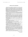
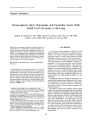
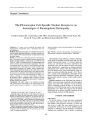
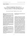
| OCR Text |
Show Journal of Neuro- Ophthalmology 21( 3): 173- 187, 2001. © 2001 Lippincott Williams & Wilkins, Inc., Philadelphia Original Contribution Clinical and Immunologic Characteristics of Melanoma- Associated Retinopathy Syndrome: Eleven New Cases and a Review of 51 Previously Published Cases John L. Keltner, MD, Charles E. ThirkiU, PhD, and Peter T. Yip, MD Objective: To evaluate the signs, symptoms, and immune responses of patients with melanoma- associated retinopathy ( MAR) syndrome. Materials and Methods: We reviewed the clinical and immunologic findings of 62 MAR syndrome patients. They include 25 patients from our institution ( 11 not previously reported) and 37 patients reported from other institutions. Results: There were 33 men and seven women ( no gender information is available for the remaining 22 cases). Age at onset of the visual disturbance averaged 57.5 years ( range, 30- 78). Visual acuity of 20/ 60 or better was initially present in 82%. Fundus examination was normal in 44%, optic disc pallor was present in 23%, and retinal vessel attenuation was present in 30%. Vitreous cells were present in 30%. The latency from melanoma diagnosis to recognition of MAR syndrome averaged 3.6 years ( range, 2 months to 19 years). Seven patients sustained visual improvement with various treatment regimens, especially with intravenous immunoglobulin and cytoreductive surgery ( metastasectomy). Indirect immunohistochemical staining of the bipolar layer was typical, but several other retinal elements were also reactive. Tissue from a metastatic melanoma excised from one of the patients expressed antigens that reacted with antiretinal antibodies. Conclusion: MAR syndrome demonstrates diverse clinical and immunologic features. Treatment, especially intravenous immunoglobulin and cytoreductive surgery ( metastasectomy), improves vision in some cases. Manuscript received May 15, 2001; accepted August 2, 2001. Supported by National Eye Institute ROl EYO9063, National Eye Institute Core Grant 1 P30 EY12576- 01and an unrestricted grant from Research to Prevent Blindness. Presented in part as a presentation at the 24th Annual North American Neuro- Ophthalmology Society ( NANOS) Meeting, March 22- 26, 1998, Buena Vista Palace Resort, Orlando, Florida, and at the 27th Annual NANOS Meeting, February 18- 22, 2001, Westin Mission Hills Resort, Palm Springs, California. Departments of Ophthalmology ( ILK, CET, PTY) and Neurology and Neurological Surgery ( ILK), University of California, Davis, Sacramento, California. Address correspondence and reprint requests to lohn L. Keltner, MD, University of California, Davis, 4860 Y Street, Suite 2400, Sacramento, CA 95817, USA; e- mail: jlkeltner@ ucdavis. edu The authors have no proprietary interest in any product or procedure described in this article. Key Words: Melanoma- associated retinopathy- Immunology- Photopsia- Antiretinal antibodies- Intravenous immunoglobulin treatment. Paraneoplastic syndromes result from the immunologic effects of cancer located remote from the affected organ. With respect to vision loss, cancer- associated retinopathy ( CAR) ( 1- 9) and melanoma- associated retinopathy ( MAR) ( 1,10- 39) are now well- recognized entities. A lesser recognized entity is autoimmune- related retinopathy and optic neuropathy ( ARRON) syndrome, in which patients have no evidence of cancer ( 40- 45). CAR syndrome is a paraneoplastic retinal degeneration often associated with small cell carcinoma of the lung but is also described in patients with gynecologic, breast, endocrine, and other malignancies. It involves antibodies against retinal elements and causes both rod and cone dysfunction. Although the 23- kd photoreceptor protein recoverin was the first and the most common retinal antigen linked to CAR syndrome, more than 15 other proteins expressed by rods, cones, and ganglion cells of the retina are now thought to act as potential autoantigens ( 45). In many cases, the CAR antigen is also expressed by the patient's cancer. Visual loss in CAR syndrome is marked by photopsias, progresses over several months, and frequently precedes the discovery of the primary cancer. Features associated with cone dysfunction are photosensitivity, abnormal visual acuity, color vision abnormalities, central scotomas, and an abnormal cone- mediated electroretinogram ( ERG). Features associated with rod dysfunction are nyctalopia, prolonged dark adaptation, peripheral or ring scotomas, and an abnormal rod- mediated ERG ( 1- 9). ARRON syndrome describes patients with autoimmune- related retinopathy and optic neuropathy who do not have evidence of cancer. They may have optic disc pallor, retinopathic changes, progressive visual field and visual acuity loss, and an abnormal ERG resembling 173 174 J. L. KELTNERETAL. that of patients with CAR syndrome. However, these features develop more slowly. ARRON patients often have systemic autoimmune disorders. They may develop one or multiple autoantibodies against elements in the retina and/ or optic nerve. Various investigators have reported ARRON patients with autoantibodies directed against the inner plexiform layer- 35- kd retinal Mtiller cell antigen, 23- kd recoverin protein ( not related to cancer), and a 22- kd neuronal antigen in the retina and optic nerve ( 40- 45). Other investigators have found noncancer- related, progressive panretinal degeneration with CAR- like clinical changes and antibodies to several retinal proteins ( 44). It is unclear whether the autoimmune serologic reactions cause visual loss or are simply epiphenomena. In the retinal degeneration associated with cutaneous melanoma- MAR syndrome- patients frequently have an established diagnosis of cutaneous melanoma and develop vision problems years later, usually associated with nonocular metastasis. Patients frequently describe the sudden onset of shimmering, flickering, or pulsating photopsias and difficulty seeing in the dark, together with progressive visual loss over several months ( 1,10- 39). The ERG reveals a characteristic pattern of a markedly reduced B wave, indicating compromised bipolar cell function, and a normal dark- adapted A wave ( negative appearance), indicating normal photoreceptor cell function. Similar ERG findings are observed in congenital stationary night blindness ( CSNB). The characteristic immunohistochemical autoimmune response involves antigens located within the bipolar layer, where horizontal and amacrine cells intermingle with axons of Muller's glia ( 1,10- 39). This immunologic reaction differs from that of CAR in that the associated antigens are not readily identifiable in Western blot reactions on extracts of retina. The antigens involved are either small quantities of proteins, proteoglycans ( 16), lipids ( 16,20), or carbohydrates. As with other paraneoplastic retinopathies, MAR syndrome is rare ( 10- 39). Because some authors have reported the same case more than once, the exact number of reported cases is difficult to determine. We have serum samples from 25 MAR patients, collected from 1992 to 2000. Features of 14 of these cases have been previously documented; 11 cases are described here for the first time. In this report, we add the features of these 11 cases to the 51 cases previously reported. METHODS We evaluated the 11 previously unreported MAR patients at the University of California, Davis Ophthalmology Research Laboratory by Western blot analyses on an extract of whole rhesus monkey retina and indirect immunohistochemistry on sectioned rhesus monkey eyes. The details of these tests were previously described ( 5,46). Inclusion criteria for MAR syndrome were a history of malignant melanoma and antibody reactions to retinal cells in the bipolar layer. Clinical symptoms and ERG changes suggestive of MAR syndrome were used to confirm the diagnosis when available. We compared the clinical and immunologic features of our new cases to those of previously reported cases fulfilling the same inclusion criteria. A formalin- fixed and paraffin- embedded metastatic melanoma removed from the left axilla of patient 58 ( Table 1) was evaluated for immunologic activity with rabbit anti- whole bovine retina serum. Six- micron sections of the melanoma were made. These sections were deparaffinized in xylene and brought through ethanol to saline where they were exposed to a 1: 10 dilution of rabbit anti- whole bovine retina serum overnight at 4° C. The rabbit anti- retina antibody localization on specific components of the melanoma was visualized using fluo-rescein- isothiocyanate conjugated goat anti- rabbit polyvalent gamma globulins, at a dilution of 1: 200. After a thorough wash in phosphate- buffered saline- Tween, sections were mounted in 50% glycerol in saline and then examined and photographed at a final magnification of 200x. Because the information in this study is recorded and presented such that subjects cannot be identified, exemption from review was approved by the Human Subjects Review Committee, Office of the Vice Chancellor for Research, the University of California, Davis Medical Center. RESULTS Clinical information from all known cases of MAR patients is summarized chronologically in Tables 1 and 2. Clinical characteristics are tabulated in Tables 3 through 10. Previously reported cases are listed as patients 1 through 51. The newly reported patients from our institution are patients 52 through 62. In the tables, we have lumped the previously reported and newly reported cases from our institution as UC Davis ( 25 serum samples) cases and referred to all other patients as patients from other institutions. Age and gender The average age was 57.5 years ( range, 30- 78). There were 33 men and seven women ( no gender information is available for the remaining cases) ( Table 3). Presenting visual acuity Visual acuity at presentation was 20/ 60 or better in 28 ( 82%) of 34 patients for whom such data were available. In six of these 34 patients at presentation, moderate to severe visual loss ( 20/ 200 or worse) was present in one or both eyes. In nine of 20 patients, the last recorded visual acuity demonstrated moderate to severe visual loss ( 20/ 200 or worse) in one or both eyes ( Table 4). Presenting visual fields Visual fields showed generalized constriction in 18 of 27 patients where such information was available. Eighteen of 27 had central or paracentral scotomas or depressions, and six of 27 had arcuate visual field defects ( Table 5) Presenting fundus findings The fundus was normal in 19 ( 44%) of 43 patients where such information was available ( Table 6). / Neuro- Ophthalmol, Vol. 21, No. 3, 2001 MAR SYNDROME IN 62 PATIENTS: UPDATE 2001 175 TABLE 1. Ophthalmic features of reported melanoma- associated retinopathy ( MAR) cases Age/ Patient no./ authors gender VA initial VF Fundus Vitreous ERG pattern Last recorded visual data 1/ Gass, 1984 ( 10) 71/ F 2/ Ripps et al., 1984 30/ M ( 11) 3/ DuBois et al., 1988 38/ M ( 12) 4/ Berson and Lessell, 69/ M 1988 ( 13) 5/ Alexander et al., 58/ M 1992 ( 14) 6/ MacKay et al., 63/ M 1992 ( 15) 7- 9/ Pollock et al., 3 cases 10/ Milam et al., 1993 36/ M ( 16) f 11/ Rushetal., 1993 50/ M ( 17) 12/ Andreasson et al., 48/ M 1993 ( 18) 13, 14/ Weinstein et 46/ F al., 1994 ( 19) 46/ M 15- 17/ Kim et al., 61/ M 1994 ( 21) 58/ M 78/ M 18- 27/ Milam, 1995 10 cases| ( 20)* 62/ M$ a 28/ Remulla et al., 52/ M 1995 ( 26) 29/ Singh et al., 1995 64/ M ( 27) 30/ Kellner et al, 1995 ( 23) 44/ M 31/ Okel et al., 1995 62/ M ( 22) 32/ Rougier et al, 1995 ( 24) Bret- Dibat et al, 1996 ( 25) § Wolf and Arden 1996 ( 28)!' 61/ M No new case OD LP, OS LP OD 20/ 20, OS 20/ 20 OD 20/ 20, OS 20/ 20 OD 20/ 25, OS 20/ 20 OD 20/ 25, OS 20/ 25 OD 20/ 20, OS 20/ 20 Slight depression OU Constricted peripheral VF Arterioles attenuated OU OU, OD hemianopic depression, OS paracentral scotoma Mild depression OU OD enlarged blind spot, parafoveal scotoma; OS normal ultiple yellow- white depigmentation in juxtapapillary and perimacular areas, arterioles attenuated. optic nerves swollen, OU jrmal OU 2+ aqueous cells and flare, vitreous cells OU - Extinguished OU CSNB OD 20/ 50, OS 6/ 200 Occasional window defects in RPE OU RPE mottling, slight vascular attenuation OU OD 20/ 20, OS Central depression OU 20/ 40 OD 20/ 40, OS 20/ 40 OD 20/ 25, OS 20/ 25 OU 1/ 200 OD 20/ 20, OS 20/ 20 OD 20/ 20, OS 20/ 20 OD 20/ 40, OS 20/ 30 OU 20/ 1200 OD general depression and nasal constriction, OS arcuate defects No peripheral constriction Normal OU with V: 4e on GMVF OU Central scotomas, constriction OU OD arcuate defects, OS constriction RPE mottling in the midperiphery, arteries slightly attenuated OU Arterial attenuation OU OU fine vitreous cells Optic nerve pallor, vessel attenuation OU Normal OU 9 had VF loss OD 20/ 20, OS 20/ 20 OD 20/ 40, OS 20/ 30 OD 20/ 20. OS 20/ 20 OD 20/ 40, OS 20/ 25 OD 20/ 30, OS 20/ 60 Central scotomas, constriction, enlarged blind spots OU Marked constriction and peripheral scotomas OU, OD paracentral scotoma Constriction of nasal peripheral field OU, OD interior arcuate scotoma Constriction OU, OD central scotoma, OS paracentral scotoma OD 20/ 20, OS OD normal, OS tubular 20/ 25 field OD pseudo- exfoliative glaucoma, 0.8 cupidisk, OS normal Drusen OU, otherwise normal OD ARMD with disciform scar, OS ARMD with geographic atrophy 9 had a normal fundus Optic nerves normal, vessels attenuated, RPE pigment mottling, epiretinal membrane OU Optic nerves slightly pale, vessels attenuated, extramacular RPE changes OU Normal discs and vasculature, RPE loss OU OD paracentral scar, OS CME Chorioretinal atrophy surrounding nerve heads and maculae, retinal vascular attenuation OU Normal OU Few vitreous cells OU 3 mild inflammation, 1 severe inflammation 2- 3+ vitreous cells, retinal periphlebitis OU CSNB CSNB CSNB CSNB CSNB CSNB CSNB CSNB CSNB CSNB CSNB CSNB CSNB CSNB CSNB CSNB CSNB 3+ vitreous cells OU CSNB No ERG CSNB OD 20/ 25, OS, 20/ 25, no change in ERG Decreased haze and improved GMVF OU Progressive VF loss OU OU 1/ 200 OD 20/ 30, OS 20/ 70 VA remained relatively normal in 10 patients OD 20/ 200, OS 20/ 200, photopsia resolved OD 6/ 400, OS 20/ 40 VA stable for 12 mo OD 20/ 400, OS 20/ 40 OD LP, OS LP OD 20/ 25, OS 20/ 30 No new case" J Neuro- Ophthalmol, Vol. 21, No. 3, 2001 176 J. L. KELTNERETAL. TABLE 1. Continued Patient no./ authors Age/ gender VA initial VF Fundus Vitreous ERG pattern Last recorded visual data OD normal, No improvement OS CSNB CSNB No improvement 33/ Kiratli et al., 1997 ( 29) 34/ Boeck et al., 1997 ( 30) Klopfer et al., 1997 ( 31) H 35/ Gittinger and Smith, 1999 ( 32) 36/ Potter et al., 1999 ( 33) 66/ M 51/ M No new casefl 59/ M 61/ M OD 20/ 30, OS Arcuate defects OU 20/ 60 OD 20/ 50, OS Marked constriction OU 20/ 40 OD 20/ 30, OS OD contriction, OS 6/ 200 OD 10/ 200, OS 20/ 200 constriction and central scotoma Central scotomas, peripheral constriction OU Normal OU Normal OU Optic nerves pale, blonde fundus with fine macular drusen OU Optic nerves pale, vessels attenuated, macular RPE change OU 1+ vitreous cells OD, trace cells OS CSNB CSNB No change in vision OD 4/ 200, OS 1/ 200 37/ McCoy and Hedges, 1999** 38/ Lei et al., 2000 ( 34) tt 39/ Vaphiades et al. 2000 ( 35) 40/ Feigl et al., 2000 ( 36) 41/ Flynn et al., 2000 ( 37) 42^ 8/ Holder, 2000 ( 38) 49- 51/ Haus, 2000 ( 39)** 52/ UC Davis patient 1 53/ UC Davis patient 2 54/ UC Davis patient 3 55/ UC Davis patient 4 56/ UC Davis patient 5 57/ UC Davis patient 6 58/ UC Davis patient 7 59/ UC Davis patient 8 § § 60/ UC Davis patient 9 § § 61/ UC Davis patient io § § 62/ UC Davis patient n § § 55/ M 1 casef f 57/ F 67/ M 1 case 7 cases 51/ M** 62/ M** 77/ M** 39/ F 68/ M 78/ F 47/ M 76/ F 42/ M 75/ M - M M F OD 20/ 20, OS 20/ 60 - OD 20/ 25, OS 20/ 25 OD 20/ 25, OS 20/ 32 - - - - - OD 20/ 25, OS 20/ 20 ODCF3', OS CF 3' OD 20/ 30, OS 20/ 25 OD 20/ 20, OS 20/ 20 OD 20/ 50, OS 20/ 50 OD 20/ 20, OS 20/ 20 OD 20/ 20, OS 20/ 20 - - - - OD paracentral scotoma, OS central depression - Constriction OU Constriction, central, and paracentral scotoma OU - - - - - Central scotomas OU Central scotomas, marked constriction OU Central scotomas, arcuate defects, constriction OU Constriction OS > OD Constriction OU OD arcuate defects and constriction; OS double arcuate defect and constriction Constriction and depression OU - - - - Normal OU - OD normal, OS anomalous optic disk vasculature Normal OU - - - - - Optic nerves pale, vessels attenuated OU Optic nerves pallor OU Optic nerves pallor, vessels attenuation, RPE changes OU Optic nerves pallor, vessels attenuation, macular RPE changes OU Optic nerves pallor OU Normal OU Mild optic nerve pallor, RPE changes OU - - - - - - - - - - - - - 2+ cells OD, 1+ cells OS 3+ cells OU - Trace cells OD - - - - CSNB - CSNB CSNB CSNB CSNB CSNB CSNB CSNB CSNB CSNB CSNB CSNB CSNB ERG almost Improved GMVF and ERGOD - OD 20/ 25, OS 20/ 30, improved color vision and GMVF, photopsia resolved No improvement - - - - - Return of color vision, scotomas smaller, improved VF and dark adaptation CMEOU OD 20/ 20, OS 20/ 20 OD 20/ 20, OS 20/ 20 OD 20/ 60, OS 20/ 60, VF expanded and back to baseline OD 20/ 20, OS 20/ 40, extinguished Improved VFs CSNB - - - - Improved VFs and visual symptoms - - - - * Pollock, et al., Melanoma- associated retinopathy ( MAR), presented as a poster at the NANOS meeting, 1992. f Two cases reported. One of them had been reported previously by Alexander et al., 1992 ( 14). | Twelve cases reported. Two of them had been reported previously by Alexander et al., 1992 ( 14) and Milam et al., 1993 ( 16). a One patient's clinical information was available through personal communication with Dr. Leah Levi, San Diego, California. § One case reported. It had been reported previously by Rougier et al., 1995 ( 24) " Three cases reported, all had been reported previously by Kim et al., 1994 ( 21) and Kellner et al., 1995 ( 23). fl One case reported. It had been reported previously by Boeck et al., 1997 ( 30). ** McCoy D, Hedges T. Patient presents with photopsias. Ocul Surg News 1999; Feb 1: 53, 58. ff Three cases reported; two of them had been reported by Kim et al., 1994 ( 21) and Kellner et al., 1995 ( 23); one is new and without clinical information. | | Twenty- three patients were reported to be suspicious for MAR syndrome in the ARVO 2000 abstract. Through personal communication with Dr. Arno Haus, Homburg/ Saar, Germany, we learned that only three of these patients had antiretinal antibody activities in their sera and met the inclusion criteria for MAR syndrome. § § Serum samples only, no clinical information. ARMD, age- related macular degeneration; CF, Count Fingers; CME, cystic macular edema; CSNB, congenital stationary night blindness; ERG, electroretinogram; GMVF, Goldmann visual field; PION, posterior ischemic optic neuropathy; RPE, retinal pigment epithelium; VA, visual acuity; VF, visual field. / Neuro- Ophthalmol, Vol. 21, No. 3, 2001 MAR SYNDROME IN 62 PATIENTS: UPDATE 2001 177 TABLE 2. Nonophthalmic features of reported metanoma- associated retinopathy ( MAR) cases Patient no./ authors 1/ Gass, 1984 ( 10) 2/ Ripps et al., 1984( 11) 3/ DuBois et al, 1988 ( 12) 4/ Berson and Lessell, 1988 ( 13) 5/ Alexander et al, 1992 ( 14) 6/ MacKay et al, 1992 ( 15) 7- 9/ Pollock et al, 1992* 10/ Milam et al, 1993 ( 16) f 11/ Rush etal., 1993 ( 17) 12/ Andreasson et al., 1993 ( 18) 13, 14/ Weinstein et al., 1994 ( 19) 15- 17/ Kim et al., 1994 ( 21) 18- 27/ Milam, 1995 ( 20)* 28/ Remulla et al., 1995 Melanoma to Melanoma metastasis to MAR ( y) ( y) 3 3 - 1.5 MAR syndrome presented first, then 2 y later malignant melanoma developed 3 2.5 Onset with metastatic melanoma, MAR developed 15 y later - - - - 3.6 3.7 Onset with metastatic melanoma, no primary; MAR developed 1 mo later Onset with metastatic melanoma, MAR developed 2 y later 0.8 1.3 0.2 0.2 - 2 0.8 1.5 Onset with metastatic melanoma, MAR developed a few months later 3.5 2 | a 2.2 Onset with metastatic melanoma, Melanoma to survival ( y) 5.1 ( died) 4 ( died) - - 5 ( died) 7.5 - - 3 ( died) 5 ( died) - 8 ( died) 7.3 Melanoma treatment Surgery Surgery and extensive chemo - Surgery and chemo Surgery and chemo - - Surgery Surgery Surgery Surgery Surgery Surgery Surgery, radiation and extensive chemo Surgery and chemo - Surgery, chemo, radiation, interferon, vaccine Surgery for multiple recurrent MAR treatment Prednisone 80 mg None - None Prednisone 80 mg - - Prednisone 60 mg Plasmapheresis None Oral prednisone, subtenon's Medrol injection None None None None - Radiation therapy Prednisone 80 mg Serum analyzed atUC Davis No No No No No No No No Yes No Yes No No No No No Yes Yes ( 26) 29/ Singh et al., 1995 ( 27) 30/ Kellner et al., 1995 ( 23) 31/ Okeletal., 1995 ( 22) 32/ Rougier et al., 1995 ( 24) Bret- Dibat et al., 1996 ( 25) § Wolf & Arden 1996 ( 28)" 33/ Kiratli et al., 1997 ( 29) 34/ Boeck et al., 1997 ( 30) Klopfer et al., 1997 ( 31) H 35/ Gittinger and Smith, 1999 ( 32) 36/ Potter et al., 1999 ( 33) 37/ McCoy and Hedges, 38/ Lei et al., 2000 ( 34) ff 39/ Vaphiades et al., 2000 ( 35) 40/ Feigl et al., 2000 ( 36) MAR developed 2.4 y later MAR presented first, 2 wk later a maxillary sinus malignant melanoma was found; no metastasis 0.9 1.8 metastatic lesions Surgery Surgery, interferon None Prednisone 19 1.3 19 1.1 Metastasis only, no primary cutaneous melanoma 4 4.1 4 4.5 1.6 Metastasis only, no primary cutaneous melanoma 19.5 ( died) Surgery Surgery, chemo 1 ( died) Surgery, chemo 7 Surgery, BCG, chemo 5.7 ( died) Surgery, interleukin- 2, interferon, radiation 5 Surgery, minimal chemo None None None Prednisone Surgery 3 Cytoreductive surgery metastatic melanoma, interferon- alpha postmetastatic tumor removal 2 ( died) Surgery, interferon- alpha, isotretinoin None None Prednisone, azathioprine, plasmapheresis, gabapentin IVIg 30 g/ mo x 16 None No No Yes No Yes Yes No Yes Yes No Yes 41/ Flynn et al., 2000 ( 37) 42^ 18/ Holder, 2000 ( 38) 49- 51/ Haus et al., 2000 ( 39)** 52/ UC Davis patient 1 53/ UC Davis patient 2 54/ UC Davis patient 3 55/ UC Davis patient 4 0.3 - No metastasis 6 - - - 0.5 13 2 7 - - - 3.5 15 5 8 ( died) - - - Cytoreductive surgery Surgery Surgery Surgery, interleukin, radiation, chemo - - - Prednisone 60 mg, 2 mo None None IVIg for 5 d No No Yes Yes Yes Yes Yes J Neuro- Ophthalmol, Vol. 21, No. 3, 2001 178 J. L. KELTNERETAL. TABLE 2. Continued Patient no./ authors 56/ UC Davis patient 5 57/ UC Davis patient 6 58/ UC Davis patient 7 59/ UC Davis patient 8 § § 60/ UC Davis patient 9 § § 61/ UC Davis patient io § § 62/ UC Davis patient n § § Melanoma to Melanoma metastasis to MAR ( y) ( y) No metastasis 3 MAR presented first, 4 mo later, a left axillary lymph node metastatic melanoma was found; no primary cutaneous melanoma 4 3.5 - - - - - - - - Melanoma to survival ( y) 4 4.3 4.5 - - - - Melanoma treatment Surgery Cytoreductive surgery Metastatic melanoma removal with cytoreductive surgery - - - - MAR treatment IVMP 125 mg bid, plasmapheresis x 5, oral prednisone taper None Prednisone for 10 d -> not improved. IVIg 200- 220 g/ mo x 9 -> improved - - - - Serum analyzed atUC Davis Yes Yes Yes Yes Yes Yes Yes * Pollock, et al., Melanoma- associated retinopathy ( MAR), presented as a poster at the NANOS meeting, 1992. f Two cases reported. One of them had been reported previously by Alexander et al., 1992 ( 14). | Twelve cases reported. Two of them had been reported previously by Alexander et al., 1992 ( 14) and Milam et al., 1993 ( 16). a One patient's clinical information was available through personal communication with Dr. Leah Levi, San Diego, California ( Patient 27). § One case reported. It had been reported previously by Rougier et al., 1995 ( 24) " Three cases reported, all had been reported previously by Kim et al., 1994 ( 21) and Kellner et al., 1995 ( 23). fl One case reported. It had been reported previously by Boeck et al., 1997 ( 30). ** McCoy D, Hedges T. Patient presents with photopsias. Ocul Surg News 1999; Feb 1: 53, 58. ff Three cases reported; two of them had been reported by Kim et al., 1994 ( 21) and Kellner et al., 1995 ( 23); one is new and without clinical information. $$ Twenty- three patients were reported to be suspicious for MAR syndrome in the ARVO 2000 abstract. Through personal communication with Dr. Arno Haus, Homburg/ Saar, Germany, we learned that only three of these patients had antiretinal antibody activities in their sera and met the inclusion criteria for MAR syndrome. § § Serum samples only, no clinical information. BCG, bacille Calmette- Guerin; bid, twice a day; ERG, electroretinogram; IVIg, intravenous immunoglobulin, IVMP, intravenous methylprednisolone. Optic nerve pallor, vessel attenuation, retinal pigment epithelium changes, and vitreous cells were seen in a number of patients. One patient had marked retinal periphlebitis ( 26). Electroretinography The ERGs of 54 of 56 patients for whom such data were available exhibited a markedly reduced B wave and a normal dark- adapted A wave ( Table 7). Similar ERG findings are seen in patients with congenital stationary night blindness. In one of the 54 patients, only one eye had an abnormal ERG. The remaining two patients showed diffuse loss of the A wave and B wave in both eyes. Latency from diagnosis of primary to metastatic melanoma In 17 patients, the average time from malignant melanoma diagnosis to metastatic disease was 3.5 years ( range, 2 months to 19 years) ( Table 8). However, three patients ( two UC Davis, one other institutions) had no metastasis, and four patients ( four UC Davis) were found TABLE 3. Available age and gender information UC Davis patients ( n = 24) Patients from other institutions ( n = 16) Total ( n = 40) Average age, ( range) No. of men No. of women 59.2 ( 39- 78) 18 6 55.3 ( 30- 78) 15 1 57.5 ( 30- 78) 33 7 to have metastatic disease but no history of primary cutaneous melanoma. Four patients ( one UC Davis, three other institutions) presented with metastatic cutaneous melanoma. Latency from diagnosis of primary melanoma to MAR syndrome In 32 patients, the time from the diagnosis of skin melanoma to diagnosis of MAR syndrome averaged 3.6 years ( range, 2 months to 19 years) ( Table 8). One patient ( one other institutions) presented with MAR syndrome first, then 2 weeks later was found to have a maxillary sinus malignant melanoma. Another patient ( one other institutions) also presented with MAR syndrome first, then 2 years later developed malignant melanoma. Latency from diagnosis of metastatic melanoma to MAR syndrome In 13 patients, the elapsed time from diagnosis of metastatic disease to diagnosis of MAR syndrome averaged 1.9 years ( range, 1 month to 15 years) ( Table 8). MAR syndrome appeared within 1 year after recognition of metastasis in 10 patients ( ten other institutions, not in averages because time of MAR presentation is unknown) ( 20). Five patients ( three UC Davis, two other institutions) were found to have metastatic disease and MAR simultaneously. Two of the UC Davis patients had no history of primary melanoma. Six patients ( four UC Davis, two other institutions) were discovered to have metastatic disease after the diagnosis of MAR, with an / Neuro- Ophthalmol, Vol. 21, No. 3, 2001 MAR SYNDROME IN 62 TABLE 4. UC Davis patients Visual acuity 2= 20/ 60 < 20/ 60 At presentation 15 3 Last recorded 6 6 average latency of 6.8 months ( range, 2 months to 1.5 years). Three patients ( two UC Davis, one other institutions) had no evidence of metastatic disease. Survival time after diagnosis of cutaneous melanoma Survival time after diagnosis of cutaneous melanoma averaged 5.9 years ( range, 1- 19.5) ( Table 9). Five patients from UC Davis died after 1,2, 8 ( two patients), and 19 years; six patients from other institutions died after 3, 4, and 5 years ( four patients). Treatment of melanoma Eighteen patients had treatment limited to excision of melanoma or metastatic disease. Nine patients are alive, and three patients have died ( average lifespan, 6.9 years) from progression of metastatic disease. In six patients, there was no survival information. Six UC Davis patients and seven patients from other institutions had a variety of other treatments for the cutaneous melanoma, which included cytoreductive surgery, chemotherapy, radiation, and immunotherapy. Treatment of MAR syndrome Treatment consisted of oral, subtenon's, or intravenous corticosteroids, plasmapheresis, intravenous immunoglobulin ( IVIg), azathioprine, gabapentin, and x-irradiation of metastases or cytoreductive surgery. Oral prednisone alone was not beneficial in six of seven patients treated; one patient ( patient 1) had such dramatic visual improvement that the diagnosis must be questioned ( 10) ( Table 10). Plasmapheresis alone was not beneficial in one patient ( patient 11) ( 17). The combination of oral prednisone, plasmapheresis, azathioprine, and gabapentin helped one patient with slight improvement in Goldmann visual fields and ERG ( patient 37). X- irradiation improved the vision of one patient when metastases shrank ( patient 27). IVIg treatment improved visual acuity in one patient ( patient 55, see details that follow). The combination of intravenous methylprednisolone and plasmapheresis improved the visual acuity and field of one patient ( patient TABLE 5. Visual field abnormalities Visual field abnormalities Overall constriction Central or paracentral scotoma or depression Arcuate defects UC Davis patients ( n = 17) 13 11 4 Patients from other institutions ( n = 10) 5 7 2 Total ( n = 27) 18 18 6 PATIENTS: UPDATE 2001 179 Visual acuity Patients from other institutions Total > 20/ 60 < 20/ 60 > 20/ 60 < 20/ 60 13 3 28 6 4 4 10 10 56, see details that follow). Cytoreductive surgery improved-color vision and visual field in one patient ( patient 52) two years after removal of the metastatic tumor ( Dr. Rafael Caruso, National Eye Institute, personal communication) and visual acuity and field in another patient ( patient 57, see details that follow). The combination of cytoreductive surgery and IVIg improved some aspects of visual function in two patients ( patients 39 and 58, see details that follow) ( Dr. Michael Vaphiades, Depart ments of Ophthalmology and Neurology, University of Arkansas, personal communication regarding patient 39). Clinical details of the following four newly reported patients ( patients 55- 58) illustrate the beneficial effect of treatment on visual function. Patient 55. A 47- year- old man developed metastatic melanoma to bone, skin, lung, and brain 6 years after the initial diagnosis of a cutaneous malignant melanoma of the left leg. He developed MAR 1 year after the onset of metastasis. Treatment of metastatic melanoma consisted of chemotherapy with interferon, tamoxifen, and inter-leukin- 2, as well as x- irradiation to brain, back, and neck. When seen at the University of California, Davis, Neuro- Ophthalmology Service on July 14, 1997, he had visual acuities of 20/ 20 + 1 OU and constricted visual fields. However, on August 29, 1997, he complained of decreased visual acuity OS. Finger counting visual acuity was present OS with a new left relative afferent pupillary defect. He underwent treatment with IVIg ( 400 mg/ kg per day for 5 days). When reexamined 4 days later, his OS visual acuity had improved to 20/ 200. By September 30, it had improved to 20/ 25 - 2 . However, he developed further metastatic disease and died in December 1997. Patient 56. A 76- year- old woman underwent removal of a malignant melanoma from her right shoulder 3 years before developing MAR syndrome. There were no known metastases. Before the melanoma diagnosis, she had developed posterior ischemic optic neuropathy in TABLE 6. Vitreoretinal abnormalities Patients Vitreoretinal abnormalities None Optic disc pallor Retinal vessel attenuation RPE changes Vitreous cells ERM UC Davis patients ( n = 18) 6 9 9 6 5* 1 from other institutions ( n = 25) 13 1 4 6 8 0 Total ( n = 43) 19 10 13 12 13* 1 * One patient also had a marked retinal periphlebitis ( 26). RPE, retinal pigment epithelium; ERM, epiretinal membranes. J Neuro- Ophthalmol, Vol. 21, No. 3, 2001 180 J. L. KELTNERETAL. TABLE 7. Electroretinographic abnormalities Electroretinographic abnormalities CSNB pattern Almost extinguished OU UC Davis patients ( n = 20) 19 1 Patients from other institutions ( n = 36) 35* 1 Total ( n = 56) 54 2 * One of these patients had a normal ERG in the right eye and a CSNB type in the left eye29. CSNB, congenital stationary night blindness. both eyes, with superior altitudinal visual field loss OU. Best- corrected visual acuities were 20/ 50 OU and visual fields were constricted. Over the subsequent 5 months, she developed severe visual field constriction and a reduction in visual acuity to 20/ 60 OD and 20/ 200 OS. She was treated with four courses of plasmapheresis and 125 mg intravenous methylprednisolone twice daily over a 10- day period. After the first plasmapheresis and 2 days of intravenous corticosteroid treatment, visual acuity had improved to 20/ 60 OU. After the third plasmapheresis treatment and 7 days of intravenous methylprednisolone, near vision had improved to Jaeger 2 in both eyes. Gold-mann visual fields eventually expanded to baseline. Patient 57. A 42- year- old man was examined on March 18, 1996, with a one- week complaint of shimmering and blurry vision OS and difficulty with night vision. Visual acuity was 20/ 20 OU, and the rest of the ophthalmic examination was normal. Over the next 10 days, he developed similar symptoms OD and more blurriness in his vision. On April 3, 1996, his visual acuity was 20/ 15 OD and 20/ 30 OS. A left relative afferent pupillary defect appeared. Computed tomography and magnetic resonance imaging of the head were normal. Mitochondrial genetic analysis for Leber's hereditary optic neuropathy and a fluorescein angiogram were normal. An ERG showed extinguished responses in both eyes. Visual field analysis showed extensive and progressive field loss in both eyes ( Fig. la and b). Over the next 3 months, he experienced further visual acuity reduction to 20/ 30 OU and further visual field loss. On July 3, 1996, a biopsy of a left axillary lymph node showed metastatic melanoma; TABLE 8. Latency from diagnosis of primary melanoma and melanoma to metastatic disease. Latency from diagnosis of metastatic disease to MAR. Patients UC Davis from other Latency patients institutions Total Primary melanoma to metastatic disease Average in years ( range) Primary melanoma to MAR Average in years ( range) Metastatic melanoma to MAR Average in years ( range) n = 9 4.7 ( 0.33- 19) n = 13 4.7 ( 0.50- 19) n = 7 0.64 ( 0.09- 2.4) n = 8 2.1 ( 0.17- 4) n = 19 2.8 ( 0.17- 15) n = 6 3.3 ( 0.09- 15) n = 17 3.5 ( 0.17- 19) n = 32 3.6 ( 0.17- 19) n = 13 1.9 ( 0.09- 15) TABLE 9. Survival after diagnosis of cutaneous melanoma Living Died Average survival, y ( range) UC Davis patients n = 11 n = 5 6.4 ( 1- 19.5) Patients from other institutions n = 1 n = 6 4.7 ( 3- 5.7) Total n = 12 n = 11 5.9 ( 1- 19.5) no primary cutaneous melanoma was discovered. On July 5,1996, he underwent cytoreductive surgery involving a left axillary lymph node dissection. On January 22, 1997, his visual acuity had improved to 20/ 15 OD and 20/ 20 OS; no relative afferent pupillary defect was seen. Visual fields continued to improve in both eyes ( Fig. lc). At his latest follow- up clinic visit in October 2000, he had no known recurrence or metastasis. Although he continued to have difficulty with night vision, his visual acuity remained stable, and his visual fields continued to improve ( Fig. Id). Patient 58. A 75- year- old man complained of vibrating day vision and poor night vision in 1998. The visual disturbance consisted of constantly vibrating, sparkling bubbles in both eyes, exacerbated by exposure to light. He had had a malignant melanoma removed from his chest wall 3 years earlier. On his initial visit to the University of California, Davis, Neuro- Ophthalmology Service on February 11, 1999, he had 20/ 20 visual acuity OU but visual field constriction. Humphrey visual field testing showed mean deviations of - 28.85 dB OD and - 27.72 dB OS ( Fig. 2a). A PET scan showed a lump in his left axilla, which proved on resection to be a metastatic melanoma. From August 1999 to May 2000, he received a total of nine IVIg treatments, approximately 200 to 220 g each time. On September 20, 2000, Humphrey visual field testing showed improvement in visual field mean deviations to - 13.00 dB OU ( Fig. 2b). Pathology The eyes of one patient ( patient 27), studied postmortem by conventional histopathologic techniques, showed no pathologic abnormalities. Immunology All UC Davis patients and those reported by other institutions produced autoantibodies reactive to components of the bipolar layer by indirect immunohistochem-ical technique. However, additional variations in immunologic features were apparent in those examined here and involved a variety of different retinal cell populations in addition to the bipolar cells, including the optic nerve, nerve fiber layer, and photoreceptors. An example of such immunologic variation is shown in patient 27 ( Fig. 3). Indirect immunohistochemical activity of a normal control subject's serum on a section of rhesus monkey retina is shown for comparison ( Fig. 3). We previously reported that patient 13 was found to have autoantibody reaction with a 22- kd neuronal antigen in the retina ( 43). However, no other consistent MAR-specific antibody responses were seen on Western blot / Neuro- Ophthalmol, Vol. 21, No. 3, 2001 MAR SYNDROME IN 62 PATIENTS: UPDATE 2001 181 TABLE 10. Visual outcome with melanoma- associated retinopathy treatment UC Davis patients Patients from other institutions Therapy performed Patient no.* 13 34 28 Outcome No improvement No improvement Improved retinal phlebitis, but no change in VA, VF, or ERGf Patient no.* 5 30 10 1 Outcome No improvement Decreased haze, but further visual deteriorationf VA not improved, but decreased haze and improved GMVF| Dramatic improvement in VA § Oral prednisone ( n = 7) Plasmapheresis ( n = 1) Oral prednisone, plasmapheresis, azathioprine, gabapentin ( n = 1) Radiation therapy ( n = 1) IVlg alone ( n = 1) IVMP and plasmapheresis ( n = 1) Cytoreductive surgery ( n = 2) IVlg and cytoreductive surgery ( n = 2) 11 37 27 55 56 52 57 39 58 No improvement, progressive VF loss Mild improvement in GMVF and ERG, but visual symptoms remained" Vision worsened with metastasis recurrence, improved after shrinkage of metastasis with radiation therapy Improved in VA Improved in both VA and VF Spontaneously improved in color vision and VF 2 y after removal of metastatic tumor Improved in both VA and VF Improved in color vision, GMVF, and visual symptoms Improved in VF * Numbers refer to patient numbers on Tables 1 and 2. f Improvement in cellular response but no change in visual function. $ Improvement in cellular response and GMVF but no change in VA. § VA improved from light perception OU to 20/ 50 OD, 6/ 200 OS. This result is atypical from all the other cases with oral prednisone treatment. " McCoy D, Hedges T. Patient presents with photopsias. Ocul Surg News 1999; Feb 1: 53, 58. ERG, electroretinogram; GMVF, Goldmann visual field; IVlg, intravenous immunoglobulin; VA, visual acuity; VF, visual field; IVMP, intravenous methylprednisolone. 04- 03- 96 OD, TOTAL PAITLKK DEVIATION hl- VIATJOM • t • • • •: t m i A MD: - 29.77 dB P< 0.5% TOTAL DEVIATION [> ATTI; R^ DEVIATION « • r B » • • • MD: - 10.03 dB P< 03aA TOTAL TATTLKN DEVIATION ni- VLVTION • I I , I 1P I • « • • « •• B MD: - 29.77 dB P< 0.5% rO'i'Ai DIATAHOK KAT. T1 : K.\ DEVIATION" I I I Nil MD: - 27.01 dB P< 0.5% c os MM <• TOTAL DF. VTATION • • • » • * • « • • • ( • M m M o • • * . « » •£ MD: - 15,54 dB PATTERN DEVIATION • * • F< 0.5% 01- 22- 97 TOTAL nrviATiox [ P i n .- • • • ttm m • • mm • *• PAlTTRN nnviATiox p • • m a m * i > • * m m MD: - 6.55 dB P< 0.5% OS , 10- 25- 00 OD TOTAL DEVIATION PATTERN DEVIATION MD: - 13.59 dB P< 0.5% TOTAL ULV] AI10\' PATLHKN DFVIATION MD: - 2.35 dB P< 0.5% FIG. 1. Visual fields of patient 57. A: Initial Humphrey visual fields on April 3, 1996. B: Humphrey visual fields on July 5, 1996, before left axillary lymph nodes dissection. C: Humphrey visual fields on January 22, 1997, 6 months after left axillary lymph nodes dissection. D: Humphrey visual fields on October 25, 2000. J Neuro- Ophthalmol, Vol. 21, No. 3, 2001 182 J. L. KELTNERETAL. 02- 11- 99 iOTAL DEVIATION ! • • • » • • • P • • | B I • £ MD: - 28. B5 dB P< 0.5% TOTAL DEVIATION [' ATTLKN DEVIATION MD: 2772 dB ?< 0- 5' Ir. lOVAi. DEVIATION • • • • • • 09- 20- 00 OD jm* 1' ATTEKN I1KVIATIOM j i ill • I • • '• I S l l l l l l l l g MD: - 13.00 dB P< 0.5% ' % ; • TOT. AI. DEVIATION" DnVIATION • • I I & a l i i • • • • • • t i l l • • • . • | | I • . • I MD: - 13,06 dB P< 0.5% FIG. 2. Visual fields of patient 58. A: Initial Humphrey visual fields on February 11, 1999. B: Humphrey visual fields on September 20, 2000, after nine intravenous immunoglobulin treatments. analysis of MAR sera on extracts of whole rhesus monkey retina. Sections of the metastatic melanoma from patient 58 reacted with rabbit anti- whole bovine retina serum. Some of the cells that composed the various cell types of that melanoma reacted with the rabbit anti- whole bovine retina serum, indicating a possible source of his retinal sensitization ( Fig. 4). This finding is presumptive evidence of retinal immune hypersensitivity in MAR syndrome. DISCUSSION The earliest report of a patient with MAR- like syndrome is credited to Gass in 1984 ( 10). The patient, who had profound visual loss together with a diagnosis of metastatic melanoma, was believed to have an acute Vogt- Koyanagi- Harada- like syndrome. Vision improved dramatically after steroid treatment, a response very different from subsequently reported MAR cases. The patient also had a prominent vitreous and anterior chamber cellular reaction, lymphocytic cerebrospinal fluid pleo-cytosis, and depigmentation of the retina and skin. These signs suggest a uveomeningitic syndrome rather than MAR syndrome. Two more cases of patients with a MAR- like syndrome were described before the recognition of MAR as a distinct clinical entity. The first, reported by Ripps et al. ( 11) in 1984, was attributed to vincristine toxicity. The second, reported by DuBois et al. ( 12) in 1988, initially attributed night blindness to migraine. In 1991, the same authors reported in a letter ( 47) that the patient had developed cutaneous malignant melanoma 2 years after the onset of night blindness and acknowledged the similarities between their case and a case of MAR reported in 1988 by Berson and Lessell ( 13). The seminal paper on MAR syndrome by Berson and Lessell ( 13) postulated a paraneoplastic cause of night blindness in a patient with malignant melanoma. Subsequently, Milam et al. ( 16) recognized that patients with MAR have circulating IgG autoantibodies showing specific immunofluorescent staining of some human rod bipolar cells. The bipolar cell antigens on which these antibodies react remain unknown; some evidence suggests that they are made of polar lipid ( 20). MAR syndrome affects predominantly males, as seen in Table 3. The male: female ratio of 4.7: 1 far exceeds the 5: 4 incidence of malignant cutaneous melanoma in the United States ( 48). MAR patients have been believed to retain near-normal visual acuity, color vision, and central visual field ( 1), but our review clearly shows that a minority loses central vision. Visual acuity may deteriorate later on, as seen in Table 4 and exemplified by patients 55 and 56. Visual fields have shown central and peripheral loss with progression ( patients 57 and 58). Visual symptoms include shimmering, flickering, or pulsating photopsias and difficulty with night vision. The ERG shows the typical features of a markedly reduced dark- adapted B wave and preservation of an A wave, resembling that seen in congenital stationary night blindness ( 1). Defects in the function of cone " on" center bipolar cells and blue- sensitive cones occur in some patients ( 14,16,23). There is evidence that the damage is restricted to cells of the magnocellular pathway ( 28). The " off," or hyperpolarizing, bipolar cells seem to be spared ( 14,23). Other abnormal electrophysiologic findings reported in some MAR patients are reduced A wave amplitude in the photopic or scotopic ERG ( 19,35), reduced amplitudes of the photopic ERG ( 14,19,23,24), reduced amplitudes of oscillatory potentials ( 14,16,19,21), maximum stimulus intensity amplitude reduction ( 23), abnormalities in the 30- Hz flicker- response latency ( 16,19,26, 33) or amplitude ( 19,23), and an abnormal pattern ERG ( 38). In this study, two patients showed an almost extinguished ERG pattern with diffuse loss of both A and B waves, and one patient had an abnormal ERG in only one eye ( Table 7). MAR syndrome is generally believed to occur only after patients have developed metastatic melanoma ( 20), but our review disclosed two patients who presented with MAR before the diagnosis of a primary melanoma, three who had no evidence of metastatic disease, five patients who presented with a simultaneous diagnosis of metastatic cutaneous melanoma and MAR, and six whose / Neuro- Ophthalmol, Vol. 21, No. 3, 2001 MAR SYNDROME IN 62 PATIENTS: UPDATE 2001 183 Nerve Fiber Layer Inner Plexiform Layer ( riner Nuclear Layer FIG. 3. Indirect immunohistochemistry. A: Rhesus monkey retina control. The relative inactivity of normal serum on sectioned rhesus monkey retina is shown. There is banana- yellow autofluorescence emanating from the lipofuchsin granules of the retinal pigment epithelium ( RPE) and inherent autofluorescence of the photoreceptor outer segments. B: Rhesus monkey retina: patient 27. The antibody activity of patient 27 on sectioned rhesus monkey retina is identified by the apple- green fluorescence of isothiocyanate conjugated-labeled goat anti- human polyvalent gamma globulins. The most intense antibody activity appears within the inner nuclear layer ( bipolar, horizontal, and amacrine cells), the outer plexiform layer, and the nerve fiber layer. Lesser reactivity is apparent within the outer segments of photoreceptor cells and the RPE. C: Rhesus monkey optic nerve. The antibody activity of patient 27 is demonstrated by the diffuse staining within the stroma and sheath of the optic nerve. FIG. 4. Indirect immunohistochemistry on 6- um sections of formalin- fixed metastatic melanoma from patient 58. Sections were probed with rabbit anti- whole bovine retina serum and were found to contain cells expressing antigens that bind these antibodies. This immunologic reaction is proposed as presumptive evidence of the retinal sensitization of melanoma- associated retinopathy syndrome. metastasis was diagnosed after the onset of MAR. In addition, one case of MAR- like syndrome was described in a patient with anti- retinal bipolar cell antibodies with no cutaneous melanoma after 4 years of follow- up ( 49). The immunologic and electrophysiologic abnormalities in patients with MAR syndrome suggest that the underlying pathogenic mechanism is molecular mimicry, such as has been described in other paraneoplastic syndromes ( 50). According to this mechanism, MAR syndrome occurs when susceptible individuals produce an immune response that cross- reacts with retinal rod bipolar cells, with which the melanoma cells share antigenic J Neuro- Ophthalmol, Vol. 21, No. 3, 2001 184 J. L. KELTNERETAL. epitopes. Neuroretinal transmission from the photoreceptors through the inner retina is then disrupted ( 16,20). Our finding that the metastatic melanoma removed from patient 58 expressed antigens that react with rabbit anti-whole bovine retina antibodies supports the proposal of molecular mimicry as the underlying pathogenic mechanism in MAR syndrome ( Fig. 4). Immunologic heterogeneity is recognized in MAR. Involvement of " on" bipolar cells has been implicated ( 14, 20,34). We previously reported that patient 13 had an autoantibody reaction with a 22- kd neuronal antigen found in the retina but not in the optic nerve ( 43). Other studies suggested that a novel membrane- associated 33- kd protein and a 35- kd retinal Miiller cell protein could be the MAR antigens ( 37,51). In our study, the variety of retinal antigens involved in indirect immunohistochem-ical staining ( Fig. 3b and c) and Western blot analysis strongly suggests that several antigens, shared by the retina and the neoplasm, may be involved. In addition, anti- bipolar cell antibodies have been demonstrated in a patient with CAR syndrome from adenocarcinoma of the colon ( 52). Although our patients' sera specifically recognized retinal bipolar cells with indirect immunohistochemistry, we were unable to identify MAR- specific retinal antigens by Western blot technique. It has been postulated that the absence of specific staining by MAR sera on Western blots could be due to modification of amino groups by paraformaldehyde, denaturation by sodium dodecyl sulfate, and obscuration of MAR- specific antigen by a nonspecifically stained component ( 16). It is possible that the MAR- specific retinal antigens may not be proteins, but gangliosides, proteoglycans ( 16), lipids ( 20), carbohydrates, or a combination of these substances. Milam et al. ( 16) reported that sera from MAR patients and some normal subjects produced nonspecific background labeling of all parts of the human retina and specific staining of nerve fiber layer. A pattern of diffuse staining of human MAR IgG throughout the retina in rhesus monkey eyes was recently reported ( 34). We found that the analysis of MAR sera by indirect immunohistochemistry on sectioned rhesus monkey eyes disclosed diffuse antibody involvement with optic nerve, retinal nerve fiber layer, and photoreceptors as well as bipolar cells ( Fig. 3). Notably, more than half of the UC Davis patients showed optic disc pallor ( Table 6). The staining of nerve fiber layer, optic nerve, and ganglion cells may be related to damaged neurotransmission or merely a harmless epiphenomenon. Histopathologic studies of postmortem retinas from MAR patients have been previously reported ( 22,32). In the first case, Okel et al. ( 22) found considerable loss of macular anatomy, with marked degeneration of photoreceptor cells and extensive destruction of the neurosensory retina beyond the bipolar layer. In the second case, Gittinger and Smith ( 32) observed marked reduction in the density of bipolar neurons in the inner nuclear layer. Photoreceptor cell neurons in the outer nuclear layer were normal. Ganglion cells were present, although many showed evidence of transsynaptic atrophy. These anatomic changes are consistent with the clinical, immunologic, and electrophysiologic data that implicate the bipolar cells as the primary site in MAR syndrome. In contrast, in patient 27, from whom eyes were made available in our study, histopathologic examination revealed no apparent anatomic abnormalities by light microscopy, even though indirect immunohistochemistry showed strong antibody activity on nerve fiber layer, inner nuclear layer, outer plexiform layer, outer nuclear layer, outer segment of photoreceptor cells, and the retinal pigment epithelium ( Fig. 3). Thus, the three cases of histopathology discussed above ( two previously reported cases and the current study) showed the diverse findings of normal retinal structure to widely destroyed retinal structure. Treatment of the visual loss of MAR has been largely ineffective. However, occasionally combinations of cy-toreductive surgery, x- irradiation, intravenous corticosteroids, plasma exchange, and IVIg have shown some benefit. IVIg has been shown to be efficacious in the treatment of two of three patients with paraneoplastic visual loss associated with CAR syndrome ( 53). It has been useful in other autoimmune neurologic diseases, including Guillain- Barre syndrome, myasthenia gravis, chronic inflammatory demyelinating polyneuropathy, multifocal motor neuropathy, multiple sclerosis, dermatomyositis ( 54- 57), and in other immunologic ophthalmologic conditions, such as ocular cicatricial pemphigoid ( 58), refractory uveitis ( 59), and linear IgA bullous disease limited to the eye ( 60). All preparations of IVIg are comparable in safety, efficacy, and cost. Although the different pools of human donors used by the various manufacturers contain a wide range of anti- idiotypic antibody specificities, there are no documented differences in the efficacy of certain products or lots for a given patient or a specific disease. The empirical therapeutic dose of IVIg is 2 g/ kg. Whereas the past practice has been to divide the total dose for infusion into five daily doses of 400 mg/ kg each, the current recommendation is to divide the total dose into two daily doses of 1 g/ kg each. The rate of infusion should not exceed 200 mL/ h or 0.08 mL/ kg per minute. Because of the drug's rapid diffusion to the extravascular space, achieving a high concentration of IVIg within 2 days may enhance its efficacy ( 54,61). There is concern that immunomodulatory therapy such as IVIg, although decreasing the titer of circulating autoantibodies, may increase the cancer mortality because MAR patients may have antibodies that are protective against tumor spread. However, in the patients whom we studied, there is no difference in survival between MAR patients, treated or untreated, and those without MAR. There are experimental studies that show the efficacy of IVIg as an antitumor agent ( 62- 64). Current research suggests that cytoreductive surgery ( complete metastasectomy) and adjuvant immunotherapy / Neuro- Ophthalmol, Vol. 21, No. 3, 2001 MAR SYNDROME IN 62 PATIENTS: UPDATE 2001 185 should be the initial treatment of most patients with melanoma metastatic to distant sites since 90% of such patients have only one to three metastatic sites detectable with modern scanning technology ( 65). Adjuvant immunotherapy can be used after the induction of a complete clinical remission by cytoreductive surgery ( 65). In a recent case of CAR syndrome secondary to adenocarcinoma of the colon with retinal anti- bipolar cell antibodies, several months after resection of the tumor and chemotherapy, there was no evidence of cancer or anti-bipolar cell antibodies, and the ERG and visual fields were markedly improved. Thus, effective treatment of cancer may result in elimination of associated anti- retinal antibodies and improved retinal function ( 52). Cancer cells, including those of malignant melanoma, generate factors that facilitate tumor growth by suppressing the immune system ( 66- 69). The degree of general immunosuppression correlates with the total burden of melanoma cells in the body ( 70). Melanoma cells also express multiple melanoma- associated antigens that may induce the production of circulating autoantibodies or other factors that block the ability of lymphocytes to kill melanoma cells ( 71), or activate T cells to suppress the anti- tumor response ( 67). These autoantibodies may cross- react with retinal rod bipolar cells in susceptible hosts, resulting in MAR syndrome. Therefore, by removing most of the immunogenic activities of melanoma cells and decreasing the tumor burden, cytoreductive surgery not only facilitates the resolution of MAR but also allows the recovery of the host's anti- tumor immune response. Among the seven MAR patients who experienced visual improvement, four ( patients 39, 52, 57, and 58) had cytoreductive surgery. Patients 52 and 57 improved with cytoreductive surgery alone, whereas patients 39 and 58 received cytoreductive surgery in addition to IVIg. The importance of decreasing the melanoma tumor burden in the treatment of MAR syndrome is further demonstrated by the clinical course of patient 27. This individual experienced a worsening of visual acuity every time the metastasis recurred, and improvement in visual acuity when the metastatic disease was reduced with radiation therapy. In MAR, as well as other central nervous system paraneoplastic syndromes, other therapies have been generally less effective than cytoreductive surgery and IVIg ( 3,9,72- 78). Results of a randomized trial in the adjuvant treatment of metastatic malignant melanoma have shown promise. Patients receiving adjuvant immunotherapies such as CancerVax/ bacille Calmette- Guerin ( BCG) and GM2 ganglioside/ BCG vaccine had prolonged disease- free intervals and increased survival rate compared with those who received BCG alone ( 79). Vaccination with treated autologous cells has been attempted ( 80). Dendritic cell-based vaccine and gene therapy have also been promising ( 81- 84). Another emerging therapy involves the transfection of cutaneous malignant melanoma nodules with retrovirus- mediated herpes simplex type I thymidine kinase suicide genes, rendering the transfected cells susceptible to ganciclovir. In another study, autologous tumor cells transfected with interleukin- 2 genes were injected back into patients to generate an immune response ( 85). Yet another approach involves injection of vaccinia/ GM- cerebrospinal fluid constructs directly into subcutaneous metastases ( 86). The treatment of patients with malignant melanoma and MAR syndrome should be directed at decreasing the tumor burden, with resection of visible metastatic tumor masses by cytoreductive surgery so that any adjuvant immunotherapy can become more effective. Other medical treatments are reserved for patients whose ophthalmic manifestations are not relieved by these approaches. Grateful acknowledgments for serum samples, clinical information and photographs: Eliot L. Berson, MD, Boston, Massachusetts* Kerstin Boeck, MD, Munich, Germany* Thomas J. Carlow, MD, Albuquerque, New Mexico ( Patient 53) Donald C. Falgoust, MD, Baltimore, Maryland ( Patient 52) Beatrix Feigl, MD, Graz, Austria* James T. Handa, MD, Sacramento, California ( Patient 55) Arno H. Haus, MD, Homburg/ Saar, Germany* Gerard L. Hershewe, DO, Reno, Nevada ( Patient 54) William F. Hoyt, MD, San Francisco, California ( Patient 55) Hayyam Kiratli, MD, Ankara, Turkey* Carol Munch, MD, Santa Clara, California ( Patient 58) Michael Potter, MD, Vancouver, British Columbia* Johannes Ring, MD, Munich, Germany* Michael S. Vaphiades, DO, Little Rock, Arkansas* Joel M. Weinstein, MD, Madison, Wisconsin* * Their patients have previously been reported. Special acknowledgments: Michael A. Beam, FRCS, FRCOphth, Carlisle, England, who provided extensive clinical information on his patient ( Patient 57). Susan Benes, MD, Columbus, OH, who provided extensive clinical information on her patient ( Patient 56) Leah Levi, MD, San Diego, California, who not only sent serum samples but also provided extensive clinical information and autopsy eyes for study ( Patient 27) Alan Roth, MD, Sacramento, California, who prepared and reviewed the histopathologic specimen for Patient 27' s eyes Barbara Holderreed and Daniel Redline, Sacramento, California, for their administrative assistance. This paper is dedicated to Ronald M. Burde, MD. ADDENDUM Since our manuscript was submitted for publication, we are aware of two additional cases of melanoma-associated retinopathy with unusual fundus findings. Case 1 was a 47- year old female with MAR syndrome with round and oval, white lesions that involved the outer retina pigment epithelium. The lesions affected the macula, reducing the patient's visual acuity. Case 2 was a 57- year old male with MAR syndrome with diffuse retinal pigment epithelial loss of pigment and numerous small, atrophic lesions which involved the retinal pigment epithelium and choroid. This patient also developed J Neuro- Ophthalmol, Vol. 21, No. 3, 2001 186 J. L. KELTNERETAL. vitiligo. Both patients had antibiodies reactive against retinal bipolar cells. These fundus lesions are unusual for MAR syndrome. Thus, the total number of melanoma- associated retinopathy patients reported to date is 64. Borkowski LM, Grover S, Fishman GA, et al. Retinal findings in Melanoma- Associated Retinopathy. Am J Ophthalmol 2001; 132: 273- 75. REFERENCES 1. Jacobson DM. Paraneoplastic diseases of neuro- ophthalmology interest. In: Miller NR, Newman NJ, eds. Walsh & Hoyt's Clinical Neuro- ophthalmology. Baltimore: Williams & Wilkins, 1998: 2497- 551. 2. Sawyer RA, Selhorst JB, Zimmerman LE, et al. Blindness caused by photoreceptor degeneration as a remote effect of cancer. Am J Ophthalmol 1976; 81: 606- 13. 3. Keltner JL, Roth AM, Chang RS. Photoreceptor degeneration. Possible autoimmune disorder. Arch Ophthalmol 1983; 101: 564- 9. 4. Kornguth SE, Kalinke T, Grunwald GB, et al. Anti- neurofilament antibodies in the sera of patients with small cell carcinoma of the lung and with visual paraneoplastic syndrome. Cancer Res 1986; 46: 2588- 95. 5. Thirkill CE, Roth AM, Keltner JL. Cancer- associated retinopathy. Arch Ophthalmol 1987; 105: 372- 5. 6. Grunwald GB, Kornguth SE, Towfighi J, et al. Autoimmune basis for visual paraneoplastic syndrome in patients with small cell lung carcinoma. Retinal immune deposits and ablation of retinal ganglion cells. Cancer 1987; 60: 780- 6. 7. Thirkill CE, FitzGerald P, Sergott RC, et al. Cancer- associated retinopathy ( CAR syndrome) with antibodies reacting with retinal, optic- nerve, and cancer cells [ see comments]. NEngl J Med 1989; 321: 1589- 94. 8. Thirkill CE, Tait RC, Tyler NK, et al. The cancer- associated retinopathy antigen is a recoverin- like protein. Invest Ophthalmol Vis Sci 1992; 33: 2768- 72. 9. Keltner JL, Thirkill CE, Tyler NK, et al. Management and monitoring of cancer- associated retinopathy. Arch Ophthalmol 1992; 110: 48- 53. 10. Gass JDM. Acute Vogt- Koyanagi- Harada- like syndrome occurring in a patient with metastatic cutaneous melanoma. In: Uveitis Update. Proceedings of the International Symposium on Uveitis- Hanasaari. 1984. Espoo, Finland. Amsterdam: Elsevier Science Publishers, 1984; 407- 408. 11. Ripps H, Carr RE, Siegel IM, et al. Functional abnormalities in vincristine- induced night blindness. Invest Ophthalmol Vis Sci 1984; 25: 787- 94. 12. DuBois L, Sadun AA, Lawton TB. Inner retinal layer loss in complicated migraine. Case report [ see comments]. Arch Ophthalmol 1988; 106: 1035- 7. 13. Berson EL, Lessell S. Paraneoplastic night blindness with malignant melanoma. Am J Ophthalmol 1988; 106: 307- 11. 14. Alexander KR, Fishman GA, Peachey NS, et al. ' On' response defect in paraneoplastic night blindness with cutaneous malignant melanoma. Invest Ophthalmol Vis Sci 1992; 33: 477- 83. 15. MacKay CJ, Gouras P, Yamamoto S. S- cone and rod ERGs reduced in paraneoplastic retinal degeneration [ abstract]. Invest Ophthalmol Vis Sci 1992; 33: 1074. 16. Milam AH, Saari JC, Jacobson SG, et al. Autoantibodies against retinal bipolar cells in cutaneous melanoma- associated retinopathy. Invest Ophthalmol Vis Sci 1993; 34: 91- 100. 17. Rush JA. Paraneoplastic retinopathy in malignant melanoma [ letter]. Am J Ophthalmol 1993; 115: 390- 1. 18. Andreasson S, Ponjavic V, Ehinger B. Full- field electroretinogram in a patient with cutaneous melanoma- associated retinopathy. Acta Ophthalmol ( Copenh) 1993; 71: 487- 90. 19. Weinstein JM, Kelman SE, Bresnick GH, et al. Paraneoplastic retinopathy associated with antiretinal bipolar cell antibodies in cutaneous malignant melanoma. Ophthalmology 1994; 101: 1236- 43. 20. Milam AH. Clinical aspects: paraneoplastic retinopathy. In: Djamgoz MBA, Archer SN, Vallerga S, eds. Neurobiology and Clinical Aspects of the Outer Retina. London: Chapman & Hall, 1995: 461- 71. 21. Kim RY, Retsas S, Fitzke FW, et al. Cutaneous melanoma-associated retinopathy. Ophthalmology 1994; 101: 1837- 43. 22. Okel BB, Thirkill CE, Anderson K. An unusual case of melanoma-associated retinopathy. Ocul Immunol Inflamm 1995; 3: 121- 7. 23. Kellner U, Bornfeld N, Foerster MH. Severe course of cutaneous melanoma associated paraneoplastic retinopathy. Br J Ophthalmol 1995; 79: 746- 52. 24. Rougier MB, Hostyn P, Bret- Dibat C, et al. [ Paraneoplastic retinopathy associated with cutaneous melanoma. An update apropos of a case]. J Fr Ophtalmol 1995; 18: 396- 403. 25. Bret- Dibat C, Rougier MB, Le Rebeller MJ, et al. [ Melanoma-associated retinopathy. Apropos of a case and review of the literature]. Bull Cancer 1996; 83: 1019- 22. 26. Remulla JF, Pineda R, Gaudio AR, et al. Cutaneous melanoma-associated retinopathy with retinal periphlebitis [ letter]. Arch Ophthalmol 1995; 113: 854- 5. 27. Singh AD, Milam AH, Shields CL, et al. Melanoma- associated retinopathy. Am J Ophthalmol 1995; 119: 369- 70. 28. Wolf JE, Arden GB. Selective magnocellular damage in melanoma- associated retinopathy: comparison with congenital stationary nightblindness. Vision Res 1996; 36: 2369- 79. 29. Kiratli H, Thirkill CE, Bilgic S, et al. Paraneoplastic retinopathy associated with metastatic cutaneous melanoma of unknown primary site. Eye 1997; ll( pt 6): 889- 92. 30. Boeck K, Hofmann S, Klopfer M, et al. Melanoma- associated paraneoplastic retinopathy: case report and review of the literature. Br J Dermatol 1997; 137: 457- 60. 31. Klopfer M, Schmidt T, Leipert KP, et al. [ Melanoma- associated retinopathy with night blindness. Case report]. Ophthalmologe 1997; 94: 563- 7. 32. Gittinger JW Jr, Smith TW. Cutaneous melanoma- associated paraneoplastic retinopathy: histopathologic observations. Am J Ophthalmol 1999; \ 21: 6\ 2- A. 33. Potter MJ, Thirkill CE, Dam OM, et al. Clinical and immunocytochemical findings in a case of melanoma- associated retinopathy. Ophthalmology 1999; 106: 2121- 5. 34. Lei B, Bush RA, Milam AH, et al. Human melanoma- associated retinopathy ( MAR) antibodies alter the retinal ON- response of the monkey ERG in vivo. Invest Ophthalmol Vis Sci 2000; 41: 262- 6. 35. Vaphiades MS, Brown H, Whitcup SM. Node way out. Surv Ophthalmol 2000; 45: 77- 83. 36. Feigl B, Faschinger C, Soyer P. Melanoma- associated retinopathy versus abnormal retinal function due to interferon-alpha/ isotretinoin therapy in cutaneous malignant melanoma. Oph-thalmologica 2000; 214: 271- 6. 37. Flynn MF, Fisherman GA, Adamus G. Antiretinal Muller cell antibodies in patients with melanoma associated and autoimmune retinopathy [ abstract]. Invest Ophthalmol Vis Sci 2000; 41: s567. 38. Holder GE. Electrophysiological features of melanoma associated retinopathy ( MAR) [ abstract]. Invest Ophthalmol Vis Sci 2000; 41: s568. 39. Haus AH, Palmowski AM, Gantenbein C, et al. Ocular findings in 28 patients with cutaneous malignant melanoma ( CMM) [ abstract]. Invest Ophthalmol Vis Sci 2000; 41: s568. 40. Mizener JB, Kimura AE, Adamus G, et al. Autoimmune retinopathy in the absence of cancer. Am J Ophthalmol 1997; 123: 607- 18. 41. Peek R, Verbraak F, Coevoet HM, et al. Muller cell- specific autoantibodies in a patient with progressive loss of vision. Invest Ophthalmol Vis Sci 1998; 39: 1976- 9. 42. Whitcuo SM, Vistica BP, Milam AH, et al. Recoverin- associated retinopathy: a clinically and immunologically distinctive disease. Am J Ophthalmol 1998; 126: 230- 7. 43. Keltner JL, Thirkill CE. The 22- kDa antigen in optic nerve and retinal diseases. J Neuroophthalmol 1999; 19: 71- 83. 44. Heckenlively JR, Fawzi AA, Oversier J, et al. Autoimmune retinopathy: patients with antirecoverin immunoreactivity and panretinal degeneration. Arch Ophthalmol 2000; 118: 1525- 33. 45. Keltner JL, Thirkill CE. Cancer- associated retinopathy vs recoverin- associated retinopathy [ editorial] [ published erratum appears / Neuro- Ophthalmol, Vol. 21, No. 3, 2001 MAR SYNDROME IN 62 PATIENTS: UPDATE 2001 187 in Am J Ophthalmol 1998; 126: 866]. Am J Ophthalmol 1998; 126: 296- 302. 46. Thirkill CE. Retinal pigment epithelial hypersensitivity, an association with vision loss: RPE hypersensitivity complicating paraneoplastic retinopathies. Ocul Immunol Inflamm 2000; 8: 25- 37. 47. DuBois L, Sadun AA. Retinitis pigmentosa misdiagnosed as complicated migraine. In reply. Arch Ophthalmol 1991; 109: 175. 48. Chang AE, Karnell LH, Menck HR. The National Cancer Data Base report on cutaneous and noncutaneous melanoma: a summary of 84,836 cases from the past decade. The American College of Surgeons Commission on Cancer and the American Cancer Society. Cancer 1998; 83: 1664- 78. 49. Collur S, Saffra N, Harooni M, et al. Antiretinal bipolar cell antibodies of melanoma- associated retinopathy in the absence of melanoma [ abstract]. Invest Ophthalmol Vis Sci 1999; 40: S153. 50. Albert LJ, Inman RD. Molecular mimicry and autoimmunity. N Engl J Med 1999; 341: 2068- 74. 51. Ohguro H, Palczewski K, Milam AH. A retinal bipolar cell protein is recognized by autoantibodies from patients with melanoma-associated retinopathy [ abstract]. Invest Ophthalmol Vis Sci 1999; 40: sl53. 52. Jacobson DM, Adamus G. Retinal anti- bipolar cell antibodies in a patient with paraneoplastic retinopathy and colon carcinoma. Am J Ophthalmol 2001; 131: 806- 8. 53. Guy J, Aptsiauri N. Treatment of paraneoplastic visual loss with intravenous immunoglobulin: report of 3 cases. Arch Ophthalmol 1999; 117: 471- 7. 54. Dalakas MC. The use of intravenous immunoglobulin for neurologic diseases. Neurology 1998; 51( 6 suppl 5): S1^ 45. 55. Qureshi AI, Choudhry MA, Akbar MS, et al. Plasma exchange versus intravenous immunoglobulin treatment in myasthenic crisis. Neurology 1999; 52: 629- 32. 56. Visser LH, Schmitz PI, Meulstee J, et al. Prognostic factors of Guillain- Barre syndrome after intravenous immunoglobulin or plasma exchange. Dutch Guillain- Barre Study Group. Neurology 1999; 53: 598- 604. 57. Federico P, Zochodne DW, Hahn AF, et al. Multifocal motor neuropathy improved by IVIg: randomized, double- blind, placebo-controlled study. Neurology 2000; 55: 1256- 62. 58. Foster CS, Ahmed AR. Intravenous immunoglobulin therapy for ocular cicatricial pemphigoid: a preliminary study. Ophthalmology 1999; 106: 2136^ t3. 59. Rosenbaum JT, George RK, Gordon C. The treatment of refractory uveitis with intravenous immunoglobulin. Am J Ophthalmol 1999; 127: 545- 9. 60. Letko E, Bhol K, Foster CS, et al. Linear IgA bullous disease limited to the eye: a diagnostic dilemma: response to intravenous immunoglobulin therapy. Ophthalmology 2000; 107: 1524- 8. 61. Dalakas MC. Mechanism of action of intravenous immunoglobulin and therapeutic considerations in the treatment of autoimmune neurologic diseases. Neurology 1998; 51( 6 suppl 5): S2- 8. 62. Shoenfeld Y, Fishman P. Gamma- globulin inhibits tumor spread in mice. Int Immunol 1999; 11: 1247- 52. 63. Besa EC, Klumpe D. Prophylactic immune globulin in chronic lymphocytic leukemia. N Engl J Med 1992; 326( 2): 139. 64. Carmeli Y, Mevorach D, Kaminski N, et al. Regression of Kaposi's sarcoma after intravenous immunoglobulin treatment of polymyositis. Cancer 1994; 73: 2859- 61. 65. Morton DL, Ollila DW, Hsueh EC. Cytoreductive surgery and adjuvant immunotherapy: a new management paradigm for metastatic melanoma. CA Cancer J Clin 1999; 49: 101- 16. 66. Mukherji B, Nashed AL, Guha A, et al. Regulation of cellular immune response against autologous human melanoma. II. Mechanism of induction and specificity of suppression. J Immunol 1986; 136: 1893- 8. 67. Chakraborty NG, Twardzik DR, Sivanandham M, et al. Autologous melanoma- induced activation of regulatory T cells that suppress cytotoxic response. J Immunol 1990; 145: 2359- 64. 68. Roth JA, Grimm EA, Osborne BS, et al. Suppressive immunoreg-ulatory factors produced by tumors. Lymphokine Res 1983; 2: 67- 73. 69. Wojtowicz- Praga S. Reversal of tumor- induced immunosuppression: a new approach to cancer therapy. J Immunother 1997; 20: 165- 77. 70. Rangel DM, Golub SH, Morton DL. Demonstration of lymphocyte blastogenesis- inhibiting factors in sera of melanoma patients. Surgery 1977; 82: 224- 32. 71. Merimsky O, Shoenfeld Y, Chaitchik S, et al. Antigens and antibodies in malignant melanoma. Tumor Biol 1994; 15: 188- 202. 72. Khatri BO. Therapeutic apheresis in neurologic disorders. Ther Apher 1999; 3: 161- 71. 73. Das A, Hochberg FH, McNelis S. A review of the therapy of paraneoplastic neurologic syndromes. J Neurooncol 1999; 41: 181- 94. 74. Cocconi G, Ceci G, Juvarra G, et al. Successful treatment of subacute cerebellar degeneration in ovarian carcinoma with plasmapheresis. A case report. Cancer 1985; 56: 2318- 20. 75. Ciavarella D, Wuest D, Strauss RG, et al. Management of neurologic disorders. J Clin Apheresis 1993; 8: 242- 57. 76. Klingele TG, Burde RM, Rappazzo JA, et al. Paraneoplastic retinopathy. J Clin Neuroophthalmol 1984; 4: 239- 45. 77. Jacobson DM, Thirkill CE, Tipping SJ. A clinical triad to diagnose paraneoplastic retinopathy. Ann Neurol 1990; 28: 162- 7. 78. Adamus G, Amundson D, MacKay C, et al. Long- term persistence of antirecoverin antibodies in endometrial cancer- associated retinopathy [ letter]. Arch Ophthalmol 1998; 116: 251- 3. 79. Livingston PO, Wong HY, Adluri S, et al. Improved survival in stage III melanoma patients with GM2 antibodies: a randomized trial of adjuvant vaccination with GM2 ganglioside. J Clin Oncol 1994; 12: 1036- 44. 80. Eton O, Kharkevitch DD, Gianan DD, et al. Active immunotherapy with ultraviolet B- irradiated autologous whole melanoma cells plus DETOX in patients with metastatic melanoma. Clin Cancer Res 1998; 4: 619- 27. 81. Timmerman JM, Levy R. Dendritic cell vaccines for cancer immunotherapy. Annu Rev Med 1999; 50: 507- 29. 82. Nestle FO, Alijagic S, Gilliet M, et al. Vaccination of melanoma patients with peptide- or tumor lysate- pulsed dendritic cells [ see comments]. Nat Med 1998; 4: 328- 32. 83. Smith KJ, Skelton H. Immune and gene therapy for melanoma, and the immunobiology of melanoma. Int J Dermatol 1999; 38: 490- 508. 84. Jantscheff P, Herrmann R, Rochlitz C. Cancer gene and immunotherapy: recent developments. Med Oncol 1999; 16: 78- 85. 85. Palmer K, Moore J, Everard M, et al. Gene therapy with autologous, interleukin 2- secreting tumor cells in patients with malignant melanoma. Hum Gene Ther 1999; 10: 1261- 8. 86. Mastrangelo MJ, Maguire HC Jr, Eisenlohr LC, et al. Intratumoral recombinant GM- CSF- encoding virus as gene therapy in patients with cutaneous melanoma. Cancer Gene Ther 1999; 6: 409- 22. J Neuro- Ophthalmol, Vol. 21, No. 3, 2001 |