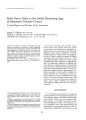
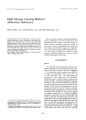
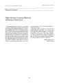
| OCR Text |
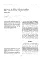
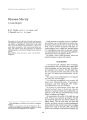
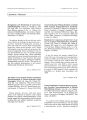
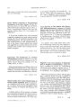
Show ! ollnlal of Clll" cal Nellro- ophtllllimology 12( 3): 192- 197, 1992. Suprasellar Tumors of Maldevelopmental Origin in Klinefelter's Syndrome A Report of Two Cases Latif M. Hamed, M. D., Bernard L. Maria, M. D., Ronald Quisling, M. D., Maher M. Fanous, M. D., and Parker Mickle, M. D. '" 1992 Raven Press, Ltd., ~ Patients with Klinefelter's syndrome may have a predisposition for the development of neoplasia, particularly extragonadal germ- cell tumors, but a suprasellar location is rarely reported. The clinical and neuroradiologic features in two patients with Klinefelter's syndrome and dysmorphic suprasellar masses of maldevelopmental origin ( presumably lipomas or lipodermoids) are described. One patient had bilateral optic atrophy and decreased vision. To our knowledge, only one similar case ( a suprasellar hamartoma) has been previously reported in association with Klinefelter's syndrome. Key Words: Klinefelter's syndrome- Suprasellar tumors. From the Departments of Ophthalmology ( L. M. H., M. M. F.). Pediatrics ( L. M. H., B. L. M.). Radiology ( R. Q.), and Neurosurgery ( P. M.), University of Florida College of Medicine, Gainesville, Florida, U. S. A. This work was supported in part by an unrestricted departmental grant from Research to Prevent Blindness, Inc., New York, NY ( to the Department of Ophthalmology). Address correspondence and reprint requests to Dr. Latif M. Hamed, University of Florida College of Medicine, Box 100284, jHMHSC, Gainesville, FL 32610- 0284, U. SA Klinefelter's syndrome is a chromosomal abnormality characterized by 47XXY karyotype that is estimated to occur in 0.6% of live male births. Patients have sterility, testicular atrophy with hyalinization of the seminiferous tubules, gynecomastia, a eunuchoid habitus, and an increased level of follicle- stimulating hormone. Patients with Klinefelter's syndrome have an increased incidence of neoplasia, with breast carcinoma ( 1) and extragonadal germ- cell tumors ( 2) being most frequently described. The site of predilection for the extragonadal germ cell tumors is the mediastinum ( 3), but lesions involving the retroperitoneum and the central nervous system ( 4- 7) have been reported. Other sporadic reports have described patients with Klinefelter's syndrome and lymphoma ( 7), leukemia ( 8), transitional cell carcinoma of bladder ( 9), prostatic carcinoma ( 10), adrenal carcinoma ( 11), bronchogenic carcinoma ( 12), reticulum cell sarcoma ( 13), and interstitial cell tumor of the testes ( 14). Involvement of the central nervous system in patients with Klinefelter's syndrome is rare, with only three previously reported cases involving the hypothalamus or suprasellar region ( 15- 17). We report two children with Klinefelter's syndrome and radiologically documented suprasellar masses, CASE REPORTS Case 1 A three- year- old boy was referred for evaluation of developmental delay and dysmorphic features. 192 SUPRASELLAR TUMORS IN KLINEFELTER'S SYNDROME 193 He was a product of a full- term, uncomplicated pregnancy and normal vaginal delivery, weighing 61/ 2 lb at birth. The umbilical cord was wrapped around the neck at delivery. He was initially cyanotic with poor Apgar scores, but the cyanosis resolved quickly after the umbilical cord was unwrapped. There was a mild delay in achieving normal developmental milestones. Specifically, he showed a social smile at 6 months, began rolling over at 1 year, was able to sit at 12- 14 months, stand and cruise at 1112 years, and babble at 15 months of age. There was no family history of developmental delay or genetic syndromes. Physical examination disclosed a hirsute child with microcephaly. His height of 82 cm, weight of 11.56 kg, and head circumference of 46.5 cm were all less than the 5th percentile for age. He had a low hairline, an anomalous right ear with a fold of the upper helix, and bilateral preauricular tags. The lungs and heart were normal. The liver edge was palpable 2- 3 cm below the right costal margin. The testes were normal and descended. Neurologic evaluation showed generalized hypotonia with normal reflexes. Eye examination disclosed central, steady, and maintained vision in both eyes with good fixation. Krimsky examination showed 30 prism diopters of exotropia at near fixation. Cycloplegic retinoscopy showed high myopia in the range of - 9.00 diopters OU. Ocular pursuit was saccadic. There was no afferent pupillary defect. Fundus examination showed a prominent choroidal pattern with normal optic nerves, macula, and vessels. Visual evoked response showed low normal amplitudes, suggesting intact projections to the primary visual cortex. The child had been previously diagnosed on the basis of clinical findings and chromosomal analysis as having Klinefelter's syndrome with a karyotype of 47XXY. Laboratory studies showed normal electrolytes, protein, albumin, and liver function tests. Magnetic resonance imaging ( MRI) of the brain revealed a large heterogeneous mass in the suprasellar space deforming the hypothalamus, the optic chiasm, the pituitary stalk, and causing obstructive hydrocephalus at the third ventricular ( foramen of Monro) level ( Fig. 1). The mass is largely cerebrospinal fluid ( CSF) intensity but is not completely homogeneous. The inhomogeneity is related in part to pulsatile effects of CSF within a closed space creating flow related enhancement. This subtle hyperintensity is not reproduced in the same location on images in other planes. The an-terior aspect of the mass contains a lipoid nodule located adjacent to the hypothalamus. The cystic part has no distinguishable capsule and does not arise from within the sella. These magnetic resonance findings are consistent with a combination of lipoma ( or lipodermoid) and a cyst, either arachnoid or epidermoid. Other cystic lesions of the suprasellar space ( Rathke's cyst or cystic craniopharyngioma) are less likely. Whether the nodular part of the lesion is a lipodermoid ( cholesterolcontaining) or whether it is a lipoma ( mature adipose tissue) is difficult to distinguish by magnetic resonance. No soft tissue components were evident to suggest teratoma, hamartoma, or malignancy. Case 2 A 51J2- year- old white boy was referred for evaluation of a brain tumor. He is the product of a full- term pregnancy and breech extraction spontaneous delivery. He weighed 6.0 lb at birth. Gestation history was unremarkable. Developmentally, the patient was able to walk at 2 years of age, but was still unable to talk. He put puzzles together and stacked blocks. He understood well and listened to commands. The patient was admitted to a hospital for management of dehydration secondary to diarrhea. During the hospitalization, an EEG, computed tomography, and MRI of the head were performed to evaluate his developmental delay. The EEG was normal. Computed tomography and magnetic resonance studies revealed a shottytype lesion with a fatlike consistency, 1 cm in size in the area of the tuber cinereum, most consistent with the diagnosis of intracranial lipoma or lipodermoid ( Fig. 2). Chromosomal analysis showed a karyotype of 47XXY consistent with Klinefelter's syndrome. Physical examination revealed a height of 113.5 cm ( 75th percentile), weight of 18 kg, and a head circumference of 51.5 cm. The nasal bridge was broad. Eye examination disclosed a visual acuitv of 20/ 400 on the right and 20/ 80 on the left. The - patient preferentially fixated with the left eve. He had 16 prism diopters of right esotropia in primary gaze. The pupils showed no afferent pupillary defect. Dilated fundus examination revealed marked temporal pallor of the optic nerve heads, more on the right. The lungs, heart, and abdomen were normal. Neurological examination was normal. The testes were descended and of normal size. Thyroid panel and cortisol level were normal for age. J Clil! Nellro- ophthaimol. Vol. 12. No. 3. 1992 194 L. M. HAMED ET AL. B c A FIG. 1. Magnetic resonance sections illustrating the radiographic features of a large suprasellar mass characterized as two lipoid nodules and a large cyst of cerebrospinal fluid intensity. ( A) Axial, T1- weight~ d, magnetic resonance image illustrating the def? rmlng effects of this mass on brain structures forming the suprasellar space including the ventral mesencephalon ( M) and the mesial left temporal lobe ( T). The mass contains a nodule with lipoid intensity and an apparent cystic component with cerebrospinal fluid intensity. The rostral aspect of the cyst stretches the optic chiasm ( Ch). The heterogeneous appearance of the cyst is likely related to cerebrospinal fluid pulsatility, since there is flow- related enhancement on the T1weighted image. which is not reproduced in the coronal plate. However, it is possible that all or a portion of the cyst is actually an epidermoid cyst with nearcerebrospinal fluid intensity. Such lesions can be indistinguishable from arachnoid cysts. ( 8) Axial T2weighted magnetic resonance image reveals heterogeneous signal from the cyst. Such heterogeneity is related in part to the lipoma ( the low- intensity nodule, arrow) and in part to phase dispersion related to cerebrospinal fluid pulsatility during the scanning sequence. ( C) Coronal, T1- weighted, Magnetic resonance section illustrates the stretched but otherwise normal pituitary stalk and a normal pituitary gland. The rostral aspect ot the hypothalamic lipoma is also apparent ( arrow). The cyst within the suprasellar space is cerebrospinal fluid density and causes no apparent displacement of the stretched but normally placed pituitary stalk and supraclinoid segments of the carotid arteries. DISCUSSION Miller ( 18) subdivides intracranial lesions of possible maldevelopmental origin into neoplastic, choristomatous, and hamartomatous categories. According to this subdivision, the neoplasms include germ- cell tumors, craniopharyngiomas ( and the closely related Rathke's cleft cyst), and lipomas. Each of these tumors has a predilection for specific midline structures. For instance, the gonads, retroperitoneum, mediastinum, pineal gland, 3rd ventricle, and hypothalamus are the preferred sites for germ- cell tumors, while the corpus callosum, tuber cinereum, quadrigeminal 10111 Neuro- opl,''' allllol. Vol. 12. No. 3. 1992 SUPRASELLAR TUMORS IN KLINEFELTER'S SYNDROME 195 A ' 8 FIG. 2. Magnetic resonance sections illustrating the radiographic features of a small lipoid nodular mass located along the ventral surface of the hypothalamus. ( A) Nongadolinium- enhanced sagittal magnetic resonance section with T1 weighting demonstrates a lipomatous mass ( arrow) adjacent to the inferior aspect of the hypothalamus involving the base of the pituitary stalk ( confirmed in multiple planes). ( B) Coronal T1- weighted magnetic resonance image with gadolinium illustrates the relationship of the lipomatous mass to the hypothalamus. A normal pituitary gland, accentuated by the gadolinium, and a normal distal segment of the pituitary stalk are evident in this section. plate, and ambient cisterns are the preferred sites for intracranial lipomas ( 18). In the absence of histopathological verification, the specific lesion may be pinpointed on the basis of clinical behavior, location, and neuroradiologic characteristics. Other investigators argue against considering lipomas as neoplastic lesions ( 19). They provide clinical and neuroradiological evidence to support the concept that intracranial lipomas are best considered as congenital malformations, as opposed to neoplasms. Lipomas, according to this concept, arise as a result of abnormal differentiation of the primitive meninx, which normally gives rise to the subarachnoid space ( 19). JClin Neuro- ophthalmol, Vol. 12. No. 3. 1992 196 L. M. HAMED ET AL. Chromosomal alteration appears to playa significant role in neoplastic disorders. Two of the relatively common trisomies, Down's syndrome and Klinefelter's syndrome, show a higher than normal risk for neoplasia. Patients with Down's syndrome, for instance, show a propensity for the development of reticuloendothelial tumors ( 20). Patients with Klinefelter's syndrome are at a greater risk for the development of breast cancer, germ cell tumors, and, to a lesser extent, other types of neoplasms ( 1- 14). The reason for the higher incidence of neoplasia in Klinefelter's syndrome is unknown, although hormonal changes and chromosomal abnormalities have been offered as explanations. It has been demonstrated that the XXY cells were transformed 3 to 10 times more frequently by the simian papovavirus 40 in patients with Klinefelter's syndrome and tumors than in a normal control population ( 12). Central nervous system neoplasms rarely occur in patients with Klinefelter's syndrome. To date, only seven cases of Klinefelter's syndrome with central nervous system tumors have been reported ( 4- 7,15- 17) ( Table 1). Of the seven cases, five were pathologically proven germinoma ( three affecting the pineal region, one affecting the posterior hypothalamus, and one affecting the supra~ ellar region). The sixth case is the first reported mstance of a radiologically documented suprasellar hamartoma in Klinefelter's syndrome ( 15). The seventh case is the first case with a pathologically proven primary B- celllymphoma involving the right frontal and parietal lobes ( 7). The magnetic resonance features of the first case appear to be those of a dysraphic mass partIy lipoma but largely a nonencapsulated cyst. Ahagon et al. ( 16) described the computed tomography findings on a suprasellar germinoma with peripheral calcification and a low- density area, suggesting a cyst formation that led to the misdiagnosis of a craniopharyngioma preoperatively. In addition, our patient did not have any clinical evidence of diabetes insipidus. The magnetic resonance findings in Case 2 suggest fatlike material in the region of the tuber cinereum that most likely represents a lipoma. The tuber cinereum area is one of the preferred sites for intracranial lipomas ( 18). Patient 2 presented with bilateral optic atrophy, TABLE 1. Central nervous system tumors in Klinefelter's syndrome: Summary of clinical data Reference ( no.) Age Site of ( years) lesion Histopathology Neuroradlology Ophthalmological findings Systemic findings Liang et al. ( 7) 33 Frontal and parietal lobes Present 3 Suprasellar Case 1 area Present 5V2 Suprasellar Case 2 area Rubenstein ( 4) 16 Pineal gland Chaussain et al. ( 15) Ahagon et al. 20 ( 16) Ellis et al. ( 17) 12 Arens et al. ( 5) 15 Oki et al. ( 6) 17 Hypothalamus Suprasellar area Posterior hypothalamus Pineal gland Pineal gland Germinoma Germinoma Germinoma Germinoma Germinoma B- cell lymphoma Circular mass in interpeduncular cistern. hamartoma of floor of third ventricle Low- density area with peripheral calcification suggesting cyst formation Irregular mass of mixed attenuation arising from midline III- defined 3- cm density dorsal and inferior to pineal gland Tumor shadow around calcified pineal extending to suprasellar region Hypodense lesion ar right frontoparietal area Large cystic mass in suprasellar cistern 1 em fatlike mass Enlarged blind spot. 00: inferotemporal visual field defect. OS Bilateral papilledema, skeletal deformities Dorsal midbrain syndrome Bilateral optic atrophy 00 > OS 3 years later developed primary mediastinal choriocarcinoma and dermoid cyst of testes Sexual precocity Polyuria, polydipsia Scoliosis, skeletal deformities Grand mal seizures Diabetes insipidUS Left hemiparesis, HIV- negative Developmental delay Developmental delay HIV. human immunOdeficiency VirUS SUPRASELLAR TUMORS IN KLINEFELTER'S SYNDROME 197 more marked on the right. Patients with Klinefelter's syndrome and optic atrophy may need to undergo neuroimaging of the head to rule out a perichiasmal mass lesion. Alternatively, it is possible that some patients with similar suprasellar lesions have undiagnosed Klinefelter's syndrome, as the diagnosis may be missed if chromosomal analysis is not performed. This is particularly relevant, since an associated hypogonadism in a patient with a suprasellar lesion may be attributed to secondary disruption of the pituitary- hypothalamic axis rather than to an underlying Klinefelter's syndrome ( in which hypogonadism is a characteristic feature). The two disorders may therefore by confused with one another clinically. In a prospective study of 22 patients presenting with mediastinal germ- cell tumor, Nichols and associates ( 3) found five of them to have previously unsuspected Klinefelter's syndrome upon chromosomal analysis. A similar study addressing the prevalence of Klinefelter's syndrome in patients with intracranial germinoma may be useful. REFERENCES 1. jackson AW, Muldal S, Ockey CH, O'Conner Pj. Carcinoma of male breast in association with the Klinefelter syndrome. Br Med ] 1965; 1: 223- 5. 2. Lee MW, Stephens RL. Klinefelter's syndrome and extragonadal germ cell tumors. Calleer 1987; 60: 1053- 5. 3. Nichols CR, Heerema NA, Palmer C. Loehrer Pj Sr, Williams SO, Einhorn LH: Klinefelter's syndrome associated with mediastinal germ cell neoplasms. f Clin Oneal 1987; 5: 1290-- 4. 4. Rubinstein I). Tumors of the central nervous system. Atlas of tumor pathology. Ser. 2, Fasc. 6. Washington DC: Armed Forces Institute of Pathology, 1972: 270. 5. Arens R. Marcus 0, Engelberg S, Findler G, Goodman R, Passwell j. Cerebral germinoma and Klinefelter syndrome: a review. Cancer 1988; 61: 1228- 31. 6. Oki S, Kakao K, Kuno S, Imura H. A case of Klinefelter's syndrome associated with hypothalamic- pituitary dysfunction caused by an intracranial germ cell tumor. Endocrinol fpn 1987; 34( 1): 145- 51. 7. Liang R, Woo E, Ho F, Collins R, Choy 0, Ma j. Klinefelter's syndrome and primary central nervous system lymphoma. Med Pediatr Oneal 1990; 18: 236- 9. 8. Mamunes P, Lapidus PH, Abbott JA, Roath S. Acute leukaemia and Klinefelter's syndrome. Lancet 1961; 2: 26- 7. 9. Fujita K. Fujita HM. Klinefelter's syndrome and bladder cancer. JUral 1976; 116: 836- 7. 10. Arduino LJ. Carcinoma of the prostate in sex chromatin positive ( XXY/ XYj Klinefelter's syndrome. f Ural 1967; 98: 234- 40. 11. Pascual J, Liano F, Garcia- Villaneuva A, Salvador JL, Herrero JA, Ortuno j. Isolated primary aldosteronism in a patient with adrenal carcinoma and XY/ XXY mosaic Klinefelter's syndrome. f Ural 1990; 144: 1454- Q. 12. Mukerjee 0, Bowen J, Anderson DE. Simian papovavirus40 transformation of cells from cancer patient with XY- XXY mosaic Klinefelter's syndrome. Cancer Res 1970; 30: 1769- 72. 13. MacSween RNM. Reticulum- cell sarcoma and rheumatoid arthritis in a patient with XY/ XXY/ XXXY Klinefelter's syndrome and normal intelligence. Lancet 1965; 1: 46~ 1. 14. Arduion LJ, Glucksman MA. Interstitial cell tumor of the testis associated with Klinefelter's syndrome: a case report. f Ural 1963; 89: 246- 8. 15. Chaussain jl. Lemerle j, Roger M, Canlorbe P, job jc. Klinefelter syndrome, tumor and sexual precocity. f Pediatr 1980; 97: 607- 9. 16. Ahagon A, Yoshida Y, Kusuna K. Uno T. Suprasellar germinoma in association with Klinefelter syndrome. ] Neurosurgery 1983; 58: 136- 8. 17. Ellis Sj, Crockard A, Barnard RO. Klinefelter's syndrome, cerebral germinoma, Chiari malformation and syrinx. Neurosurgery 1986; 18: 22~ 2. 18. Miller NR. Tumors of maldevelopmental origin and related lesions. In: Walsh and Hoyt's clinical neuro- ophthalmology. 4th Ed. Baltimore: Williams & Wilkins, 1988: 138~ 1425. 19. Truwit CL, Barkovich AJ. Pathogenesis of intracranial lipoma: an MR study in 42 patients. AfNR 1990; 11: 665- 74. 20. Harnden DG, O'Riordan ML. Down's syndrome and leukaemia. Lancet 1973; 1: 26~ 1. JClin Neuro- ophthalmol. Vol. 12. No. 3, 1992 |