| OCR Text |
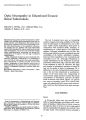
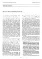
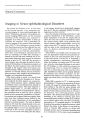
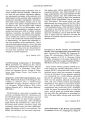
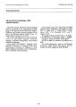
Show JouTtlJJI of Clinical Neuro-ophthalmology 7(2): 93-95, 1987. Unilateral Third Nerve Palsy and Somnolent Mutism Fernando Zermeno, M.D., Oscar H. Del Brutto, M.D., Guillermo Van Wielink, M.D., and Juan Nader, M.D. © 1987 Raven Press, New York A 64-year-old man developed unilateral third nerve palsy and somnolent mutism. Computed tomography (CT) revealed a small unilateral midbrain infarct. Previous reports of somnolent mutism placed the lesion in both sides of the midbrain tegmentum, usually extending into the thalamus. The unilateral third nerve palsy, and high-resolution 4-mm contiguous tomographic sections of the brainstern, helped localize the lesion. To our knowledge, this is the first cr clinical report of somnolent mutism induced by unilateral midbrain infarct. Key Words: Brain infarct-Mutism-Somnolent mutism- Third nerve palsy. From the Divisi6n de Neurologia, Instituto Nacional de Neurologia y Neurocirugia, Mexico, D.F. Address correspondence and reprint requests to Fernando Zermeno, M.D., Instituto Nadonal de Neurologia y Neurodrugia, Insurgentes Sur No 3877, CP: 14410, Mexico 22, D.F. 93 When midbrain infarcts result in third nerve palsy and somnolence, the former is usually bilateral and associated with visual hallucinations, behavioral abnormalities, and bilateral motor dysfunction (1-7). This syndrome almost always results from embolic occlusion of the paramedian thalamopeduncular branches of the basilar communicating artery (1-3), which has been defined as the segment of the posterior cerebral artery proximal to the ostium of the posterior communicating artery (8). In such cases, the responsible lesion usually involves the paramedian regions of the midbrain, thalamus, and subthalamus bilaterally, and produces a most striking syndrome, which has been called the "top of the basilar syndrome" (1), the "syndrome of the mesencephalic artery" (2), and the "mesencephalothalamic syndrome" (9). By contrast, when unilateral third nerve palsy is the result of more localized midbrain infarcts, somnolence and behavioral abnormalities are not part of the syndrome (10). We report a patient with a unilateral midbrain infarct in whom profound somnolence was observed in association with unilateral third nerve palsy. We also discuss the possible mechanism responsible for this uncommon association and emphasize the value of high-resolution, 4-mm contiguous tomographic sections of the brainstem when studying a patient with clinical evidence of midbrain infarct. CASE REPORT A 64-year-old hypertensive man was admitted 24 h after the abrupt onset of dizziness, right arm weakness, and somnolence. In the emergency room he was lethargic but rousable. He lay immobile and did not respond to commands. The blood pressure was 190/120 mm Hg. The right pupil was 94 F. ZERMENO ET AL. 3 mm, round, and reactive; the left pupil was 6 mm and did not react to light. The left eye was deviated outward and there was ptosis of the left upper lid. The right eye rested in a normal position. Motor testing I'howed a right paresis, which involved the arm and the lower half of the face. The right plantar response was extensor. The other results of physical examination were unremarkable. A computed tomographic (CT) scan showed mild cortical atrophy but was otherwise considered normal. The patient experienced little improvement. He opened his eyes when powerful stimuli were applied, but he soon closed them and sank back into a state of lethargic inertia alternating with bouts of excitement and motor agitation. A control CT scan performed with 4-mm contiguous sections of the brainstem showed an area of decreased attenuation in the left upper paramedian midbrain tegmentum, which extended into the medial portion of the ipsilateral cerebral peduncle (Fig. 1). Four weeks later, he showed only slight paresis of the right arm and face, but the left FIG 1. Plain computed tomographic scan done 1 week after the onset of symptoms, showing a well-defined, area ,of decreased attenuation in the left upper l"ii;c_tJr~tn·.;~~g""entlJm 1Clin Neuro-ophthalmol, Vol. 7, No.2, 1987 third nerve palsy and somnolence persisted. He was discharged on a program of physical therapy, and 6-month follow-up showed no change in either his oculomotor palsy or his behavioral abnormality. DISCUSSION Somnolent mutism is a term coined by Segarra (2) to describe the special type of apathy in which patients with extensive paramedian midbrain infarcts rest most of the time. This apathy is interrupted by brief bouts of excitement, restlessness, and motor agitation, and is usually associated with bilateral oculomotor, visual, and motor dysfunction (1-7). These manifestations are the result of embolic occlusion of the paramedian thalamopeduncular branches of the basilar communicating artery and the subsequent necrosis in the oculomotor nuclear complex, periaqueductal gray matter, red nucleus, and mesencephalothalamic reticular activating substance (3). Unilateral midbrain infarcts may result when embolic occlusion affects only one of two or more paramedian thalamopeduncular branches arising independently from the basilar communicating artery (3). In such cases, the lesion is too small to produce marked alteration in the level of consciousness, as the mesencephalothalamic reticular activating substance has a bilateral distribution (3). Syndromes that may result from unilateral midbrain infarcts are the well-known Benedikt's, Claude's, and Weber's syndromes (10) and isolated oculomotor disturbances (10-12). Neither somnolence nor bouts of psychomotor agitation are prominent features of these syndromes (10-12). The behavioral pattern of our patient closely resembled that described by Segarra (2) in patients with extensive paramedian thalamopeduncular infarcts. However, the extent of the infarct in our patient differs significantly, as he had a small unilateral infarct limited to the upper midbrain tegmentum. Why such different lesions result in similar forms of pathologic behavior is not completely understood, but it seems logical to assume that in our case this alteration was related to indirect compression of the reticulothalamic pathways by perilesional edema. As the patient is still alive, we lack pathologiC correlation. However, the unilateral third nerve palsy with contralateral motor weakness, and the CT evidence of unilateral area of decreased attenu- UNILATERAL THIRD NERVE PALSY AND SOMNOLENT MUTISM 95 ation in the midbrain (Fig. 1), clearly support the presence of unilateral midbrain infarct, a condition not previously described in association with somnolent mutism. Conventional cr studies usually fail to demonstrate small midbrain infarcts (7,9). Partial volume effect and artifacts resulting from dense osseous structures at the base of the skull are major determinants in CT failures (10). Timing is another factor affecting the efficiency of CT in identifying such infarcts (13). Time lapses of 8-12 hare needed before the earliest changes can be visualized on CT. In patients with clinical evidence of midbrain infarcts, repeated cr scans with 4-mm contiguous sections of the brainstem and a special scanning plane will reduce the partial volume effect and osseous artifacts, increasing the accuracy of CT in such cases (7). To the best of our knowledge, there has been no previous cr clinical correlation of unilateral midbrain infarcts in which a classic Weber's syndrome is associated with a behavioral pattern that approximates the somnolent mutism described by Segarra (2). This report reflects a similar clinical picture, which can induce midbrain infarcts with a different pathologic basis, and points out the importance of 4-mm contiguous tomographic sec~ tions of the brainstem in the study of a patient with clinical evidence of midbrain infarcts and routine cr scans showing no abnormalities. REFERENCES 1. Caplan LR. Top of the basilar syndrome. Neurology 1980;30:72-9. 2. Segarra JM. Cerebral vascular disease and behavior. I: The syndrome of the mesencephalic artery. Arch Neurol 1970;22:408-18. 3. Castaigne P, Lherrnitte F, Buge A, et al. Paramedian thalamic and midbrain infarcts: clinical and neuropathologic study. Ann NeuroI1981;10:127-48. 4. Guberman A, Stuss D. The syndrome of bilateral paramedian thalamic infarction. Neurology 1983;33:540-6. 5. Lepore FE, Gulli V, Miller DC. Neuro-ophthalmological findings with neuropathological correlation in bilateral thalamic- mesencephalic infarction. J Clin Neuro-ophthalmol 1985;5:224-8. 6. Biller J, Sand JJ, Corbett JJ, et al. Syndrome of the paramedian thalamic arteries: clinical and neuroimaging correlation. JClin NeurlHJPhthalmoI1985;5:217-23. 7. Hochman MS, Sowers JJ, Bruce-Gregorios J. Syndrome of the mesencephalic artery: report of a case with CT and necropsy findings. JNeurol Neurosurg Psychiatry 1985;48:117981. 8. Percheron G. Les arteres du thalamus humain. Arteres et territoires thalamiques paramedians de l'artere basilaire communicante. Rev NeuroI1976;132:309-24. 9. Fisher eM. Lacunar strokes and infarcts: a review. Neurology 1982;32:871-6. 10. Fog M, Hein-Sorensen O. Mesencephalic syndromes. In: Vinken PJ, Bruyn GW, eds. Handbook of clinical neurology, vol 2. Amsterdam: North-Holland, 1969:272-85. 11. Smith MS, Laguna JF. Upward gaze paralysis following unilateral pretectal infarction. Computerized tomographic correlation. Arch NeuroI1981;38:127-9. 12. Pierrot-Deseilligny Ch. Circuits oculomoteurs centraux. Rev NeuroI1985;141:349-70. 13. Bonafe A, Manelfe C, Scotto B, et al. Role of computed tomography in vertebrobasilar ischemia. Neuroradiology 1985;27:484-93. JClin Neuro-ophthalmol, Vol. 7, No.2, 1987 |