| OCR Text |
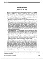
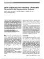
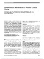
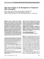
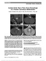
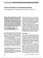
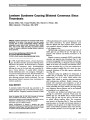
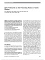
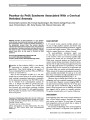
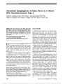
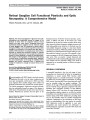
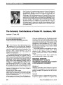
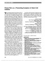
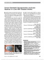
Show Progressive Diplopia and Facial Weakness in a 62-Year-Old Woman Monika R. Kolloori, MD, Luis J. Mejico, MD, Joseph Corbo, MD, PhD, Aseem Sharma, MD, Melissa W. Ko, MD Dr. Kolloori: A 62-year-old woman presented to a local emergency department with horizontal diplopia that she first noted upon awakening the previous day. She had experienced a mild headache the prior evening but was otherwise well and denied any constitutional symptoms, concurrent or recent illnesses, jaw claudication, or eye pain. She had a history of hypercholesterolemia. Physical examination revealed normal vital signs. The patient's visual acuity, intraocular pressures, and funduscopy were normal, and she had a mild abduction deficit of the left eye. Hematologic studies including complete blood count, metabolic panel, and sedimentation rate were normal. Noncontrast computed tomography (CT) of the head and magnetic resonance imaging (MRI) of the brain with and without contrast were performed. Dr. Sharma: Axial MRI with fluid-attenuated inversion recovery (FLAIR) technique demonstrates nonspecific hyperintense foci scattered in the white matter, without mass effect or loss of volume (Fig. 1). No enhancement or restricted diffusion was noted (scans not shown). Dr. Kolloori: The patient was diagnosed with left sixth nerve palsy, most likely ischemic in origin. Six days later, her diplopia worsened, and she was referred for neuro-ophthalmic evaluation. Visual acuity, color vision, pupillary reactions, and visual fields were normal. She had no ptosis, and extraocular movements revealed a complete left abduction defect. Corneal and facial sensations were equal and normal bilaterally. Further testing was done, including C-reactive protein, reactive plasma regain, and Lyme titers. Four days later, the patient developed left facial weakness consistent with a left seventh nerve palsy, and she was admitted to the hospital. Lumbar puncture demonstrated a cerebrospinal fluid (CSF) lymphocytic pleocytosis with an elevated protein concentration (Table 1, sample 1). Cytologic assessment revealed abundant atypical lymphocytes with plasmacytoid characteristics and mitotic figures. CSF electrophoresis was sus-picious for a faint monoclonal band, but the sample was not viable for flow cytometry. Autoimmune panel and serological testing for fluorescent treponemal antibody absorption, crypto-coccus, and herpes simplex virus were negative or gave normal results. Blood cultures were negative. Repeat brain MRI was unchanged, and magnetic resonance angiography was normal. The patient was given 1 gm of intravenous (IV) methylpred-nisolone for a possible inflammatory disorder of the central nervous system (CNS). The following evening, the enzyme-linked immunosorbent assay andWestern blot IgM for Borrelia burgdorferi returned positive (2.81 Lyme Index Value and 41 and 23 kDa bands, respectively), with a negative IgG pattern. The patient was treated with 2 g of IV ceftriaxone and dis-charged home on 21 days of IV antibiotic therapy. FIG. 1. Axial fluid-attenuated inversion recovery (FLAIR) images show scattered foci of hyperintensity in the sub-cortical and deep white matter. No enhancement was seen in the corresponding contrast-enhanced T1 images (not shown). Department of Medical Education (MRK), St. Joseph's Hospital and Health Center, Syracuse, New York; Departments of Neurology (LJM, MWK) and; Ophthalmology (LJM, MWK), Upstate Medical University, Syracuse, New York; Department of Pathology and Immunology (JC), Washington University School of Medicine, St Louis, Missouri; and Mallinckrodt Institute of Radiology (AS), Washington University School of Medicine, St Louis, Missouri. Supported by unrestricted grants from the Research to Prevent Blindness, Inc, New York, NY, and Lions District 20-Y1. The authors report no conflict of interest. Address correspondence to Melissa W. Ko, MD, Upstate Medical University, Department of Neurology, 90 Presidential Plaza, Syracuse, NY 13202; E-mail: kom@upstate.edu Kolloori et al: J Neuro-Ophthalmol 2012; 32: 359-362 359 Clinical-Pathological Case Study Section Editor: Neil R. Miller, MD Copyright © North American Neuro-Ophthalmology Society. Unauthorized reproduction of this article is prohibited. Three days following discharge from hospital, she underwent repeat lumbar puncture that demonstrated further increase in the CSF pleocytosis (Table 1, sample 2). Flow cytometry showed 84% T cells with a normal CD4:CD8 ratio and 11% B cells with a decreased kappa:lambda ratio of 0.4. Cytologic assessment of the CSF again was performed. Dr. Corbo: As in the first CSF sample, numerous atypical lymphocytes are present. The cells have plasmacytoid features including prominent juxtanuclear areas of clearing that correspond to the Golgi apparatus (Fig. 2A). Dr. Kolloori: Immunoglobulin heavy chain polymerase chain reaction (PCR) of the CSF revealed a monoclonal band of approx-imately 240 base pairs using primers for the framework 2 region (Fig. 2B). Because these findings raised the concern of a B-cell lymphoma, the patient was referred to hematol-ogy- oncology, and a bone marrow biopsy was performed. Dr. Corbo: In contrast to the CSF samples, the bone marrow biopsy shows normal trilineage hematopoiesis without evidence of malignancy (Fig. 3). Dr. Kolloori: On serum protein electrophoresis, a vague clonal band of similar size to that found in the patient's second CSF sam-ple was detected (Fig. 4). CT of the neck, chest, abdomen, and pelvis revealed a thyroid nodule but were otherwise negative for malignancy. Ultrasound-guided biopsy of the thyroid nodule was not performed because of the nodule's close proximity to the patient's internal carotid artery. With completion of a 3-week course of antibiotics, the patient's diplopia and left peripheral seventh nerve palsy improved, such that the treatment for a possible lympho-proliferative disorder was withheld. A third spinal tap performed 10 days following the completion of therapy revealed a reduced lymphocytic pleocytosis, small lympho-cytes, and monocytes of normal appearance, with no evidence of malignancy (Table 1, sample 3). Two months following the patient's initial presentation, the Western blot IgG returned positive (58, 45, 41, 39, and 23 kDa) for B. burgdorferi. Final Diagnosis Neuroborreliosis associated with a reactive lymphocytosis mimicking CNS lymphoma. Dr. Kolloori: Five months after presentation, the patient had complete clinical resolution of her left sixth and seventh nerve palsies. CSF analysis at this time was entirely normal (Table 1, sample 4). DISCUSSION Lyme disease is the most common tick-borne illness in Europe and North America (1). In North America, only 1 strain, B. burgdorferi sensu stricto, is recognized as pathogenic (2). Approx-imately 10%-15% of all B. burgdorferi infections progress to neuroborreliosis (3). Symptoms may include radiculopathy, cranial neuropathies, with the seventh cranial nerve most often affected, and mononeuropathy multiplex (3). Involvement of the nervous system may occur anywhere from 3 to 12 weeks following infection, often in the presence of the classic TABLE 1. Analysis of CSF samples Sample 1 Sample 2 Sample 3 Sample 4 WBC/mm3 310 550 39 0 Lymphocytes, % 99 100 100 0 Neutrophils, % 1 0 0 0 RBC/mm3 6 24 6 26 Protein (normal: 15-45 mg/dL), mg/dL 101 81 37 31 Glucose (normal: 30-70 mg/dL), mg/dL 51 61 67 65 Cytology Atypical, plasmacytoid characteristics, abnormal mitotic figures Atypical, plasmacytoid characteristics, binucleated cells Small lymphocytes, monocytes, smudge cells; no evidence of malignancy None Flow cytometry No longer viable 84% T cells; normal CD4:CD8 ratio; 11% B cells; 0.4 kappa:lambda Not performed Not performed CSF, cerebrospinal fluid; RBC, red blood cells; WBC, white blood cells. 360 Kolloori et al: J Neuro-Ophthalmol 2012; 32: 359-362 Clinical-Pathological Case Study Copyright © North American Neuro-Ophthalmology Society. Unauthorized reproduction of this article is prohibited. erythema migrans (EM). Additional findings including pe-ripheral neuropathy, encephalopathy, or encephalomyelitis may develop later (4). The patient's MRI findings of nonspe-cific white matter FLAIR hyperintensity have been described in Lyme disease. Prior case reports have documented nonspe-cific demyelinating lesions with brain, nerve root, and spinal cord imaging that may be clinically asymptomatic (3,5 7). The possibility of coexisting lymphoma with positive B. burgdorferi serology is an important diagnostic consider-ation. Of particular interest in our case was the possibility of malignancy versus infection raised by the patient's initial CSF and PCR findings. There are reports of neuroborreliosis mim-icking lymphoma, but flow cytometric analyses were not performed in those cases (5,8). In neuroborreliosis, alteration of the blood-brain barrier allows for intrathecal antibody for-mation, which may appear as oligoclonal bands on PCR. Typical CSF findings in neuroborreliosis include elevated pro-tein, normal glucose, and a lymphocytic pleocytosis (9). Cel-lular atypia suggestive of non-Hodgkin lymphoma may simply represent a lymphocytic response to antigenic stimu-lation by B. burgdorferi (5). In rare cases, B. burgdorferi causes a persistent antigenic stimulation that leads to malignant transformation of cells (10), although in our patient, the monoclonal B-cell expansion was self-limited. An association between Lyme disease and B-cell lymphoma may result from specific B-cell lymphocyte chemoattractants released in Borre-lia infections. CXCL13, a B-cell chemoattractant, has been found to be increased in the CSF of neuroborreliosis patients (11). Upon exposure to Borrelia garinii antigen (a pathogenic European strain causing Lyme disease), monocytes release FIG. 3. Bone marrow biopsy showing normal morphology, with small lymphocytes and megakaryocytes (hematoxylin and eosin, ·200). FIG. 4. Serum protein electrophoresis demonstrating a mono-clonal band (arrow) of comparable size and location to that detected in CSF. FIG. 2. A. Cerebrospinal fluid cytology showing atypical lymphocytes with abundant cytoplasm, prominent Golgi regions, and visible nucleoli (Wright-Giemsa, ·1,000). B. Cerebrospinal fluid protein electrophoresis revealing a dis-tinct band of approximately 240 base pairs in the framework 2 region (arrow), suggesting monoclonality in the heavy chain (IgH) of B lymphocytes. Kolloori et al: J Neuro-Ophthalmol 2012; 32: 359-362 361 Clinical-Pathological Case Study Copyright © North American Neuro-Ophthalmology Society. Unauthorized reproduction of this article is prohibited. CXCL13, which may be a significant chemoattractant for B-cell migration to the CSF (12,13). In distinguishing between neuroborreliosis and lym-phoma, high-grade cellular atypia, monomorphism, and large numbers of lymphoid cells are characteristics of lymphoma, whereas polyclonality and elevated protein fractions are more likely because of infection (5). Utilizing these criteria, our patient's initial cytology and CSF analysis had components consistent with either diagnosis. A further confounding factor in our case was the use of methylprednisolone, leading to potential steroid suppression of lymphoma. Our patient exhibited signs of monoclonality on PCR analysis of serum and CSF. Although the B-cell kappa: lambda ratio was abnormal, consistent with monoclonal expansion, the absolute number of B cells was small compared with the number of T lymphocytes. This mono-clonality diminished in the patient's bone marrow 10 days following antibiotic administration. As a result, the final pathologic interpretation of these findings was one of the infections causing a reactive CSF lymphocytosis with tran-sient monoclonal B-cells production. B-cell lymphocytosis with monoclonal or oligoclonal expansion in blood and bone marrow occurs in approximately 3.5% of individuals older than 40 years (14). There are no known clinical consequen-ces if the following criteria are met: lack of cytopenias, lym-phocytosis with monoclonal B cells of chronic lymphocytic leukemia phenotype (CD5+) numbering less than 5,000 per microliter, or lymphocytosis lasting less than a 3-month duration. However, it remains unclear if this lymphocytosis is a precursor to lymphoma (14). Although our patient clearly had neuroborreliosis, initially she denied a history of EM or tick exposure. Although EM is the most common symptom reported in B. burgdorferi infection, it is found in only 60%-80% of all infections (6). Despite absent history of EM on initial visit, the patient's transient headache the night before symptom onset may have been significant. In 1 clinical trial, patients with sixth or seventh nerve palsies and neuroborreliosis reported pain including headache more often than patients with cranial neuropathies of other etiologies (15). Possible tick exposure in our patient became apparent when her husband recalled that she frequently cleared brush in a local state park, known to be endemic for Lyme disease. Her husband remembered that the patient had complained of a red rash 3 months before the initial presentation, but his description was not typical EM. During the year of the patient's illness, 50% of Lyme disease cases reported in her county of residence were within the patient's home ZIP code. The greatest number occurred during the month of the patient's presentation (Morrow C, personal communication, November 2010). In summary, Lyme neuroborreliosis can simulate CNS lymphoma. Despite concern for neoplastic process, clinical and diagnostic abnormalities can completely resolve with appropriate antibiotic treatment for neuroborreliosis. The reasons for overlap between Lyme infection and CNS lymphoma may be purely coincidental or because of a common etiology. In clinical practice, given the distinctive treatment for each condition, thoroughly pursuing evalua-tion for both etiologies would be our recommended diagnostic approach. ACKNOWLEDGMENTS The authors thank Barbara Henriquez, MD, and Robert Hutchinson, MD, for their assistance and preparation of the neuroimaging and histopathologic figures, respectively. REFERENCES 1. Huppertz H. Lyme disease in children. Curr Opin Rheumatol. 2001;13:434-439. 2. Steere AC. Lyme disease. N Engl J Med. 2001;345: 115-125. 3. Greer DM, Schaefer PW, Plotkin SR, Hasserjian RP, Steere AC. Case records of the Massachusetts General Hospital. Case 11- 2007. A 59-year-old man with neck pain, weakness in the arms, and cranial-nerve palsies. N Engl J Med. 2007;356:1561-1570. 4. Ogden NH, Artsob H, Lindsay LR, Sockett PN. Lyme disease. Can Fam Physician. 2008;54:1381-1384. 5. Kieslich M, Fiedler A, Driever PH, Weis R, Schwabe D, Jacobi G. Lyme borreliosis mimicking central nervous system malignancy: the diagnostic pitfall of cerebrospinal fluid cytology. Brain Dev. 2000;22:403-406. 6. Hurley RA, Taber KH. Acute and chronic Lyme disease: controversies for neuropsychiatry. J Neuropsychiatry Clin Neurosci. 2008;20:1-6. 7. Fernandez RE, Rothberg M, Ferencz G, Wujack D. Lyme disease of the CNS: MR imaging findings in 14 cases. AJNR Am J Neuroradiol. 1990;11:479-481. 8. Walther EU, Seelos K, Bise K, Mayer M, Straube A. Lyme neuroborreliosis mimicking primary CNS lymphoma. Eur Neurol. 2004;51:43-45. 9. Mikkila HO, Seppala IJ, Viljanen MK, Peltomaa MP, Karma A. The expanding clinical spectrum of ocular lyme borreliosis. Ophthalmology. 2000;107:581-587. 10. Batinac T, Petranovic D, Zamolo G, Petranovic D, Ruzic A. Lyme borreliosis and multiple sclerosis are associated with primary effusion lymphoma. Med Hypotheses. 2007;69: 117-119. 11. Rupprecht TA, Plate A, Adam M, Wick M, Kastenbauer S, Schmidt C, Klein M, Pfister HW, Koedel U. The chemokine CXCL13 is a key regulator of B cell recruitment to the cerebrospinal fluid in acute Lyme neuroborreliosis. J Neuroinflammation. 2009;6:42. 12. Rupprecht TA, Kirschning CJ, Popp B, Kastenbauer S, Fingerle V, Pfister HW, Koedel U. Borrelia garinii induces CXCL13 production in human monocytes through Toll-like receptor 2. Infect Immun. 2007;75: 4351-4356. 13. Rupprecht TA, Pfister HW, Angele B, Kastenbauer S, Wilske B, Koedel U. The chemokine CXCL13 (BLC): a putative diagnostic marker for neuroborreliosis. Neurology. 2005;65:448-450. 14. Muller-Hermelink HK, Montserrat E, Catovsky D, Campo E, Harris NL, Stein H. Chronic lymphocytic leukemia/small lymphocytic lymphoma. In: Swerdlow SH, Campo E, Harris NL, Jaffe ES, Pileri SA, Stein H, Thiele J, Vardiman JW, eds. WHO Classification of Tumours of Haematopoietic and Lymphoid Tissues. 4th ed. Lyon, France: IARC, 2008. 15. Kindstrand E. Lyme borreliosis and cranial neuropathy. J Neurol. 1995;242:658-663. 362 Kolloori et al: J Neuro-Ophthalmol 2012; 32: 359-362 Clinical-Pathological Case Study Copyright © North American Neuro-Ophthalmology Society. Unauthorized reproduction of this article is prohibited. |