| OCR Text |
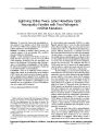
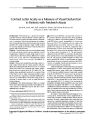
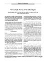
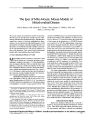
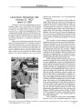
Show ORIGINAL CONTRIBUTION Lightning Strikes Twice: Leber Hereditary Optic Neuropathy Families with Two Pathogenic mtDNA Mutations Neil Howell, PhD, Neil R. Miller, MD, David A. Mackey, MD, Anthony Arnold, MD, Corinna Herrnstadt, PhD, Isla M. Williams, MD, and Iwona Kubacka, MS Objective: To report the clinical and mitochondrial genetic analyses of two families, each of which carries both the 11778 and 14484 Leber hereditary optic neuropathy ( LHON) mutations in mitochondrial DNA. Methods: In addition to detailed clinical histories, the complete sequence of the mitochondrial DNA ( mtDNA) from each family was determined. Results: A small Australian LHON family ( Vic20) and a family from the United States carry the 11778 and 14484 LHON mutations. In addition to the optic neuropathy, one branch of the Baltimore LHON pedigree had a high incidence of a fatal infantile encephalopathy. In both families, the 14484 LHON mutation was homoplasmic, whereas the 11778 LHON mutation was heteroplasmic. Conclusions: There are no additional mtDNA sequence changes that explain the encephalopathy in the Baltimore LHON family, and a nuclear gene involvement is an alternative explanation that is supported by the available data. The ophthalmological characteristics and penetrance in the 11778 and 14484 " two- mutation" LHON families are not markedly more severe than those of classic LHON families who carry a single mtDNA mutation. ( JNeuro- Ophthalmol 2002; 22: 262- 269) MitoKor, San Diego, California ( NH, CH); Department of Radiation Oncology, The University of Texas Medical Branch, Galveston, Texas ( NH, IK); Neuro- Ophthalmology Unit, The Wilmer Eye Institute, The Johns Hopkins Hospital, Baltimore, Maryland ( NRM); Center for Eye Research, Melbourne, Australia ( DAM), and Royal Victoria Eye and Ear Hospital, Melbourne, Australia ( DAM, IMW); and Department of Ophthalmology, UCLA Medical Center, Los Angeles, California ( AA). Address correspondence to Neil Howell, PhD, MitoKor, 11494 Sorrento Valley Road, San Diego, CA 92121- 1318, USA; E- mail: howelln@ mitokor. com NH is supported in part by a research grant from the Eierman Foundation. DAM acknowledges support from the Ophthalmic Research Institute of Australia. Leber hereditary optic neuropathy ( LHON) is a maternally inherited form of vision loss that predominantly affects otherwise healthy males, most often in the second to fourth decades of life ( 1- 5). LHON typically manifests as a painless, bilateral, simultaneous, or sequential vision loss that is associated with reduced color vision ( dyschromatop-sia) and central or cecocentral scotomas. The optic discs may initially appear normal during the acute phase, but they are often slightly elevated with peripapillary telangiectasia. As vision is lost, the telangiectasia ( if present) disappears, and the discs become pale. The symptoms and signs of LHON result from loss of function of the retinal ganglion cells that subserve central visual acuity and field, followed by death of some of these cells and loss of their axons. In 1988, Wallace et al. ( 6) first showed that the maternal inheritance of LHON reflects the fact that the primary pathogenic event is a single mutation in the mitochondrial genome ( mtDNA). More than a dozen LHON mutations have been identified ( 4,5,7,8), but mutations at mtDNA nucleotide positions 3460, 11778, and 14484 account for approximately 95% of LHON cases in individuals of European ( 9) or Asian ( 10,11) descent. In the vast majority of LHON patients, and specifically in those that carry one of these three mutations, the optic neuropathy is an isolated manifestation that is not associated with systemic disturbances or neurologic deficits. A small proportion of LHON patients manifest neurologic abnormalities, particularly a multiple sclerosis- like syndrome ( 12,13), and there are rare LHON families in which the optic neuropathy is accompanied by a variety of severe neurologic manifestations. For example, a single mutation at nucleotide position 14459 causes a syndrome in which the optic neuropathy is accompanied by dystonia ( 14). The present report deals with two unusual phenomena. First, we describe two independent families, both of which carry two primary LHON mutations. Our observations complement those of Brown et al. ( 15), who have studied another " two- mutation" LHON family. Second, a single branch of one LHON pedigree has a high incidence Copyright © Lippincott Williams & Wilkins. Unauthorized reproduction of this article is prohibited. 262 J Neuro- Ophthalmol, Vol. 22, No. 4, 2002 LHONFAMILIES WITH TWO PATHOGENICMTDNA MUTATIONS JNeuro- Ophthalmol, Vol. 22, No. 4, 2002 of a fatal infantile encephalopathy. These results are discussed in view of our current understanding of the pathogenesis of LHON and other mitochondrial diseases. METHODS DNA Sequencing For both LHON pedigrees, the mtDNA sequence was determined for the WBC/ platelet fraction of venous blood samples that had been obtained with informed consent from family members. Total DNA was isolated using standard procedures. The one exception was that a paraffin-embedded brain section was obtained for family member IV- 6. DNA was extracted from this section using an EX-WAX DNA Extraction Kit ( Serologicals Corp., Norcross, Georgia) after the procedure of the supplier and then used for polymerase chain reaction ( PCR) amplification of mtDNA fragments. The complete mtDNA sequence of the Vic20 LHON pedigree was determined by the MitoKor group as detailed elsewhere ( 16). This approach entails PCR amplification of mtDNA as a series of 68 overlapping fragments of approximately 500 basepairs each. Sequencing reactions were then carried out using these PCR primers for initiation and Big- Dye Terminator chemistry ( Applied Biosystems, Foster City, California). Electrophoresis and base calling were performed using a 3700 DNA Analyzer ( Applied Biosystems, Foster City, California). Sequence data for the PCR fragments were assembled into contiguous mtDNA sequences and aligned with revised Cambridge Reference Sequence ( rCRS) ( 17). The PCR primers were designed to provide approximately 50% sequence overlap with adjacent mtDNA fragments, and both strands of the mtDNA were sequenced. As a result of these operations, each base-pair is sequenced up to four times to provide a high degree of quality control on the sequences. The complete sequence of the mtDNA from a member of the Baltimore LHON pedigree was determined by the UTMB group as described previously ( 16). The mtDNA was PCR amplified as a series of 66 overlapping mtDNA fragments of approximately 300 basepairs in length ( 30- 50 basepairs overlap at each end with the adjacent fragments). These PCR fragments were ligated into Ml3 cloning vectors that, after bacterial transformation and isolation of recombinant clones, are used for manual dideoxy chain termination single strand DNA sequencing. Again, the resulting mtDNA sequences were compared with the rCRS to detect sequence differences. A number of comparative and blinded quality control experiments have been carried out and the two DNA sequencing approaches produce sequences of very high accuracy. For some members of the Baltimore LHON family, the complete mtDNA sequence was also determined at MitoKor with the automated sequencing procedure described above. In addition, the electropherograms were visually inspected to detect heteroplasmy as described elsewhere ( 16). We estimate that this procedure can detect heteroplasmic alleles even if the proportion of the minor mtDNA allele is no more than 15% to 20%. In addition to the complete sequence of the mtDNA from these two LHON families, we also ascertained the degree of heteroplasmy at nucleotides 11778 and 14484, the sites of the LHON mutations. The proportions of the mutant alleles were determined by manual sequencing of multiple independent M13 recombinant clones that carried the appropriate mtDNA fragment. RESULTS The Baltimore LHON Family The pedigree for this family is shown in Figure 1. The proband ( IV- 2) is a 23- year- old woman who experienced bilateral vision loss in 1996 and who was subsequently diagnosed with a bilateral optic neuropathy that was compatible with LHON. ( Her history also includes a congenitally dislocated hip.) Visual acuity at the time of vision loss decreased to 20/ 100 OD and 20/ 50 OS, and this decrease was accompanied by diminished color vision and by the development of bilateral optic disc pallor. However, visual acuity has subsequently improved to 20/ 30 OU. The proband's color vision, using Hardy- Rand- Rittler pseudoisochro-matic plates, is 3/ 10 OD and 5/ 10 OS. The latest visual field testing reveals full peripheral fields associated with small cecocentral scotomas. Both discs continue to be pale. o u i •- C LTT- 0 0 r ;- © n U i , •;- c o -€> i>-^- 3 *# k* n - j 6 - C m D Fig. 1. Pedigree of the Baltimore LHON Family. The half-filled symbols indicate manifestation of optic neuropathy, whereas the filled symbols indicate manifestation of fatal infantile encephalopathy. The Roman numerals at the right indicate the generations through which the pedigree has been established. Note that family members 11- 4 and 11- 5 were twins. Copyright © Lippincott Williams & Wilkins. Unauthorized reproduction of this article is prohibited. 263 JNeuro- Ophthalmol, Vol. 22, No. 4, 2002 Howell et al. The first instance of LHON in this pedigree occurred in the proband's great aunt ( II- 7). This individual was visually normal until age 37, when she noted rapid, painless loss of vision in both eyes. She eventually visited an ophthalmologist who noted pale discs, but details of the level of visual acuity are not available. She eventually stabilized, but she never regained vision in either eye. No further details are available. This family member had a son ( III- 8) who also developed the characteristic optic neuropathy. He was visually normal until age 24 when he developed visual loss in both eyes. It is unclear if loss was simultaneous or sequential, but both eyes were affected within two months. Visual acuity eventually dropped to 20/ 400 OU and was associated with pale optic discs. In addition to the optic neuropathy, there were multiple instances of severe neurologic abnormalities in this family. Pedigree members II- 4 and II- 5 were twin sisters, the second of whom died at age 1 from unknown causes. The surviving sister, who is not affected with LHON, has given birth to two daughters ( III- 4 and III- 5), both of whom are visually affected. Family member III- 4 was visually normal until age 21, when she noted simultaneous progressive vision loss in both eyes. She was evaluated by an ophthalmologist, who noted decreased vision in both eyes and normal- appearing optic discs. The ophthalmologist initially thought that she was " faking." However, he then learned of the family history of optic atrophy. He observed her over the next 6 months, and it was noted that her vision dropped to 20/ 100 OU, after which it stabilized. Her most recent visual acuity is 20/ 200. She currently has bilateral central scotomas with full peripheral fields and pale, somewhat small optic discs. Family member III- 5 was visually normal until age 29, when she lost vision rapidly and painlessly in both eyes. She did not visit an ophthalmologist for several months. Examination eventually disclosed decreased vision associated with pale optic discs. Her vision later stabilized and she currently has a visual acuity of 20/ 400 OU with diminished color perception, cecocentral scotomas, full peripheral fields, and pale optic discs with shallow cupping. Family member III- 5 gave birth to three children ( IV- 6, IV- 7, and IV- 8), all of whom died before 3 years of age. The daughter ( IV- 6) crawled at 6 months, talked at 11 months, sat up and rolled around at 11 months, and walked at 22 months. Her dexterity was never good, although she was well until about 30 months of age, when she began to experience tonic- clonic seizures. She was placed on diphe-nylhydantoin without improvement. She became increasingly lethargic over the next 3 weeks. She then developed difficulty in walking, spasmodic breathing, and a gradual unresponsiveness to external stimuli. She was admitted to a local hospital, at which time she was lethargic and barely responsive. In contrast to her brother ( IV- 8), her optic discs were described as " extremely pale." She had hypotonic extremities, and the plantar reflex was extensor on the left. The results of a lumbar puncture were normal. She was initially thought to have diphenylhydantoin toxicity, but serum levels were normal. Over the next 36 hours after hospital admission, she had several apneic episodes and tonic-clonic seizures. She died 6 days later. Upon postmortem examination, her brain was grossly normal. Microscopic examination revealed that all cortical areas showed moderate edema with increased perineuronal spaces without neuronal change. There was no evidence of demyelination. These changes were thought to be most consistent with recurrent hypoxia and anoxia. The second daughter ( IV- 7) was well until 9 months of age, when she developed sudden difficulty with breathing. She died shortly thereafter, but no details of the illness are known. The son ( IV- 8) was normal until the age of 12 months, when he began to experience generalized seizures that were associated with a deterioration of developmental milestones, including speech, coordination, equilibrium, and general intellect. His optic discs appeared normal throughout the deterioration of his neurologic functions. One morning, his mother found him unresponsive in his crib. He was admitted to a local hospital where he was intubated and given assisted ventilation. He died about 2 days after admission at the age of 19 months, or about 7 months after the onset of his neurologic dysfunction. A postmortem examination revealed only nonspecific congestion of the cerebral cortex. Family member III- 4, sister of the mother with the three children who died early in life, had childhood asthma, LHON, and a neurologic disorder that began in childhood and that was characterized in part by a resting tremor and ataxia. She underwent electroencephalography, pneumoencephalography, and evaluation of cerebrospinal fluid, all of which gave normal results. She gave birth to two children, IV- 4 and IV- 5, both of whom also developed a fatal infantile encephalopathy. The affected son ( IV- 4) was well until 16 months of age. He had previously been evaluated by an ophthalmologist, who noted myopia but no other ophthalmological abnormalities. At this time, he had an episode in which he seemed unaware of his surroundings for a few minutes. One week later, he suddenly became confused and developed difficulty breathing. He was admitted to a local hospital, where he was found to be afebrile, cyanotic, and pale. His breathing was in irregular, deep gasps, and an evaluation revealed pulmonary edema. He underwent a tracheotomy and was placed on assisted ventilation, but he remained hypotonic and unresponsive. He died 20 hours after admission. Postmortem examination revealed interstitial pneumonia. His brain weighed 1000 g with normal appearing Copyright © Lippincott Williams & Wilkins. Unauthorized reproduction of this article is prohibited. 264 © 2002 Lippincott Williams & Wilkins LHONFAMILIES WITH TWO PATHOGENICMTDNA MUTATIONS JNeuro- Ophthalmol, Vol. 22, No. 4, 2002 meninges, sulci, and convolutions. Coronal sections through the brain revealed no abnormalities of the gray or white matter. The brain stem and spinal cord also appeared grossly normal. Microscopic sections of the brain revealed only a mild congestion that was consistent with hypoxia. The affected daughter ( IV- 5) had slightly delayed developmental milestones. She did not walk until 19 months of age, and she was slow to talk. She was otherwise well until 25 months of age, when she began to have noisy, croupy breathing and irregular respirations. She then became increasingly lethargic and seemed to have more difficulty walking. After eating dinner one night, she suddenly became limp and had difficulty breathing. She was admitted to a local hospital where she was found to be afebrile, comatose, cyanotic, and unresponsive to pain. Her respirations were noisy and irregular, but her ocular fundi were said to be normal. Her extremities were flaccid and areflexic. Plantar responses were bilaterally extensor. She underwent a tracheotomy and subsequently improved over the next 24 hours to the point where she was awake, alert, talkative, and responsive to her mother, although she could not breathe on her own. Over the next 2 days, however, she became increasingly lethargic and then comatose with no spontaneous respirations. The results of a lumbar puncture revealed no abnormalities, but serial electroencephalograms became increasingly abnormal. The patient subsequently experienced several apneic spells, and she eventually had a cardiorespiratory arrest from which she could not be resuscitated. She died 10 days after admission. Postmortem examination revealed minimal congestion and edema of the lungs, results that were consistent with focal bronchopneumonia. The brain and spinal cord showed no specific lesions. The Vic20 LHON Family The proband is a woman who was born in 1957 and who was diagnosed with sarcoidosis in 1985. This diagnosis was confirmed by the results of multiple biopsies over the next 3 years. In August 1999, 14 years after this diagnosis, she complained of blurred vision OD. At that time, her visual acuity was 6/ 60, but it was 6/ 12 two days later. The right optic disc was slightly congested. Fluorescein angiography revealed slow, progressive leakage from the right optic disc. Her angiotensin converting enzyme level rose to 197 U/ L at the time vision deteriorated OD. Furthermore, a cholesterol embolus was noted in a small branch of the supertemporal retinal arteriole near the macula of this eye. On the basis of this information, it was concluded that she had right optic papillitis secondary to sarcoid. Dexa-methasone was prescribed, but the visual acuity OD fell to counting fingers at 0.5 meters. The disc congestion resolved, but the intraocular pressure rose to 33 mmHg OD and 30 mmHg OS. The visual acuity OS remained 6/ 6. Topical betaxolol 0.5% bd was prescribed to control the intraocular pressures and the dose of dexamethasone was reduced, and then stopped. In June 2000, the patient noted blurred vision OS, and the optic disc in this eye was observed to be swollen. Over the next 30 days, the visual acuity OS fell to 6/ 24, whereas the visual acuity OD was 6/ 9. By August 2000, the visual acuity OS was reduced to 1/ 60, and the optic disc remained swollen. Dexamethasone was prescribed for 4 days, but there was no significant improvement in the visual acuity OS. In September 2000, the visual acuity OD dropped from 6/ 9 to 6/ 12. Methotrexate and dexamethasone were prescribed, but the visual acuity of this eye had fallen to 3/ 60 by December 2000. In early 2001, the patient was given two courses of intravenous methylprednisolone with apparent but unsustained improvement in vision. However, the systemic corticosteroid treatment increased her intraocular pressure to the high 20s ( mmHg), and she was treated with betaxolol hydrochloride and, later, timolol maleate eye drops. At this time, she also developed diabetes mellitus, and her current medications include sertraline hydrochloride, glipizide, insulin, and diclofenac sodium. With the removal of the corticosteroid treatment and the prescription of timolol maleate 0.5%, her intraocular pressures returned to normal. At the present time, the visual acuity OD is 6/ 60 for distance, whereas visual acuity OS is counting fingers at 0.5 meters. With the OD, she was able to achieve an acuity of 20/ 200 for near vision and she is now able to complete correctly the D15 Farnsworth Munsell test. On the 24- plate Ishihara color chart test, she missed only plates 16, 20, and 21 OD, but she is unable to see the test plate with the OS . The visual field OD is impaired by a cecocentral scotoma that just includes the point of fixation. The visual field OS has a dense central scotoma, and she has a relative afferent defect in this eye. Both optic discs are pale, but there was no evidence of cupping ( Fig. 2). In May 2001, the cerebral angiogram and examination of the cerebrospinal fluid indicated no evidence of neurosarcoidosis. In July 2001, dedicated optic nerve imaging revealed no evidence of cerebral sarcoid and no evidence of sarcoid affecting the optic nerves. At that time, she was tested for the presence of LHON mutations. The severe chronic vision loss is consistent with LHON, but the color vision is atypically good. Her sister had completely normal vision upon examination, and there are no reports of vision loss in other family members. As detailed below, both sisters carry pathogenic LHON mutations and they were designated as the Vic20 LHON family. Mitochondrial Genetic Analysis Preliminary genetic screening revealed that the probands of these two pedigrees carried both the 11778 and Copyright © Lippincott Williams & Wilkins. Unauthorized reproduction of this article is prohibited. 265 JNeuro- Ophthalmol, Vol. 22, No. 4, 2002 Howell et al. Fig. 2. Optic discs of Vic20 Proband show pallor without cupping ( OD, left; OS, right). 14484 LHON mutations. Because LHON patients can be heteroplasmic ( both wild- type and mutant alleles are present), and because the load of these pathogenic mtDNA mutations is a significant determinant of manifesting the optic neuropathy ( 18), we investigated whether either, or both, LHON mutations were heteroplasmic in five members of the Baltimore pedigree ( Fig. 1, Table 1). It is clear that the 14484 LHON mutation is homoplasmic in all family members in the white blood cell/ platelet fraction of blood, and it is likely to be homoplasmic in all tissues including the target tissue ( the retinal ganglion cell layer). In contrast, all five family members are heteroplasmic for the 11778 LHON mutation with the load ranging from 23% to 77%. The numbers of family members analyzed are small, and penetrance is a complex and poorly understood phenomenon ( 1- 5), but the relationship between 11778 mutation load and manifestation of the optic neuropathy is similar to that observed for LHON family members who carry only the 11778 LHON mutation ( 18). Therefore, the two TABLE 1. mutations Family member II- 4 III- 2 III- 5 III- 7 III- 8 IV- 2 Mutation loads of the 11778 and 14484 LHON in the Baltimore pedigree 11778 mutation* 10/ 44( 23%) 19/ 54( 35%) 35/ 44 ( 77%) NDf 20/ 48 ( 42%) 17/ 24 ( 71%) 14484 mutation 23/ 23 ( 100%) 24/ 24( 100%) 19/ 19( 100%) 11/ 11 ( 100%) 63/ 63 ( 100%) 68/ 68( 100%) * Mutation load was determined by DNA sequencing of multiple clones that carried the relevant region of the mtDNA. The denominator is the total number of clones that were sequences, whereas the numerator is the number of clones that carried the LHON mutation. f Because of the low yield and poor quality of the DNA, the allele proportions at nucleotide 11778 could not be determined. family members with the lowest loads of the 11778 mutation are unaffected, whereas the two family members with loads > 70% are affected. The occurrence of the optic neuropathy in the family member ( III- 8) with a load of 42% is unusual ( 18), but not without precedent ( 19). We were able to recover some DNA from a paraffin- embedded section of the brain from family member IV- 6, but analysis was extremely difficult. It could be confirmed that the 14484 mutation was homoplasmic, but we were unable to obtain reliable data for the 11778 mutation. Results for the two members of the Vic20 LHON pedigree show a similar relationship between 11778 mutation load and penetrance. By analysis of the DNA sequencing electropherograms ( 16), we observed that the 14484 mutation was homoplasmic in the proband. As was found for the Baltimore pedigree, however, the 11778 LHON mutation was heteroplasmic, and the load was about 65% in the visually affected proband. Her unaffected sister was also homoplasmic forthe 14484 mutation and heteroplasmic for the 11778 mutation, although the load of the latter was approximately 40% ( data not shown). For purposes of comparison, the similar results that were obtained for the 11778 + 14484 LHON pedigree of Brown et al. ( 15) should be noted. Both the affected proband and her unaffected mother were homoplasmic for the 14484 mutation and heteroplasmic for the 11778 mutation. The 11778 mutation load was 94% in the proband, but only 31% in her mother. The most striking clinical feature of the Baltimore LHON family is the high incidence of a fatal infantile encephalopathy in the maternal descendants of II- 4. To ascertain if there were additional pathogenic mutations in this pedigree, and to determine if these two LHON families were simply branches of a single lineage, the complete mtDNA sequences of both pedigrees were determined ( Table 2). The mtDNA from the Baltimore LHON family belongs to the U/ K superhaplogroup, and the polymorphisms at nucleotides 1811 and 9055 indicate an assignment to Copyright © Lippincott Williams & Wilkins. Unauthorized reproduction of this article is prohibited. 266 © 2002 Lippincott Williams & Wilkins LHON FAMILIES WITH TWO PATHOGENIC MTDNA MUTATIONS JNeuro- Ophthalmol, Vol. 22, No. 4, 2002 TABLE 2. mtDNA polymorphisms in the 11778/ 14484 LHON pedigrees Vic20 LHON pedigree Baltimore LHON pedigree mtDNA basepair 73 146 152 263 512 709 750 1438 1700 1811 2706 3197 3480 4561 4769 5495 7028 7988 8697 8860 9055 9254 9477 9698 9716 10550 11299 11348 11467 11719 11914 12308 12372 12771 13617 14167 14766 14793 14798 15218 15326 15924 16224 16231 16256 16270 16311 16399 16519 Basepair change' A: G - i T: C A: G - - A: G A: G T: C - A: G T: C - - A: G T: C C: T - - A: G - - G: A - - - - - A: G G: A - A: G G: A G: A T: C - C: T A: G - A: G A: G A: G - T: C C: T C: T - A: G - Gene and amino acid changef Basepair change A: G T: C T: C A: G A: C G: A A: G A: G A: G A: G A: G T: C A: G C: T C: T G: A A: G G: A A: G T: C T: C A: G T: C C: T A: G G: A G: A A: G G: A C: T C: T T: C A: G T: C Gene and amino acid change D- Loop D- Loop D- Loop D- Loop D- Loop 12S rRNA 12S rRNA 12S rRNA 16S rRNA 16S rRNA ND1/ K58K ND2/ V31A ND2/ M100M COX1/ A375A COX2/ L135L AT6/ M57M AT6/ T112A AT6/ A177T COX3/ W16W COX3/ L164L COX3/ G170G ND4/ M27M ND4/ T180T ND4/ L197L ND4/ L236L ND4/ G320G ND4/ T385T tRNAleu ND5/ L12L ND6/ E170E CYB/ T7I CYB/ F18L CYB/ T194A D- Loop D- Loop D- Loop D- Loop 12S rRNA 12S rRNA 16S rRNA 16S rRNA 16S rRNA ND2/ M100M ND2/ F342F COX1/ A375A AT6/ T112A COX3/ V91I ND4/ L236L ND4/ G320G tRNAleu ND5/ L12L ND5/ E145E ND5/ I427I CYB/ T7I CYB/ H16R CYB/ T158A CYB/ T194A TRNAthr D- Loop D- Loop D- Loop D- Loop T: C A: G D- Loop D- Loop * Sequence changes in the L- strand. f In those genes that encode proteins, the amino acid position affected by the predicted sequence change is shown. In addition, the first letter is the one- letter code for the amino acid in the wildtype protein, whereas the second letter is the amino acid that results from the sequence change. In the case of synonymous substitutions, the first and second letters are the same. J A dash (-) indicates that this mtDNA does not carry the sequence change that occurs in the other mtDNA. For example, the polymorphism at nucleotide 146 occurs in only the Baltimore LHON mtDNA. Several sequence changes are carried by both mtDN As. For example, the D- loop polymorphism at nucleotide 73 occurs in both mtDNAs. Copyright © Lippincott Williams & Wilkins. Unauthorized reproduction of this article is prohibited. 267 JNeuro- Ophthalmol, Vol. 22, No. 4, 2002 Howell et al. haplogroup K of European mtDNA sequences. There are no unusual features to this mtDNA, other than the presence of the two pathogenic LHON mutations, and all of the nonsyn-onymous substitutions are common polymorphisms, as are the sequence changes in the rRNA and tRNA genes. To investigate the possibility that a heteroplasmic pathogenic mtDNA mutation may cause or contribute to the fatal infantile encephalopathy, the complete mtDNA sequences of family members III- 4, III- 5, and IV- 2 were determined. The first two family members are the mothers of the children who died of the infantile encephalopathy ( Fig. 1). The sequencing electropherograms were analyzed, and there was no indication of heteroplasmic mtDNA mutations that went undetected in the initial sequencing analysis ( data not shown). Therefore, there is no evidence for pathogenic mtDNA mutations that, in addition to the 11778 and 14484 LHON mutations, may explain the fatal infantile encephalopathy in this pedigree. The presence of polymorphisms at nucleotides 12308 and 12372 indicates that the mtDNA from the Vic20 LHON pedigree also belongs to European superhaplogroup U/ K. This phylogenetic group of sequences accounts for approximately 20% of all European mtDNAs ( 20). Furthermore, the presence of the 14793 polymorphism allows us to assign this mtDNA sequence to subgroup U5al ( 20). Relative to the rCRS, there are five nonsynonymous substitutions in this mtDNA in addition to the 11778 and 14484 LHON mutations, but all of them are common polymorphisms. The same conclusion can be drawn with regard to the polymorphisms that occur in the mitochondrial tRNA and rRNA genes ( Table 2). The mtDNA sequences from these two pedigrees are only distally related in terms of human evolution, and it can therefore be asserted with confidence that the 11778 and 14484 LHON mutations arose independently in these two pedigrees. The fact that the 11778 mutation is heteroplasmic in both pedigrees indicates that these mutations arose subsequent to the 14484 mutations. DISCUSSION We report the clinical and genetic analyses of two 11778+ 14484 LHON pedigrees. These are the third and fourth examples of such " two- mutation" LHON families, a result that is noteworthy in view of the assumed rarity of pathogenic LHON mutations. As was the case for our two pedigrees, that of Brown et al. ( 15) is homoplasmic for the 14484 mutation and heteroplasmic for the 11778 mutation. In contrast, the LHON pedigree of Riordan- Eva et al. ( 1) was homoplasmic for the 11778 mutation and heteroplasmic for the 14484 mutation. The most striking aspect of our study is the occurrence of a fatal infantile encephalopathy in one branch of the Baltimore LHON pedigree. One explanation for this severe clinical phenotype is the presence of two pathogenic LHON mutations at relatively high loads in the affected individuals. This explanation is appealing because of the occurrence of a clinically similar fatal encephalopathy in a Queensland 14484 LHON pedigree that carries a second pathogenic mutation at nucleotide 4160 ( 21). The available data, however, do not support this hypothesis. In the first place, severe neurologic abnormalities are not found in two other 11778+ 14484 LHON pedigrees ( 1,15), in the Vic20 pedigree, or in the other branches of the Baltimore pedigree. In addition, the mtDNA sequences of three members of the Baltimore LHON pedigree did not reveal any other mtDNA mutations, homoplasmic or heteroplasmic, that are candidates to be pathogenic. It remains possible that a pathogenic mtDNA mutation is present at high levels in target tissue, but that it is at undetectably low levels in blood. Although such an explanation cannot be ruled out, and although some pathogenic mtDNA mutations are present at lower levels in blood ( 22), there is no precedent, to our knowledge, for such an etiology. On the basis of the available data, therefore, we suggest that the fatal infantile encephalopathy in the Baltimore LHON family results from a dominant nuclear gene mutation in one branch of the pedigree. Even that explanation is not entirely satisfactory, however, because a highly penetrant, autosomal- dominant mutation would not be predicted to affect all five offspring and to spare both mothers ( Fig. 1). Some sort of phenotypic interaction between a nuclear mutation and one of the LHON mutations is a possible scenario. The ophthalmological abnormalities in the " two-mutation" LHON families reported here or in the published reports of the other " two- mutation" LHON families ( 1,15) do not appear to differ markedly in terms of the severity of initial vision loss, the eventual recovery in some affected individuals, or the overall penetrance in the pedigrees from classic " one- mutation" LHON pedigrees ( 1- 5). For example, we observed recovery of vision in at least two members of the Baltimore LHON pedigree. Brown et al. ( 15) also noted spontaneous improvement in vision after its nadir. However, robust conclusions are difficult to draw because of the complexity and variability of the presentation and because of the small number of " two- mutation" family members. Historically, LHON families show a penetrance of about 50% in males and about 10% in females, although overall penetrance does seem to be decreasing over the last century ( 1- 5). The Baltimore LHON family does show a high proportion of affected females. One unusual finding of this study is the ophthalmological presentation in the Vic20 proband. The presentation was not typical of LHON ( for example, the retention of good color vision OD and in the cholesterol embolus), and one can make clinical arguments that the presentation was Copyright © Lippincott Williams & Wilkins. Unauthorized reproduction of this article is prohibited. 268 © 2002 Lippincott Williams & Wilkins LHON FAMILIES WITH TWO PATHOGENIC MTDNA MUTATIONS JNeuro- Ophthalmol, Vol. 22, No. 4, 2002 optic neuropathy associated with sarcoidosis complicated or exacerbated by the subclinical effects of the LHON mutations. Alternatively, sarcoidosis may have compromised optic nerve function, which in turn triggered an " add- on" LHON- like optic neuropathy. The optic neuropathy in the other affected individuals in this study seems similar to that of " single- mutation" families, although there is a wide range in the extent of vision loss. On the basis of biochemical assays of the mitochondrial electron transfer chain, Brown et al. ( 15) concluded that the 11778 and 14484 LHON mutations caused a deleterious synergistic interaction in their " two- mutation" LHON patients. One might, as a result, predict a correspondingly severe clinical presentation. LHON is striking because despite the fact that the mutation is carried in all tissues, the pathology is focal. Furthermore, there is evidence that several LHON mutations ( including that at 14484) affect a specific region of the ND6 subunit of complex I ( 7). The biochemical analyses indicate that the clinical abnormalities in LHON are not the simple result of a decrease in mitochondrial respiratory chain activity or energy production ( 4,23). Taken together, the previous results suggest that LHON pathogenesis involves a specific defect of complex I other than reduced electron transfer activity, such as increased free radical production ( 24). Furthermore, the course of the optic neuropathy indicates a disease process in which the LHON mutation compromises mitochondrial activity but not optic nerve function. It is only when additional secondary etiological events " push" mitochondrial dysfunction beyond some threshold that the actual pathologic cascade is triggered ( 4,5). Viewed from this perspective, it can be understood why two LHON mutations would not be synergistic in their " downstream" clinical effects. A better understanding of LHON pathogenesis, including further investigation of these rare " two- mutation" families, will be important for the development of effective therapeutic and preventive strategies. ACKNOWLEDGMENTS The authors thank the LHON family members for their cooperation in this study. REFERENCES 1. Riordan- Eva P, Sanders MD, Go van GG, et al. The clinical features of Leber's hereditary optic neuropathy defined by the presence of a pathogenic mitochondrial DNA mutation. Brain 1995; 118: 319- 37. 2. Kleiner L, Sherman J. Leber's hereditary optic neuropathy: historical and contemporary considerations. Optom Clin 1996; 5: 77- 112. 3. Nikoskelainen EK, Huoponen K, Juvonen V, et al. Ophthalmologic findings in Leber hereditary optic neuropathy, with special reference to mtDNA mutations. Ophthalmol 1996; 103: 504- 14. 4. Howell N. Leber hereditary optic neuropathy: Mitochondrial mutations and degeneration of the optic nerve. Vision Res 1997; 37: 3495- 507. 5. Howell N. Leber hereditary optic neuropathy: Potential opportunities/ potential pitfalls for drug therapy of optic nerve degenerative disorders. Drug Devel Res 1999; 46: 34- 43. 6. Wallace DC, Singh G, Lott MT, et al. Mitochondrial DNA mutation associated with Leber's hereditary optic neuropathy. Science 1988; 242: 1427- 30. 7. Chinnery PF, Brown DT, Andrews RM, et al. The mitochondrial ND6 gene is a hot spot for mutations that cause Leber's hereditary optic neuropathy. Brain 2001; 124: 209- 18. 8. Brown MD, Zhadanov S, Allen JC, et al. Novel mtDNA mutations and oxidative phosphorylation dysfunction in Russian LHON families. Hum Genet 2001; 109: 33- 9. 9. Mackey DA, Oostra RJ, Rosenberg T, et al. Primary pathogenic mtDNA mutations in multigeneration pedigrees with Leber hereditary optic neuropathy. Amer JHum Genet 1996; 59: 481- 5. 10. Hotta Y, Fujuki K, Hayakawa M, et al. Clinical features of Japanese Leber's hereditary optic neuropathy with 11778 mutation of mitochondrial DNA. Jpn J Ophthalmol 1995; 39: 96- 109. 11. Yamada K, Mashima Y, Kigasawa K, et al. High incidence of visual recovery among four Japanese patients with Leber's hereditary optic neuropathy with the 14484 mutation. JNeuro- ophthalmol 1997; 17: 103- 7. 12. Harding AE, Sweeney MG, Miller DH, et al. Occurrence of a multiple sclerosis- like illness in women who have a Leber's hereditary optic neuropathy mitochondrial DNA mutation. Brain 1992; 115: 979- 89. 13. Tran M, Bhargava R, MacDonald IM. Leber hereditary optic neuropathy, progressive visual loss, and multiple- sclerosis- like symptoms. Amer J Ophthalmol 2001; 132: 591- 3. 14. Shoffner JM, Brown MD, Stugard C, et al. Leber's hereditary optic neuropathy plus dystonia is caused by a mitochondrial DNA point mutation. Ann Neurol 1995; 38: 163- 9. 15. Brown MD, Allen JC, Van Stavern GP, et al. Clinical, genetic, and biochemical characterization of a Leber hereditary optic neuropathy family containing both the 11778 and 14484 primary mutations. Amer J Med Genet 2001; 104: 331- 8. 16. Herrnstadt C, Preston G, Andrews R, et al. A high frequency of mtDNA polymorphisms in HeLa cell sublines. MutatRes 2002; 501: 19- 28. 17. Andrews RM, Kubacka I, Chinnery PF, et al. Reanalysis and revision of the Cambridge reference sequence for human mitochondrial DNA [ letter]. Nature Genet 1999; 23: 147. 18. Chinnery PF, Andrews RM, Turnbull, DM, et al. Leber hereditary optic neuropathy: does heteroplasmy influence the inheritance and expression of the Gl 1778A mitochondrial DNA mutation? Amer J Med Genet 2001; 98: 235- 43. 19. Howell N, Xu M, Halvorson S, et al. A heteroplasmic LHON family: tissue distribution and transmission the 11778 mutation. Amer J Hum Genet 1994; 55: 203- 6. 20. Finnila S, Lehtonen MS, MajamaaK. Phylogenetic network for European mtDNA. Amer J Hum Genet 2001; 68: 1475- 84. 21. Howell N. Primary LHON mutations: Trying to separate " fruyt" from" chaf'. ClinNeurosci 1994; 2: 130- 7. 22. Chinnery PF, Zwijnenburg PJ, Walker M, et al. Nonrandom tissue distribution of mutant mtDNA. Amer J Med Genet 1999; 85: 498- 501. 23. Brown MD. The enigmatic relationship between mitochondrial dysfunction and Leber's hereditary optic neuropathy. J Neurol Sci 1999; 165: 1- 5. 24. Danielson SR, Wong A, Carelli V, et al. Cells bearing mutations causing Leber's hereditary optic neuropathy are sensitized to Fas- Induced apoptosis. J Biol Chem 2002; 277: 5810- 5. Copyright © Lippincott Williams & Wilkins. Unauthorized reproduction of this article is prohibited. 269 |