| OCR Text |
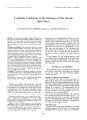
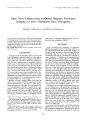
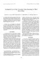
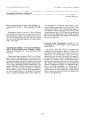
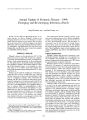
Show Journal of Neuro- Ophthalmology 19( 4): 246- 248, 1999. © 1999 Lippincott Williams & Wilkins, Inc., Philadelphia Ophthalmoplegia Associated With the Anti- Ri Antibody Richard Ohmer M. D., Karl C. Golnik, M. D., Arthur I. Richards, M. D., FACP, and Gregory S. Kosmorsky, D. o. Anti- Ri antibodies most often occur in patients with breast cancer and typically are associated with the paraneoplastic syndrome of opsoclonus- myoclonus- ataxia. This study reports a patient with diplopia and ophthalmoplegia. She had anti- Ri antibodies, and despite an exhaustive search for malignancy at presentation, breast cancer was not detected for six months. Key Words: Anti- Ri antibody- Ophthalmoplegia- Paraneoplastic. Anti- Ri is a highly specific anti- neuronal antibody that interacts with central nervous system ( CNS) neurons, but does not interact with glia or other CNS cells. The Ri antigen is thought to be homologous to an RNA- binding protein expressed in the developing motor system ( 1). The anti- Ri antibody ( anti- Ri Ab) is usually detected in patients with breast cancer ( 1), although it has been reported in patients with ovarian ( 2), fallopian ( 1), and small- cell lung cancer ( 3). Patients with anti- Ri Ab may develop the syndrome of opsoclonus- myoclonus- ataxia ( OMA) ( 1- 3). We report a patient with anti- Ri Ab who developed diplopia and ophthalmoplegia, but not nystagmus. Breast cancer was not discovered for six months, despite an exhaustive search at the onset of her disease. CASE REPORT A 60- year- old hypertensive woman sought treatment in January 1998 for dizziness and diplopia. Her physician believed she had a right abducens nerve palsy of viral etiology. The diplopia persisted, and she developed fatigue, generalized muscle aches, dysphagia, and difficulty chewing. Neurologic evaluation suggested bilateral abducens nerve palsies. Past medical history included asthma, degenerative joint disease, and endometriosis. She had undergone a hysterectomy in 1984, and a breast biopsy in 1990 showed cystic change. Medications at Manuscript received July 27, 1999; accepted September 17, 1999. From the Cincinnati Eye Institute ( R. O., K. C. G.), Cincinnati, Ohio, U. S. A., the Department of Ophthalmology and the Neuroscience Institute at the University of Cincinnati, ( K. C. G.), Cincinnati, OH, Oncology Hematology Care ( A. I. R.), Cincinnati, OH, and the Division of Ophthalmology, the Cleveland Clinic Foundation ( G. S. K.), Cleveland, Ohio, U. S. A.. Address correspondence to Karl C. Golnik, M. D., 10494 Montgomery Road, Cincinnati, OH 45243. presentation were clonidine, Cardura, Bumex, and Es-trace. Results of evaluations in February 1998, including computed tomography ( CT) scan and magnetic resonance ( MR) imaging of the brain, lumbar puncture, edrophonium test, electromyography, antiacetylcholine receptor antibody levels, chest radiograph, complete blood count, renal profile, thyroid stimulating hormone, anti-nuclear antibody ( ANA), antistriated muscle antibody, and cardiac enzymes, were normal or negative. She was referred for neuro- ophthalmologic examination. Additional symptoms noted in March 1998 included change in character of voice, a 10 pound weight loss, and occasional urinary incontinence. Results of vision and pupillary examinations were normal. Vertical ductions and versions were full. Abduction and adduction of each eye was almost completely absent, and there was a 10 prism diopter esotropia in primary position. There was no nystagmus or involuntary eye movement. Results of the Dolls maneuver were negative. There was no ptosis or proptosis, and orbicularis strength was normal. Magnetic resonance imaging with gadolinium was repeated, and the test results were normal; 1 mm sections through the pons showed no abnormalities. Treatment with 60 mg per day of prednisone was begun for possible myasthenia gravis. Within two weeks the ophthalmoplegia had improved ( Fig. 1), and by April 1998 adduction was almost normal, but abduction was still moderately abnormal. The dosage of prednisone was tapered because of fluid retention, and by May 1998 her symptoms and ophthalmoplegia were worsening. Repeat edrophonium test, single fiber electromyography, and antiacetylcholine receptor antibody levels were negative. CT scan of the chest showed no thymona. The patient developed ataxia and dysdiadokinesis. Anti- Yo and anti- Hu antibodies were negative, but anti- Ri antibody was positive. A mammogram, breast ultrasound, and Miraluna nuclear breast scan ( Dupont, Bil-lerica, MA) showed fibrocystic change. Test results for serum cancer antigen ( CA) 15- 3, CA 27- 29, carcinogenic embryonic antigen ( CEA), and the human immunodeficiency virus were negative. Results of an abdominal CT, esophageal gastroduodenoscopy, and colonoscopy were negative. A gynecologic examination in the context of her previous surgery was normal. Four sessions of plas- 246 ANTI- RIAB AND OPHTHALMOPLEGIA 247 FIG. 1. Extraocular movements demonstrating almost complete left gaze palsy, moderate right abduction deficit, and mild left adduction deficit 2 weeks after prednisone was begun. A 10 prism diopter esotropia was present in primary position at distance. mapheresis produced no improvement in symptoms or signs. In December 1998, breast MR imaging showed a suspicious area in the right breast. Repeat mammography showed no obvious lesion. A quadrantectomy was done, and pathology showed two separate lesions. One lesion, slightly less than 1 cm was identified as an intraductal carcinoma, and a second 4 mm area of infiltrating lobular carcinoma was also seen. The patient underwent four cycles of cytoxan- methotrexate- flourouracil and is now receiving radiation to the chest. There has been no significant change in her moderate bilateral abduction and adduction deficits since the discovery and treatment of her cancer. Interestingly, her ophthalmoplegia improved with repeated attempts at right and left gaze. DISCUSSION Neurologic paraneoplastic syndromes associated with autoantibodies are recognized with increasing frequency. Commonly detected antibodies include anti- Yo, anti- Hu, and anti- Ri ( 1,3). The malignancy may be occult; small-cell cancer of the lung ( anti- Hu) and ovarian or breast cancer ( anti- Yo) are most common when such antibodies are present ( 4). Syndromes include brainstem encephalitis and subacute sensory neuropathy associated with anti- Hu ( 5- 8), and anti- Yo associated subacute cerebellar degeneration ( 6,9). Neuro- ophthalmologic abnormalities are varied and dependent on the area of brain affected. Nystagmus, gaze palsy, ophthalmoplegia, and ocular misalignment are most frequently reported ( 9,10). Anti- Ri Ab is usually detected in patients with breast cancer ( 1). However, single case reports document an association with cancer of the lung ( small cell) ( 3), fallopian tube ( 1), ovarian duct ( 11), and bladder ( 1). Anti- Ri Ab is usually associated with the paraneoplastic syndrome of OMA ( 1,3). However, a variety of ophthalmologic abnormalities, including torsional nystagmus, abducens nerve palsy, abnormal pursuit, upgaze palsy, and blepharospasm, have been associated with the Anti- Ri Ab. Luque et al. ( 1) described one patient each with nystagmus and abnormal pursuit, torsional nystagmus and abducens nerve palsy, and blepharospasm. Escudero et al. ( 12) described a patient who had an upgaze palsy and eyelid apraxia but otherwise normal eye movement. Hormigo et al. ( 13) reported a patient with opsoclonus who developed impaired smooth pursuit and optokinetic nystagmus, abnormal vetibulo- ocular reflex, upgaze palsy, and bilateral abduction deficits. Concomitant neurologic and systemic abnormalities other than myoclonus and ataxia include dizziness, nausea, proximal muscle weakness, spastic quadriparesis, hyperreflexia, dysphagia, dysarthria, dementia, and progressive encephalopathy and rigidity ( 1,12,14). Our patient presented with diplopia and mild systemic symptoms. She was found to have almost no horizontal eye movements, normal vertical eye movements, and small esotropia. Results of repeated evaluations for pontine abnormality and myasthenia gravis were negative. We believe the anti- Ri Ab was responsible for her neurologic syndrome and was initially selectively affecting horizontal gaze centers in the pons. The pathogenesis of these paraneoplastic neurologic syndromes is unclear, but most investigators feel it is an autoimmune process ( 4,8,10,13). However, anti- Ri Ab may be present in the absence of cancer or neurologic disease. In three patients with OMA and anti- Ri Ab, no ./ Neuro- Ophthaliiiol, Vol. 19, No. 4, 1999 248 R. OHMERETAL. malignancy was found 20 to 42 months after presentation, despite thorough evaluation, even following the results of an autopsy in one patient ( 3,13,14). Drlicek et al. ( 2) identified anti- Ri Ab in 7 of 181 patients with ovarian cancer and none of these patients developed a paraneoplastic syndrome over 2 years of follow- up. It appears that when present, the anti- Ri antibody is not always pathogenic, and the pathogenic presence of the anti- Ri antibody is not always associated with a malignancy. Moll et al. ( 4) investigated 23 patients with paraneoplastic syndromes associated with either anti- Hu, anti- Yo, or anti- Ri Ab. These patients were compared with 66 cancer patients without paraneoplastic syndromes and with 107 age- matched controls for the presence of systemic autoantibodies ( ANA, anti- DNA, anticentromere, antiribo-nucleoprotein, antiSmith, antisingle- stranded- A, anti-single- stranded- B, antisclerodermal, antimitochondrial, antismooth muscle, antiparietal cell, antibrush border). Moll et al. found significantly more patients with the paraneoplastic syndromes to have other systemic autoantibodies, and they speculated that a genetic susceptibility may exist. Treatment of any paraneoplastic syndrome begins with removal of the cancer. Unfortunately, this removal often does not reverse the paraneoplastic abnormalities. Systemic corticosteroids, cyclophosphamide, intravenous immunoglobulin, and plasmapheresis have been used in patients with the anti- Ri Ab with mixed results ( 3,11,14). Dropcho et al. ( 3) reported resolution of opsoclonus during prednisone treatment in one patient. The dosage of corticosteroids was tapered over 30 months, and cyclophosphamide was used for the next year, during which the patient had no recurrent opsoclonus. Casado et al. ( 14) used prednisone and triazolam to treat a patient with opsoclonus, encephalomyelitis, and rigidity; eye movements were normal eleven months later. Jongen et al. ( 11) reported near resolution of OMA following resection of an ovarian duct carcinoma, radiation, chemotherapy, and plasma exchange. However, no improvement in ophthalmologic signs or symptoms has been noted after treatment with prednisone ( 13) or chemotherapy ( 12). Our patient was treated with prednisone before discovery of the cancer and experienced some improvement in ophthalmoplegia. Her condition worsened when the steroid dosage was tapered and did not improve with subsequent higher prednisone dosages, removal of the cancer, and plasmapheresis. Fortunately, she has remained stable. In conclusion, neurologic paraneoplastic syndromes are recognized more frequently. Our patient developed a unique brainstem syndrome characterized by an isolated horizontal gaze paresis. The responsible lesion was likely in the region of the parapontine reticular formation and the abducens nuclear complex, which are responsible for conjugate horizontal gaze. The normality of the results of MR images in this clinical situation should suggest the possibility of a paraneoplastic etiology. The paraneoplastic syndrome may precede the discovery of cancer by months or years, despite thorough evaluation. If the anti- Ri antibody is present and mammogram and ultrasound are negative, breast MR images should be obtained. REFERENCES 1. Luque FA, Furneaux HM, Ferziger R, et al. Anti- Ri: an antibody associated with paraneoplastic opsoclonus and breast cancer. Ann Neurol 1991; 29: 241- 51. 2. Drlicek M, Bianchi G, Bogliun G, et al. Antibodies of the anti- Yo and anti- Ri type in the absence of paraneoplastic neurological syndromes: a long- term survey of ovarian cancer patients. J Neurol 1997; 244: 85- 9. 3. Dropcho EJ, Kline LB, Riser J. Antineural ( anti- Ri) antibodies in a patient with steroid- responsive opsoclonus- myoclonus. Neurology 1993; 43: 207- 11. 4. Moll JW, Hooijkaas H, van- Goorbergh BC, Roos LG, Henzen- Longmans SC, Vecht CJ. Systemic and anti- neuronal autoantibodies in patients with paraneoplastic neurological disease. J Neurol 1996; 243: 51- 6. 5. Yamada M, Inaba A, Yamawaki M, et al. Paraneoplastic en-cephalo- myelo- ganglionitis: cellular binding sites of the antineu-ronal antibody. Acta Neuropathol ( Berl) 1994; 88: 85- 92. 6. Graus F, Rene R. Clinical and pathological advances on central nervous system paraneoplastic syndromes. Rev- Neurol ( Paris) 1992; 148: 496- 501. 7. Lovblad KO, Boucraut J, Bourdenet S, et al. Sensory neuronopathy and small cell lung cancer: antineuronal antibody reacting with neuroblastoma cells. J Neurol 1993; 240: 327- 32. 8. Brashear HR, Caccamo DV, Heck A, Keeney PM. Localization of antibody in the central nervous system of a patient with paraneoplastic encephalomyeloneuritis. Neurology 1991; 41: 1583- 7. 9. Anderson NE, Rosenblum MK, Posner JB. Paraneoplastic cerebellar degeneration: clinical- immunological correlations. Ann Neurol. 1988; 24: 559- 67. 10. Salmaggi A, Nemni R, Pozzi A, et al. Antineuronal antibody in a patient with neuroblastoma and opsoclonus- myoclonus- ataxia: a case report. Tumori 1997; 83: 709- 11. 11. Jongen JLM, Moll WJB, Sillevis Smith PAE, Vecht VJ, Tijssen CC. Anti- Ri positive opsoclonus- myoclonus- ataxia in ovarian duct cancer. J Neurol 1997; 245: 691- 2. 12. Escudero D, Barnadas A, Codina M, Fueyo J, Graus F. Anti- Ri associated paraneoplastic neurological disorder with opsoclonus in a patient with breast cancer. Neurology 1993; 43: 1605- 6. 13. Hormigo A, Dalmau J, Rosenblum MK, River ME, Posner JB. Immunological and pathological study of anti- Ri- associated en-cephalomyopathy. Ann Neurol 1994; 36: 896- 902. 14. Casado JL, Gil- Peralta A, Graus F, Arenas C, Lopez JM, Alberca R. Anti- Ri antibodies associated with opsoclonus and progressive encephalomyelitis with rigidity. Neurology 1995; 44: 1521- 2. J Neuro- Ophlhalmol, Vol. 19, No. 4, 1999 |