| OCR Text |
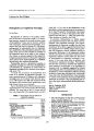
Show journal of Neuro- Ophthalmology 14( 2): 91- 94, 1994. 1994 Raven Press, Ltd., New York Spontaneous Remission of Papilledema and Sixth Nerve Palsy in Acute Lymphoblastic Leukemia Gil I. Wolfe, M. D., Steven L. Galetta, M. D., and Joan E. Mollman, M. D. Spontaneous regression of hematologic malignancies is not uncommon and occurs in a wide variety of lymphomas and leukemias. In contrast, spontaneous remission of neurologic symptoms produced by these tumors is exceedingly rare. We report a patient with central nervous system acute lymphoblastic leukemia who experienced at least one spontaneous remission of papilledema and sixth nerve palsy. This represents, to our knowledge, the first case of spontaneous remission of neuro-ophthalmologic signs in a patient with acute leukemia. We conclude that meningeal leukemia may have a protracted course, and that spontaneous remission of neuro- ophthalmologic findings should not be so readily ascribed to a benign process in a patient with preexisting leukemia. Key Words: Spontaneous remission- Acute lymphoblastic leukemia- Leptomeningeal metastasis- Papilledema- Sixth nerve palsy. From the Department of Neurology, University of Pennsylvania School of Medicine, Philadelphia, Pennsylvania, U. S. A. Address correspondence and reprint requests to Dr. Steven L. Galetta, Department of Neurology, Hospital of the University of Pennsylvania, 3400 Spruce St., Philadelphia, PA 19104. The spontaneous regression of cancer refers to the partial or complete disappearance of malignant tumor in the setting of no treatment or treatment judged inadequate to explain the observed remission ( 1,2). Spontaneous regression has been reported in a wide variety of cancers, including solid tumors and hematologic malignancies ( 1,3- 5). The duration of spontaneous regression in lymphoma can be prolonged and even permanent ( 3,6- 9). In contrast, spontaneous regression of acute leukemias tends to be short- lived ( 3,10- 14). Documented regression of hematologic malignancies that involve the central nervous system ( CNS) is exceedingly rare. Rubin and colleagues ( 15) reported a complete spontaneous regression of primary CNS lymphoma, which lasted 13 months. Prolonged survival has been described in cases of meningeal lymphoma ( 16) and neurolym-phomatosis ( 17), where the diagnosis was delayed from months to years. Spontaneous remission of neurologic symptoms produced by these tumors is equally rare. Galetta and colleagues ( 18) recently reported the spontaneous resolution of a third nerve palsy in a patient subsequently found to have meningeal lymphoma. On repeat magnetic resonance imaging ( MRI), abnormal enlargement and enhancement of the patient's third nerve also resolved. To our knowledge, spontaneous remission of neurologic symptoms has never been observed in meningeal leukemia. We report a patient with CNS acute lymphoblastic leukemia ( ALL) who had relapsing and remitting episodes of diplopia and papilledema over a 3- year period without definitive treatment. CASE REPORT The patient presented in August 1984 at age 16 with leg pain, a 5- lb weight loss, and fatigue. Phys- 91 92 G. /. WOLFE ET Ah. ical examination was significant for left anterior cervical adenopathy. Neurologic examination, including funduscopy, was normal. On complete blood count the hemoglobin was 6 g/ dl, white blood cell count was 2,500/ mm3, and platelets were 67,000/ mm3. Chest radiograph showed hilar and mediastinal adenopathy. Plain films of the lumbosacral spine were normal. Ultrasound of the abdomen was normal. A bone marrow biopsy showed ALL. All cells were CALLA- positive ( common ALL antigen, positive in greater than 80% of pediatric ALL cases), and the TdT marker ( terminal deoxynucleotidyl transferase, present in immature ALL cells) was positive. Cerebrospinal fluid ( CSF) examination showed normal protein, white blood cell count of 0/ mm3, and red blood cell count of 0/ mm3. She underwent induction chemotherapy with vincristine, prednisone, and L- asparaginase, followed by 2 years of maintenance with vincristine, methotrexate ( oral, intravenous, and intrathecal), and 6- mercaptopurine. Chemotherapy was well tolerated, and follow- up bone marrow and lumbar puncture in November 1986 showed continued remission. She did well until January 1988, when she developed horizontal diplopia and mild headache. Examination revealed visual acuity 20/ 20 OU, a right sixth nerve palsy ( 16 prism diopters of esotropia in the primary position), and bilateral papilledema with fine, flame- shaped hemorrhages interiorly. There was no evidence of an infiltrative process on funduscopic examination. Computed tomography ( CT) of the head and orbits was negative. CSF analysis showed white blood cell count of 334/ mm3 ( 72% lymphocytes, 12% atypical lymphocytes, and 14% immature cells). Flow cytometry was unrevealing. The lymphocytes were 17% CALLA- positive, with a few cells demonstrating weak, scattered TdT positivity ( see Table 1). CSF cryptococcal antigen, Lyme antibody, viral cultures, fungal cultures, and acid- fast bacteria cultures were negative. Serum Epstein- Barr virus, herpesvirus, cytomegalovirus, and Lyme titers were negative. The purified protein derivative of tuberculin ( PPD) test was negative. She received one dose of cytosine arabinoside ( ara- C) and intrathecal methotrexate. A repeat lumbar puncture in February 1988 revealed a white blood cell count of 43/ mm3 ( 97% mature lymphocytes, 3% histiocytes). CALLA and TdT markers were negative. By March 1988 she was asymptomatic. The sixth nerve palsy and papilledema had resolved and diagnosis of a viral syndrome was made. In May 1989 she developed horizontal diplopia and blurred vision. Bilateral sixth nerve palsies TABLE 1. Cerebrospinal fluid results and cell marker positivity CSF sample 1/ 88" 2/ 88 5/ 89" 5/ 89 6/ 89 4/ 91c WBCs/ mm3 334 43 62 73 45 1,200 Abnormal cells (%) 26 0 7 7 12 40 CALLA+ (%) 17 0 0 21 0 87 TdT+ (%) - 0 50 0 0 60 CSF, cerebrospinal fluid; WBCs, white blood cells; CALLA, common acute lymphoblastic leukemia antigen; TdT, terminal deoxynucleotidyl transferase. " Patient received one dose of ara- C ( cytosine arabinoside) and intrathecal methotrexate. b Patient placed on Diamox 250 mg b. i. d. for 2 months. c Patient diagnosed with central nervous system relapse of acute lymphoblastic leukemia. and papilledema were present. Visual acuity was 20/ 20 OU. Lumbar puncture revealed an opening pressure of 33 cmH20; protein, 34 mg/ dl; and white blood cells, 62/ mm3 ( 89% lymphocytes, 4% histiocytes, and 7% suspicious cells). Also, 50% of cells were TdT- positive. CALLA was negative. Cerebrospinal fluid VDRL, Lyme, viral, and acid- fast bacteria studies were negative. MRI of the brain with gadolinium enhancement was unremarkable. A repeat lumbar puncture that month showed an opening pressure of 28 cmH20 and a white blood cell count of 73/ mm3 ( 90% lymphocytes, 7% suspicious cells) with 21% CALLA and negative TdT markers. Myelin basic protein level was normal. The patient was placed on Diamox 250 mg b. i. d. for 2 months. A repeat lumbar puncture in June 1989 showed an opening pressure of 25 cmH20 and a white blood cell count of 45/ mm3 ( 84% lymphocytes, 7% immature cells, and 5% atypical cells). CALLA and TdT markers were negative. In August 1989, examination showed the papilledema had resolved, and there was no ocular motility defect. Follow- up funduscopic examination in January 1990 remained normal. In April 1991 she first presented to the Hospital of the University of Pennsylvania with headaches, sore throat, rhinorrhea, right retro- orbital pain, and numbness over the right lower face. On examination visual acuity was 20/ 20 in both eyes and ocular motility was full. She had severe bilateral papilledema with enlargement of both blind spots and decreased sensation to pinprick over the right chin. MRI of the head with gadolinium showed left mastoiditis, a soft tissue mass occupying the left posterior auricular region, preauricular adenopathy, and subtle enhancement of the meninges consistent with tumor spread. The optic nerves and chiasm were normal. CSF examination showed an / Neuw- Ophthalmol, Vol. 14, No. 2, 1994 SPONTANEOUS REMISSION OF NEURO- OPHTHALMIC SIGNS IN ALL 93 opening pressure exceeding 55 cmH20; protein, 59 mg/ dl; and white blood cell count of 1,200/ mm3 ( 16% lymphocytes, 44% monocytes, and 40% atypical lymphocytes). Cells were 87% CALLA- positive and 60% TdT- positive. Biopsy of the left posterior auricular mass showed malignant lymphocytes. CT scans of the chest, abdomen, and pelvis revealed a small right pleural soft tissue density representing leukemic mass or adenopathy. Peripheral blood smear and bone marrow biopsy from both iliac crests were normal. A diagnosis of CNS relapse of ALL was made. The patient received whole- brain radiation therapy followed by systemic DATVP ( daunorubicin, ara- C, 6- thiogua-nine, vincristine, and prednisone) and intravenous and intrathecal methotrexate. CSF cytology remained positive, and intrathecal therapy with ara- C was given from May through July 1991. Following reinduction chemotherapy, she received craniospinal radiation. The headaches and enlarged blind spots improved with this therapy, and her papilledema resolved with mild residual left optic nerve atrophy. In August 1991 she was accepted for allogeneic bone marrow transplant at another institution. DISCUSSION Despite evidence for leptomeningeal metastasis of acute lymphoblastic leukemia, our patient had at least one spontaneous remission of papilledema and sixth nerve palsy over a 3- year period. A single dose of chemotherapy administered early in her central nervous system presentation failed to clear the cerebrospinal fluid pleocytosis. However, definitive therapy was not continued, since her neuro- ophthalmologic signs rapidly improved, and a benign process was considered responsible. When the papilledema and ocular motility defect returned 1 year later, Diamox was started for the unexplained increased intracranial pressure. Although it is conceivable that Diamox played some role in the second remission, the patient remained asymptomatic for nearly 2 years after the Diamox was discontinued. The persistent CSF pleocytosis and atypical cells would also exclude a diagnosis of pseudotumor cerebri ( 19). Thus, any role Diamox played in the prolonged remission was likely negligible. We believe the clinical remissions observed in our patient reflect an inherent control of the host over tumor growth. Presumably, she was able to reverse some manifestation of the CNS leukemia but could not eliminate the entire tumor load. In instances where the host response is more robust, complete spontaneous regression would occur. Spontaneous regression of hematologic malignancy is not uncommon and may occur in up to 20% of patients with low- grade non- Hodgkin's lymphoma ( 3,8). Although the mechanism of spontaneous regression is unknown, a number of factors that enhance immunologic surveillance have been proposed. In Cole's reviews ( 1,5), " operative trauma" was the most commonly cited mechanism, present in 46% of cases of spontaneous regression ( 2). Surgical removal of part of the tumor theoretically heightens the immune system's response to the tumor that remains ( 2,5). The appearance of a second malignancy may coincide with or follow spontaneous remission of leukemia ( 3,20,21). Antigenic competition, whereby the immunologic response to one antigen enhances the immune system's response to a second antigen ( 22), has been forwarded as a possible mechanism in these remissions ( 3). Viral infections have been implicated in a wide variety of lymphomas that have undergone spontaneous regression, including Burkitt's lymphoma ( 6), non- Hodgkin's lymphoma ( 7,8), and lymphosarcoma ( 23). Dock ( 24) reviewed the therapeutic effect of infection on leukemia in 1904. Early reports documented spontaneous regression of leukemia following bacterial ( 3,10,11,25), viral ( 25), and unspecified infections ( 26). More recent cases of spontaneous regression in leukemia have followed a variety of infections. Complete and partial remissions of adult T- cell leukemia have followed cytomegalovirus pneumonia ( 12) and bacterial infections ( 13). Murakawa and coworkers ( 13) proposed that the bacterial infections induced expansion of an adult T- cell leukemia clone and secretion of cytokines, which further enlarged the clone. Antibiotic therapy suppressed cytokine production with subsequent shrinkage of the clone population. Stimulation of natural killer cell function by concurrent tuberculosis infection may have contributed to the 13- year disease- free survival of a patient with acute nonlymphoblastic leukemia ( 27). Multiple studies failed to identify a viral, bacterial, or fungal infection in our patient. Spontaneous remission of neurologic signs and symptoms produced by CNS hematologic malignancies is rare and to our knowledge has never been reported in acute leukemia. CSF cytology in our patient remained negative or showed an equivocal number of suspicious cells and markers for more than 3 years before she was diagnosed with CNS relapse of ALL. Absence of malignant cells in the CSF, however, does not exclude a di- / Neuro- Ophlhalmol. Vol. 24, No. 2, 2994 94 G. 1. WOLFE ET AL. agnosis of leptomeningeal metastasis ( 28). Initial CSF cytology may be negative in 15 to 46% of patients whose tumor has spread to the lepto-meninges ( 29). Examination of CSF cytology shortly before death failed to identify nearly half of lymphoma or leukemia patients with autopsy-proven leptomeningeal tumor ( 28). Cytology of cisternal CSF may help detect patients with suspected leptomeningeal metastasis who have unremarkable lumbar punctures ( 29). It is unclear if cisternal puncture would have led to an earlier diagnosis in our patient. This patient demonstrates that untreated CNS hematologic malignancies may have a protracted course. Spontaneous remission of neuro-ophthalmologic signs in a patient with preexisting leukemia may not represent a benign process, and careful surveillance of such patients is warranted. REFERENCES 1. Everson TC, Cole WH. Spontaneous regression of cancer. Philadelphia: WB Saunders, 1966. 2. Challis GB, Stam HJ. The spontaneous regression of cancer: a review of cases from 1900 to 1987. Acta Oncol 1990; 29: 545- 50. 3. Wiernik PH. Spontaneous regression of hematologic cancers. Natl Cancer Inst Monogr 1976; 44: 35- 8. 4. Boyd W. The spontaneous regression of cancer. Springfield, IL: Charles C Thomas, 1966. 5. Cole WH. [ Opening address]: Spontaneous regression of cancer and the importance of finding its cause. Natl Cancer Inst Monogr 1976; 44: 5- 9. 6. Bluming AZ, Ziegler JL. Regression of Burkitt's lymphoma in association with measles infection. Lancet 1971; 2: 105- 6. 7. Krikorian JG, Portlock CS, Cooney DP, Rosenberg SA. Spontaneous regression of non- Hodgkin's lymphoma: a report of nine cases. Cancer 1980; 46: 2093- 9. 8. Horning SJ, Rosenberg SA. The natural history of initially untreated low- grade non- Hodgkin's lymphomas. N Engl ] Med 1984; 311: 1471- 5. 9. Poppema S, Postma L, Brinker M, de Jong B. Spontaneous regression of a small non- cleaved cell malignant lymphoma ( non- Burkitt's lymphoblastic lymphoma): morphologic, im-munohistological, and immunoglobulin gene analysis. Cancer 1988; 62: 791- 1. 10. Diamond LK, Luhby AL. Pattern of " spontaneous" remissions of leukemia of childhood observed in 26 of 300 cases. Am J Med 1951; 10: 238- 9. 11. Southam CM, Craver LF, Dargeon HW, Burchenal JH. A study of the natural history of acute leukemia with special reference to the duration of the disease and the occurrence of remissions. Cancer 1951; 4: 39- 59. 12. Schnitzer B, Lovett EJ, Kahn LE. Adult T- cell leukemia with spontaneous remission. Lancet 1983; 11: 1030. 13. Murakawa M, Shibuya T, Teshima T, et al. Spontaneous remission from acute exacerbation of chronic adult T- cell leukemia. Blut 1990; 61: 346- 9. 14. Spadea A, Latagliata R, Martinelli E, Petti MC. Transient spontaneous remission in a case of adult acute myelogenous leukemia. Br ] Haematol 1990; 76: 154. 15. Rubin M, Libman I, Brisson M, Goldenberg M, Brem S. Spontaneous temporary remission in primary CNS lymphoma. Can J Neurol Sci 1987; 14: 175- 7. 16. Kepes JJ, Maxwell JA, Hedeman L, Slaven J. Primary diffuse malignant lymphoma of the leptomeninges presenting as " pseudotumor cerebri." Neurochirurgia 1971; 14: 188- 96. 17. Borit A, Altrocchi PH. Recurrent polyneuropathy and neu-rolymphomatosis. Arch Neurol 1971; 24: 40- 9. 18. Galetta SL, Sergott RC, Wells GB, Atlas SW, Bird SJ. Spontaneous remission of a third- nerve palsy in meningeal lymphoma. Ann Neurol 1992; 32: 100- 2. 19. Wall M, George D. Idiopathic intracranial hypertension: a prospective study of 50 patients. Brain 1991; 114: 155- 80. 20. Durant JR, Finkbeiner JA. " Spontaneous" remission in chronic lymphatic leukemia? Cancer 1964; 17: 105- 13. 21. Han T, Sokal JE. Spontaneous remission of leukemic lym-phoproliferative disease. Cancer 1971; 27: 586- 95. 22. Pross H, Eidinger D. In vitro studies of " antigenic competition." II. Reconstitution of the immune defect and the relationship between antigen- induced suppression and non- specific enhancement. Immunology 1973; 25: 269- 81. 23. Weintraub LR. Lymphosarcoma: remission associated with viral hepatitis. JAMA 1969; 210: 1590- 1. 24. Dock G. The influence of complicating diseases upon leukemia. Am J Med Sci 1904; 127: 563- 92. 25. Bierman HR, Crile DM, Dod KS, et al. Remissions in leukemia of childhood following acute infectious disease: staphylococcus and streptococcus, varicella, and feline panleukopenia. Cancer 1953; 6: 591- 605. 26. Bassen FA, Kohn JL. Multiple spontaneous remissions in a child with acute leukemia: the occurrence of agranulocytosis and aplastic anemia in acute leukemia and their relationship to remissions. Blood 1952; 7: 37- 46. 27. Ress SR, Morris CD, Jacobs P. Enhanced natural killer cell activity and long survival in acute non- lymphoblastic leukemia. S Afr Med ] 1990; 77: 648- 50. 28. Glass JP, Melamed M, Chernik NL, Posner JB. Malignant cells in cerebrospinal fluid ( CSF): the meaning of a positive CSF cytology. Neurology 1979; 29: 1369- 75. 29. Rogers LR, Duchesneau PM, Nunez C, et al. Comparison of cisternal and lumbar CSF examination in leptomeningeal metastasis. Neurology 1992; 42: 1239- 41. / Neuro- Ophthalmol, Vol. 14, No. 2, 1994 |