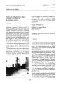
| OCR Text |
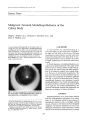
Show Journal of Clinical Neuro- ophtiullmology 10( 4): 231- 238, 1990. © 1990 Raven Press, Ltd., New York Optic Nerve Sheath Distention in Leber's Optic Neuropathy and the Significance of the " Wallace Mutation" J. Lawton Smith, M. D., David T. Tse, M. D., Sandra Frazier Byrne, Donald R. Johns, M. D., and Edwin M. Stone, M. D., ph. D. We recently encountered a 27- year- old man who presented an atypical clinical picture of Leber's hereditary optic neuropathy: His family history was negative, visual loss continued steadily for over 8 months, circumpapillary microangiopathy was equivocal, the optic discs showed large physiologic cups, and both optic nerve sheaths were notably distended with increased subarachnoid fluid. The latter was confirmed by ultrasonography, computed tomography, and magnetic resonance imaging. The patient's asymptomatic brother also showed unimpressive circumpapillary microangiopathy in the fundi. The asymptomatic mother from France was then seen, and she showed classic circumpapillary microangiopathy in the fundi. Studies of mitochondrial DNA showed the classic point mutation at position 11778 as reported by Wallace in all three family members. Another patient previously seen with classic Leber's hereditary optic neuropathy recently had mitochondrial DNA studies along with three other affected family members and five unaffected family members in the maternal lineage. All nine of these individuals were completely normal at the Wallace locus. In fact, sequencing of the entire ND- 4 gene from one affected individual revealed it to be perfectly normal at the amino acid level. The importance of obtaining quantitative ultrasonography and the 30° test, and studying mitochondrial DNA in patients suspected of having Leber's optic nerve disease is emphasized. Key Words: Leber's hereditary optic neuropathy- Optic nerve sheath distention- Wallace mutation. From the Bascom Palmer Eye Institute, Department of Ophthalmology, University of Miami School of Medicine, Miami, Florida a. L. S., D. T. T.); the Department of Neurology, Johns Hopkins University School of Medicine, Baltimore, Maryland ( D. R. J.); and the Department of Ophthalmology, University of Iowa, Iowa City, Iowa ( E. M. S.). Address correspondence and reprint requests to Dr. J. Lawton Smith, Bascom Palmer Eye Institute, P. O. Box 016880, MIami, FL 33101, U. S. A. 231 This is the first report, to our knowledge, of notable bilateral distention of the orbital optic nerve sheaths in Leber's hereditary optic neuropathy. The diagnosis was confirmed by mitochondrial DNA studies in the patient and two unaffected family members, all of whom showed the classic mutation at position 11778 as reported by Wallace and co- workers ( 1,2). A second patient, seen earlier and with more typical clinical findings of Leber's optic nerve disease and a positive family history dating back into the mid- 1800s, was not found to have the 11778 point mutation. This patient's affected family members also did not have the mutation. The importance of obtaining optic nerve ultrasonography with 30° tests and also mitochondrial DNA studies for the " Wallace mutation," and the significance of these findings, is addressed. CASE REPORTS Case One This 27- year- old Frenchman had 20/ 20 uncorrected vision at a routine pilot's physical examination in October 1988. In July 1989, he noted the sudden painless loss of central vision in the left eye. He was hospitalized in September 1989 for 1 week for investigation, and was given no treatment at the time. The vision never improved in the eye. About 10 days later, he noted the onset of blurred central vision in the right eye, and again had no pain on motion of the globe. Intravenous steroids were given for 3 weeks at home, but the vision did not improve. The patient was hospitalized for 3 weeks on a neurology service in Paris in December 1989 and studied carefully. He was given 5 days of intravenous methylprednisolone succinate and 3 weeks of 232 f. L. SMITH ET AL. concomitant oral doxycycline along with high doses of vitamins B6, B12, E, and C, but the vision continued to worsen steadily. Past history revealed no antecedent infection, Uhthoff's or L'hermitte's symptoms, or weakness, paresthesias, or other complaints. He had migraine headaches about five to six times a year since age 10. He denied use of tobacco, alcohol, or narcotic drugs. The family history revealed that the parents were both in good health. He has two brothers, age 31 and 24, respectively, both in good health and with excellent vision. The family history was totally negative for severe vision loss to their knowledge, but there had been no maternal uncle in the two previous generations. Examination at the Bascom Palmer Institute on February 2, 1990, revealed a best corrected acuity of 20/ 140 in the right eye and 20/ 320 in the left eye. The external eye examination was normal except for a 1+ afferent pupillary response on the left. Visual field studies revealed full peripheral fields but dense bilateral absolute central scotomas. Slit lamp examination was normal and no cells were seen in the vitreous of either eye. Ophthalmoscopy disclosed clear media, sharp optic discs but with large physiologic cups, and definite pallor of the papillomacular bundles in both eyes. The nerve fiber layer could still be seen in the upper and lower bundles, but the discs were definitely becoming atrophic. A careful search revealed only equivocal circumpapillary telangiectatic changes. The maculae and periphery were otherwise normal. A good quality fluorescein angiogram made January 18, 1990, in Paris and kindly provided through the courtesy of Dr. Martine Flamand was quite normal and showed no leakage of dye in the optic discs or retina ( Figs. 1- 4). These pictures document the large physiologic cups and show subtle microangiopathic changes on the fluorescein prints that could not be appreciated on the black and white prints. The patient's asymptomatic 31- year- old brother had 20/ 20 uncorrected vision in both eyes, and on ophthalmoscopy showed sharp pink discs. Again, a careful search showed only equivocal circumpapillary telangiectatic changes. Quantitative ultrasonography on the propositus revealed that both optic nerves measured 5.2 mm in diameter at the posterior orbital segment, and both decreased to 4.4 mm on 30° eccentric gaze. In this laboratory, the normal optic nerve sheath averages 2.7 mm, and the upper limit of normal is 3.3 mm. Therefore, these sheaths at the posterior orbital measurements were twice normal size. Review of the patient's computed tomography ( CT) FIG. 1. Fundus photograph, right eye, of patient one, made January 18, 1990, in Paris. Note that the microangiopathy is equivocal. ( Figs. 5- 7) and magnetic resonance imaging ( MRI) studies ( Fig. 8) confirmed the presence of bilaterally distended optic nerve sheaths. The orbital CT scans confirmed the ultrasonographic 30° test; the diameter of the optic nerve sheath was 7 mm at maximum diameter ( 4 mm behind the globe) and decreased to 6 mm on right gaze and to 5 mm on extreme left gaze. This observation was made by Dr. Robert Quencer. The CT and MR studies were otherwise normal except for the presence of an incidental small arachnoidal cyst over the tip of the left temporal lobe ( Fig. 9) considered by Dr. FIG. 2. Fluorescein angiogram. right eye. of patient one, January 18, 1990. Note the large physiologic cup. and absence of fluorescein staining. Microangiopathy is more easily seen than in Fig. 1 ( arrow). NERVE SHEATH DISTENTION, LEBER'S NEUROPATHY, AND WALLACE MUTATION 233 FIG. 3. Fundus photograph, left eye, of patient one, January 18, 1990. Again, note that the microangiopathy is unimpressive. Quencer to be of no clinical significance. He estimated that he saw such cysts on - 2% of the MR scans that he studies. A previous report discussed swollen optic disc with intracranial subarachnoid cysts ( 3), but those patients had entirely different temporal profiles than the present patient. Dr. Quencer also suggested that obtaining orbital CT coronal cuts of the optic nerves in primary gaze and right and left gaze might be an even more precise way to measure optic nerve sheath diameters with eccentric gaze; but he thought that these axial sections were of good quality and did show FIG. 4. Fluorescein angiogram, left eye, of patient one, January 18, 1990. Note the large physiologic cup, and absence of fluorescein staining. Microangiopathy is more easily seen ( arrows). FIG. 5. Computed tomographic scan, enhanced, patient one, made in France, November 21, 1989, primary gaze. Note distended optic nerve sheaths ( arrow heads) that measured 7 mm in maximum diameter 4 mm behind the globe. the changes. Other data on the patient revealed nonreactive RPR, FTA- ABS, and negative serum HIV tests; lumbar puncture revealed an opening pressure of 170 mm with a clear acellular fluid with protein 35 mgm% and showing no other abnormalities. The patient's mother visited from France and was seen a few days later. She had normal corrected acuity, but on ophthalmoscopy showed definite circumpapillary microangiopathy ( Fig. 10), which was much more impressive than that seen in either of her sons. Blood sent for mitochondrial DNA studies on the patient, his 31- year- old FIG. 6. Computed tomography, left gaze. Note maximum sheath diameter here measures 6 mm ( arrow heads). I Oill Neuro- ophlhalmol. Vol. 10, No. 4, 1990 234 f. L. SMITH ET AL. FIG. 7. Computed tomography, right gaze. Note maximum sheath diameter here measures 5 mm ( arrow heads), confirming positive echographic 30° test. brother, and the mother was reported as positive for the 11778 point mutation in all three family members. Because of the severe visual loss in the patient and due to the fact that no other patient with Leber's optic neuropathy to our knowledge has shown distended optic nerve sheaths, the patient had a left optic nerve fenestration procedure by FIG. 8. Magnetic resonance imaging made November 22, 1989, in France Note the bilateral distention of optic nerve sheaths Large while arrows, sheaths; smaller white arrows. optic I"" rves. FIG. 9. Axial computed tomogram of head showing incidental arachnoid cyst over left temporal tip ( black arrow). This section is not through the optic nerve on either side. Dr. David Tse on February 9, 1990. Dr. Tse noted that on opening the distended sheath at surgery, the underlying optic nerve appeared reddish in color, in contrast to the usual pallid appearance noted in performing this operation on patients with pseudotumor cerebri, for example. The explanation for this difference is unknown. The possibility of telangiectatic angiopathy present on the pial surface of the retrobulbar orbital optic nerve as well as on the disc and circumpapillary retina can be considered, but this warrants further documentation and study before a definite conclusion. The optic nerve sheath decompression, done 8 months after onset of visual loss, unfortunately did not improve the visual acuity in the patient's left eye. The ultrasonographic findings before and after surgery are presented in Table 1. The patient was discharged horne on oral co- enzyme Q, 270 mgrn/ day, but reported via letter in May 1990 that the left eye vision had not improved beyond the preoperative level. Comment This 27- year- old man presented an atypical clinical picture of Leber's hereditary optic neuropathy: The family history was negative, visual loss con- NERVE SHEATH DISTENTION, LEBER'S NEUROPATHY, AND WALLACE MUTATION 235 FIG. 10. Fundus photograph, right eye, of the asymptomatic mother of patient one, made February 5, 1990. Note microangiopathy ( arrows). Anterior 4.7 3.9 4.2 3.2 Posterior 5.2 4.4 5.2 4.4 Left optic nerve sheath decompression surgery 2- 2- 90 2- 12- 90 3 days postoperation Anterior 4.7 not done 5.0 not done Posterior 4.9 not done 4.9 not done 2- 22- 90 13 days postoperation Anterior 4.9 3.9 4.3 4.0 Posterior 4.9 4.0 4.3 3.9 tinued steadily for over 8 months, circumpapillary microangiopathy was equivocal, the optic discs showed large physiologic cups, and both optic nerve sheaths were notably distended with increased subarachnoid fluid. His asymptomatic brother also had unimpressive microangiopathy. However, mitochondrial DNA studies showed the Wallace 11778 point mutation in the patient, his brother, and mother. Furthermore, the mother was found to show typical circumpapillary microangiopathy, and the diagnosis of Leber's optic neuropathy was thus established beyond question. The presence of optic nerve sheath distention with increased subarachnoid fluid in this patient raises the question of arachnoiditis in the optic nerve sheath as well as in the previously reported perichiasmatic arachnoiditis reported in the Japanese literature ( 4,5) in some of these cases. Dr. Neil Miller ( 6) described a 10- year- old boy, the son of a woman with Leber's disease, who had a cranioto- TABLE 1. Ultrasound data Date 2- 2- 90 Preoperation R optic nerve l' position R optic nerve 30' gaze L optic nerve l' position L optic nerve 30' gaze my: " The intracranial optic nerves were found to be surrounded by thickened arachnoid with stagnation of cerebrospinal fluid. Postoperatively, the patient's visual acuity was unchanged in each eye." The possible significance of this finding with regard to considering optic nerve sheath decompression early in the management of a similar case certainly warrants optic nerve ultrasonography with the 30° test on patients early in the course of suspected Leber's optic neuropathy. Case Two A 25- year- old right- handed white man was seen at the Bascom Palmer Eye Institute on June 3, 1988, with a chief complaint of poor vision in both eyes. Present illness revealed that the patient had 20/ 20 vision in both eyes prior to 1985. In September 1985, he bumped into a tree on his Moped, did not lose consciousness, and thought he had no injuries. However, within a few weeks, he began to note visual difficulty in school. He did not check one eye versus the other and simply reported a painless progressive loss of vision in both eyes over the next several months. In January 1986 slight pallor of the left disc was described. On March 20,1986, visual acuity was 20/ 100 in the right eye and hand motions in the left eye, and both discs were said to be pale. An electrocardiogram ( ECG), MR brain scan, serum VORL, B- 12, and folate levels were normal. In May 1986 bilateral cecocentral scotomas were found on visual field examination. A high resolution CT scan of orbits and optic nerves in July 1986 was reported as normal. He was treated with vitamin J Clin Neuro- ophthalrnol, Vol. 10, No. 4, 1990 236 ]. L. SMITH ET AL. B- 12 injections as well as oral B12 and folate, and thought that this treatment helped stabilize his vision. He had no headaches or pain on motion of the globes, and there were no other neurologic complaints. The family history had been carefully documented by Dr. Michael Slavin, who noted that the bilateral visual loss in this patient's family had spanned 5 generations and 150 years. The disease was always transmitted through females but had clinically affected three females and four males. There was a positive history of heavy- to- moderate alcohol use in this patient as well as the other three living affected family members. Examination revealed visual acuity of 10/ 600 in each eye on the Feinbloom low vision cards. With + 26 spheres he could spell on ] 7 print. External examination was within normal limits, with both pupils reacting crisply to light. No afferent response was seen. Perimetry revealed slight concentric contraction of otherwise intact peripheral fields in both eyes, but there were large absolute 50 to 60° central scotomas in both eyes. Slit lamp examination was normal. On ophthalmoscopy both discs were diffusely pale and showed some atrophic cupping. There was some residual telangiectatic microangiopathy in both eyes. Applanation tension was 21 in both eyes. Electrocardiogram revealed a short PR interval ( 100 msec) with a ventricular rate of 84, and was considered typical of the Lown- Canong- Levine variant of pre- excitation syndrome. Serum Venereal Disease Research Laboratory test and FTAABS were nonreactive. The diagnosis was classic Leber's hereditary optic neuropathy with associated preexcitation syndrome. Dr. Edwin M. Stone of the University of Iowa has had the opportunity of studying the mitochondrial DNA of four affected and five unaffected members of this patient's family. He found that none of the nine individuals studied had any detectable mitochondrial DNA containing the Wallace mutation. Moreover, when an amplified segment of DNA bearing the entire ND- 4 gene from an affected patient was sequenced, it was found to be completely normal at the amino acid level ( 7). This is significant; in Wallace's original report ( 1), there was a suggestion that pedigrees who lacked the 11778 mutation were clinically different from " classic" Leber's families. The pedigree of case two, however, shows ( a) typical clinical findings of the disease; ( b) a clear maternal inheritance pattern dating back to the mid- 1800s; ( c) a frequently associated ECC finding [ prl'excitation syndrome as reported by !' Jil-; oskt · j( jinen ( X) I. But there was no evidence of the Wallace mutation at position 11778. One explanation for this finding could be that these patients were actually heteroplasmic for the Wallace mutation: Cells of the visual system contain the Wallace mutation, but cells of the peripheral blood do not ( 9). In our opinion, it is very unlikely that nine individuals spanning three generations would all be homoplasmic normal in the blood and yet have a substantial fraction of abnormal mitochondrial DNA in cells of the visual pathway. Although optic nerve ultrasonography was not performed when we saw the patient, both an MR scan and a high resolution CT scan of the orbits had been performed elsewhere and had been reported as normal. The patient was advised to try Coenzyme QlO, but to our knowledge had no visual improvement thereafter. Comment A 25- year- old male presented with classic historical and physical findings of Leber's optic neuropathy. The family history was strongly positive for maternally transmitted vision loss for nearly 2 centuries. The patient showed circumpapillary microangiopathy, optic atrophy, bilateral dense central scotomas, and the Lown- Canong- Levine preexcitation syndrome on ECC. Mitochondrial DNA studies showed that none of the four affected family members or five unaffected family members had the 11778 point Wallace mutation. DISCUSSION Leber's hereditary optic neuropathy, an uncommon but distressing cause of profound vision loss affecting young adults, usually males in the prime of life, has been the subject of intense clinical research in recent years. Numerous advances in our knowledge of this disorder have occurred within the past 25 years. These include ( a) photographic documentation of the ophthalmoscopic triad of the acute disease ( circumpapillary telangiectatic microangiopathy, pseudoedema of the optics discs, and absence of fluorescein staining ( 10); ( b) the realization that there is little- to- no correlation between the degree of microangiopathy and the severity of the visual loss ( 11); ( c) the recognition of the frequent association of ECC abnormalities ( e. g., preexcitation syndromes being the most common) in these families ( 8); ( d) emphasis that the second eye is virtually always involved within 1 year from the time of onset of the first eye ( 12); ( e) the discovery that a point mutation at the 11778 position of the NERVE SHEATH DISTENTION, LEBER'S NEUROPATHY, AND WALLACE MUTATION 237 ND- 4 gene is frequently found on mitochondrial DNA studies of these families ( 1,2,13,14). This paper emphasizes two additional points about the disease. In our ultrasound laboratory, about 10 to 12 cases of Leber's optic neuropathy have been studied in the past. To our knowledge, no previous case has shown distended optic nerve sheaths and a positive 30° test. Personal communications with Drs. W. F. Hoyt and Neil Miller revealed that they also were unaware of a case of Leber's optic neuropathy that had shown ultrasonographic evidence of distended optic nerve sheaths. Imachi ( 4,5) and others in Japan have reported instances of chiasmatic arachnoiditis found on exploratory craniotomy in some patients with Leber's optic neuropathy, and have reported some improvement of visual function following lysis of these adhesions. Such surgery did not gain adherence in this country. Our first case documents the fact that a patient with definite Leber's optic neuropathy did indeed show optic nerve sheaths distended to nearly twice normal size with increased subarachnoid fluid at 8 months after onset of vision loss in the first eye. This was confirmed by ultrasonography, as well as by MR and CT scans. Dr. Miller surmised that if chiasmal arachnoiditis truly can occur in the disease, then perhaps such arachnoiditis could occur in the optic nerve sheath and lead to fluid accumulation there. An optic nerve sheath decompression procedure was done on one eye but did not achieve improved visual function. The patient also reported no visual improvement after a course of oral Coenzyme Q10 therapy. It is advised that other investigators encountering patients with early- onset Leber's optic neuropathy perform optic nerve ultrasonography to help determine the frequency with which optic nerve sheath distention occurs in this disease. This may lead to additional information about the pathogenesis of the disease. The entire subject of the optic nerve sheath decompression early in the course of such cases would have to be considered, but data are as yet insufficient in this regard. Two statements can be made with regard to the mitochondrial DNA studies in these cases. First of all, the test is quickly obtained and easily performed and can be of tremendous help in diagnosis as illustrated by the finding of the Wallace mutation in all three of the studied family members of our first case. The finding of the 11778 mutation is of great help, particularly when there are atypical clinical features. This finding is said to have no false positives: When found, 100% of the families are carrying the Leber's gene. However, the converse is not true. Approximately 60% of affected families studied to date have shown the Wallace mutation. This means that one can have typical Leber's optic neuropathy and not show the finding, as was true of our second case. Thus, the absence of this mutation does not exclude the disease. It is also evident that the Wallace mutation cannot be the sole cause of the disease, if it is present in some pedigrees but absent in others. There may be other associated mutations, yet to be defined, or other associated etiologic or pathogenetic factors that must also be determined. The purpose of this report is to emphasize to the clinician that quantitative optic nerve ultrasonography can show optic nerve sheath distention early in the course of this disease and is a test that merits further investigation. The second point is that every patient suspected of having Leber's optic neuropathy not only warrants electrocardiography, but also mitochondrial DNA studies. The latter can be obtained by sending two EDTA anticoagulated (" purple top") tubes of blood by overnight express mail to either Dr. Stone or Dr. Johns, who at this time perform these studies on a research basis at no expense to the patients. However, it is advised that these investigators be contacted by phone ( Dr. D. R. Johns at 3011955- 3950; TABLE 2. Work- up suggested for Leber's optic neuropathy 1. Complete office neuro- ophthalmologic examination. Slit lamp examination for vitreous cells; Hruby lens and direct ophthalmoscopy for circumpapillary microangiopathy. 2. Careful history ( emphasis on prior trauma. neurologic symptoms- L'hermittes, Uhthoff's, pain on motion, toxic exposure, alcohol, tobacco, nutrition, and the like). Complete family history with specific questions about maternal uncles. 3. 2x stereo fundus photographs of optic discs. Fluorescein angiography of optic discs and circumpapillary retina. 4. Ophthalmoscopic examination of all available family members on maternal side. Check visual acuity and look at discs for circumpapillary microangiopathy. Fluorescein angiography in suspicious cases. 5. Electrocardiography of patient and available maternal family members. 6. Quantitative optic nerve ultrasonography with 30° tests. 7. High resolution orbital computed tomography with axial and coronal views to confirm or detect optic nerve sheath distention. Magnetic resonance imaging may also be indicated in atypical cases to rule out other intracranial pathology. 8. Serum VORL, FTA- ABS, Lyme IFA and ELISA; and other tests such as HIV. as indicated. 9. Send two " purple top" tubes of blood by overnight express mail to Dr. Johns or Stone, after calling them first, for cytoplasmic DNA studies for the Wallace mutation on patient and available maternally related family members. JClill Neuro- ophlhalmol, Vol. 10, No. 4, 1990 238 J. L. SMITH ET AL. Dr. E. M. Stone at 319/ 335- 8270) prior to sending the specimens. Finally, the workup recommended for patients suspected of having Leber's optic neuropathy, which should be done in addition to a good routine general physical and neurological examination, is seen in Table 2. Acknowledgment: We thank Dr. Martin Flamand, Paris, France, who kindly provided the fundus photos and fluorescein angiograms, and the other French physicians ( including Prof. Hamard, Prof. Coscas, and Dr. Merigynal) who graciously provided detailed notes and the CT and MR scans for case one. Grateful acknowledgment is also given to Drs. Neil Miller, William Hoyt, and Robert Quencer for help and suggestions, and to Barbara French and all the staff of the photography section of the Bascom Palmer Institute for invaluable aid. REFERENCES 1. Wallace DC, Singh G, Lott MT, et al. Mitochondrial DNA mutation associated with Leber's hereditary optic neuropathy. Science 1988; 242: 1427- 30. 2. Singh G, Lott MI, Wallace DC. A mitochondrial DNA mutation as a cause of Leber's hereditary optic neuropathy. New Engl JMed 1989: 320: 1300- 5. 3. Conn H, Ienzel RR, Smith JL. Optic disc changes with intracranial subarachnoid cysts. J ( lin Neuro- ophthalmol 1982; 2: 1B>- 92. 4. Imachi, J. Uber 50 hirnchirurgisch behandelte falle von Le- I Clin Neuro- ophlhalmol. Vol 10, No. 4, 1990 berscher opticusatrophie. Berl Dtsch aphtha/ mol Ges 1961; 64: 261>- 71. 5. Imachi, J. Neurosurgical treatment of Leber's optic atrophy and its pathogenetic relationship to arachnOiditis. In: Progress in Ophthalmology; vol. 1. Proct: edings ot the second international congress of euro- genetics and Neuroophthalmology. Montreal; Excerpta Medica, 1967: 121- 7. 6. Miller, N. In: Walsh and Hoyt's Clinical neuro- ophthalmology. 4th ed; vol 1. Baltimore, Williams and Wilkins, 1982: 316. 7. Coppinger JM, Stone EM, Slavin ML, et al. Leber hereditary optic neuropathy in a six generation pedigree With a wild type ND4 gene. Invest Ophthalmol VIS SCI 1990; 31( suppl): 296. 8. Nikoskelainen E, Wanne 0, Dahl M. Pre- excitation syndrome and Leber's hereditary optic neuroretinopathy. Lancet 1985; 1: 696. 9. Lott MI, Voljavec AS, Wallace DC. Variable genotype of Leber's hereditary optic neuropathy patients. Am JOphthalmol 1990; 109: 625- 31. 10. Smith JL, Hoyt WF, Susac JO. Ocular fundus in Leber's optic neuropathy. Arch Ophthalmol 1973; 90: 349- 54. 11. Nikoskelainen E, Hoyt WF, Nummelin K. Ophthalmoscopic findings in Leber's optic neuropathy. 1. Fundus findings in asymptomatic family members. Arch Ophthalmol 1982; 100: 1597- 1602. 12. Lopez PF, Smith JL. Leber's optic neuropathy- new observations. J Clin Neuro- ophthalmoI1986; 6: 144- 52. 13. Nikoskelainen, EK. New aspects of the genetic, etiologic, and clinical puzzle of Leber's disease. Neurology 1984; 34: 1482- 4. 14. Nikoskelainen EK, Savontaus M- L, Wanne OP, et al. Leber's hereditary optic neuroretinopathy, a maternally inherited disease; a genealogic study in four pedigrees. Arch Ophthalmol 1987; 105: 665- 71. |