| OCR Text |
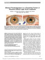
Show Diagnostic Value of Imaging in Horner Syndrome in Adults Yehoshua Almog, MD, Raz Gepstein, MD, Anat Kesler, MD Background: The yield of imaging in Horner syndrome has been explored only in children. This study evaluates the yield of imaging in adults. Methods: This was a retrospective cohort study of 52 patients with Horner syndrome examined in 2 neuro-ophthalmology hospital clinics. Patients were divided into 3 groups according to the ability to determine the etiology at the time of the first neuro-ophthalmology consultation: group I, etiology of Horner syndrome known at the initial neuro-ophthalmologic examination; group II, etiology of Horner syndrome not known at the initial neuro-ophthalmologic examination, but sufficient infor-mation obtained to allow targeted imaging; and group III, etiology of Horner syndrome not known at the initial neuro-ophthalmologic examination, and sufficient infor-mation not obtained to allow targeted imaging. The yield of investigation and the frequency of the different etiologies were evaluated. Results: In 32 (62%) patients, the etiology was already known at the initial neuro-ophthalmologic examination (group I). The most prevalent etiology was surgical trauma. In 11 (21%) patients, a targeted imaging workup was possible, revealing an etiology in 7 patients (group II). Carotid dissection and cavernous sinus mass were the most common etiologies. In 9 (17%) patients, a nontargeted imaging evaluation was necessary, revealing an etiology in only 1 patient, who had a previously undetected thyroid malignancy (group III). Conclusions: The etiology of Horner syndrome is usually known at the time of initial presentation to a neuro-ophthalmologist. When the etiology is not known and clinical information permits a targeted imaging evalua-tion, an etiology can usually be determined, most commonly a cervical carotid artery dissection or a cav-ernous sinus mass. When the etiology is not known and clinical information is insufficient to allow a targeted imaging evaluation, an etiology is rarely discovered. Even so, nontargeted imaging is warranted because life-threatening lesions, such as thyroid malignancies, may rarely be detected. Journal of Neuro-Ophthalmology 2010;30:7-11 doi: 10.1097/WNO.0b013e3181ce1a12 2010 by North American Neuro-Ophthalmology Society Horner syndrome classically presents with ipsilateral blepharoptosis, pupillary miosis, and facial anhidrosis (1) and is caused by a lesion along the oculosympathetic pathway from the hypothalamus to the eye (2). The long and complicated course of the oculosympathetic pathway predisposes it to a wide variety of pathologic processes, ranging from harmless vascular headaches to life-threatening conditions such as carotid artery dissection or malignancy (3). Yet the reported incidence of the various etiologies of Horner syndrome varies widely (4-7) and recommenda-tions for the required investigations differ as well (8-11). A comprehensive workup often includes expensive imaging tests that are unrevealing. The yield of such imaging in Horner syndrome has been explored only in children (9-11). We reviewed 53 cases of Horner syndrome in adults who were examined in the neuro-ophthalmology clinics of 2 medical centers to answer 2 questions: 1) To what extent can the etiology be determined at the initial neuro-ophthalmologic examination without further investiga-tions? 2) What is the yield of an imaging workup when the etiology is not clear at the initial clinical encounter? METHODS A retrospective chart review was performed for all patients with Horner syndrome examined in the neuro-ophthalmology clinics of the Tel Aviv Medical Center between 1992 and 2001 and Meir Medical Center between 2001 and 2006. The data from both centers were obtained from the personal databases of one of the authors (Y.A.). The study population included outpatients referred by community ophthalmologists and neurologists and those examined in the emergency room or as inpatients. Referral Department of Ophthalmology (YA, RG), Meir Medical Center and Sackler Faculty of Medicine, Tel Aviv University, Tel Aviv, Israel; and Department of Ophthalmology (AK), Tel Aviv Medical Center and Sackler Faculty of Medicine, Tel Aviv University, Tel Aviv, Israel. Address correspondence to Yehoshua Almog, MD, Neuro-Ophthal-mology Unit, Meir Medical Center, 59 Tchernichovsky Street, Kfar- Saba 44281, Israel; E-mail: shuky.almog@gmail.com Almog et al: J Neuro-Ophthalmol 2010; 30: 7-11 7 Original Contribution patterns and patient demographics were similar in both medical centers. Patients had to fulfill the following criteria to be enrolled in the study: 1) ptosis and miosis in the same eye; 2) ‘‘positive'' results on a 10% cocaine test indicated by an increase in anisocoria 30 minutes after topical instillation; 3) performance of a ‘‘targeted'' imaging evaluation when the etiology of Horner syndrome could be reasonably inferred from the clinical examination; 4) performance of a ‘‘non-targeted'' imaging evaluation (MRI of the head and neck, CT or MRI of the chest, or, when appropriate, CT angiography, MRA, or thyroid and pituitary function tests) when the etiology of Horner syndrome could not be reasonably inferred from the clinical examination. All patients received a complete neuro-ophthalmologic examination. Because our aim was to assess the yield of imaging in cases of Horner syndrome for which the etiology was not apparent at the initial neuro-ophthalmologic examination, all information obtained at the time of that examination was considered part of the history and not part of the workup. Data collected included age, gender, relevant information from the history and physical examination, laboratory tests, and imaging results. The patients were divided into 3 groups: group I, patients in whom the etiology of Horner syndrome could be determined at the initial neuro-ophthalmologic examina-tion; group II, patients in whom the etiology of Horner syndrome could not be definitely determined but in whom there was enough localizing information obtained at the initial neuro-ophthalmologic examination to permit tar-geted imaging; and group III, patients in whom the etiology of Horner syndrome could not be definitely determined at the initial neuro-ophthalmologic examination but in whom there was not enough localizing information obtained at that encounter to permit targeted imaging. Group I patients underwent no further imaging (Table 1). Group II patients underwent a targeted imaging workup based on the suspected etiology or the suspected anatomical site as specified in Table 2. Group III patients underwent a nontargeted imaging work-up; topical hydroxyamphet-amine testing was not performed. Differentiation between preganglionic and postganglionic Horner syndrome was determined by the presenting signs and symptoms or by imaging studies. Institutional review board/ethics committee approval was obtained for this study. RESULTS A total of 52 patients with the diagnosis of Horner syndrome fulfilled study entry criteria based on review of charts from the 2 participating institutions (35 patients from Meir Medical Center and 17 patients from Tel Aviv Medical Center). Among these, 24 were inpatients and TABLE 1. Group I: etiology of Horner syndrome determined at the initial neuro-ophthalmologic examination without need for further imaging (32 patients) Diagnosis Inpatients Outpatients Clinical and Pathologic Details Caused by surgery (12 patients) 2 10 Thyroid cancer, coronary artery bypass graft (3 patients each), benign lung tumor, mediastinal tumor in neurofibromatosis (1 patient each), benign thyroid mass, cervical lymph node biopsy, sympathectomy, skull base schwannoma (1 patient each) Wallenberg syndrome, other brainstem strokes (8 patients) 8 0 6 patients fulfilled clinical criteria for Wallenberg syndrome and 2 had vertebrobasilar stroke, not fulfilling criteria for Wallenberg syndrome Congenital or long-standing Horner syndrome (4 patients)* 1 3 Based on history, old photographs, and heterochromia Trauma (without carotid dissection) (3 patients) 3 0 Skull base fracture with sixth cranial nerve palsy, chest trauma with pneumothorax, neck trauma (1 patient each) Neck mass (2 patients) 2 0 Metastatic ovarian carcinoma, old carotid dissection (1 patient each) Thoracic mass (1 patient) 1 0 Upper lung carcinoma Syringomyelia (1 patient) 0 1 Developmental Cavernous sinus mass (1 patient) 0 1 Metastatic prostate cancer *Long-standing Horner syndrome-known or documented in photographs from childhood. Original Contribution 8 Almog et al: J Neuro-Ophthalmol 2010; 30: 7-11 28 were outpatients. There were 28 men and 24 women. Their mean SD age was 50 6 15 years (range 18-78 years). In group I, which included 32 patients, the etiology was reached with high probability at the initial neuro-ophthalmologic examination without a need for additional workup (Table 1). In 12 patients, the Horner syndrome was due to surgery on the chest (6 patients), neck (5 patients), or head (1 patient). In 8 patients, the cause was brainstem stroke (Wallenberg syndrome, 6 patients; other brainstem, 2 patients) In 4 patients, it was congenital or very long-standing, based on history, heterochromia, or photographs. In 3 patients, it was caused by chest, neck, or skull trauma without carotid dissection. In 5 group I patients, the etiology of Horner syndrome depended on prior imaging, which had revealed neck metastasis of ovarian carcinoma (1 patient), old carotid dissection (1 patient), upper lung carcinoma (1 patient), syringomyelia (1 patient), and cavernous sinus metastasis of prostate carcinoma (1 patient). In group II, which included 11 patients, the etiology of the Horner syndrome was not definite but could be reasonably inferred at the initial neuro-ophthalmologic examination (Table 2). Enough information was obtained at that visit to permit a targeted imaging workup. In 7 patients, the suspected diagnosis was confirmed by imaging (carotid dissection, 3 patients; cavernous sinus mass, 3 patients; and cervical spine lesion, 1 patient). The cavernous sinus masses were suspected because patients also had an ipsilateral sixth cranial nerve palsy. In 4 patients, imaging results were negative. All 4 patients were suspected to have had carotid dissection. In these 4 patients, the etiology of the Horner syndrome remained undetermined. In group III, which included 9 patients, the initial neuro-ophthalmologic examination did not furnish any localizing information in regard to the Horner syndrome, so that the imaging evaluation could not be targeted (Table 3). In 8 patients, no etiology could be determined even after thorough imaging. The Horner syndrome had been present for at least 1 year in 6 of these patients, as suggested by history or old photographs. None had any systemic disease or neurologic sign relevant to the Horner syndrome. One of the 8 patients had to have a parathyroid adenoma on imaging, but it was located in the neck contralateral to the Horner syndrome, so that its relationship to the Horner syndrome is uncertain. The remaining patient was found to have a thyroid carcinoma. We were able to localize the Horner syndrome to the central, preganglionic, or postganglionic segments of the oculosympathetic pathway in 36 patients (Table 4). A pre-ganglionic lesion was present in 44%; central or post-ganglionic lesions were each present in 28%. Neoplasms were identified as the cause of Horner syndrome in 9 (17%) patients (cavernous sinus, 4 patients; metastatic ovarian carcinoma to cervical lymph node, 1 patient; cervical schwannoma, 1 patient; cervical spine TABLE 2. Group II: etiology of Horner syndrome could not be determined at the initial neuro-ophthalmologic examination, but the examination provided enough information to permit targeted imaging (11 patients) Suspected Diagnosis Confirmation of Suspected Diagnosis after Imaging Inpatients Outpatients Helpful Clinical Signs Imaging and Etiology Carotid artery dissection (7 patients) Yes (3 patients) 3 0 Acute painful Horner syndrome (3 patients), after neck trauma (1 patient) Cervical CT angiography (2 patients), MRA (1 patient) show carotid dissection No (4 patients) 2 2 Acute painful Horner syndrome (4 patients), neck trauma (1 patient) CT angiography along common and internal carotid artery, MRI of head and neck: normal results Cavernous sinus mass (3 patients) Yes 1 2 Ipsilateral sixth cranial nerve palsy and facial hypesthesia (2 patients), ipsilateral sixth cranial nerve palsy (1 patient) CT of brain shows pituitary adenoma (1 patient), meningioma (1 patient), brain MRI shows meningioma (1 patient) Cervical spine lesion (1 patient) Yes 0 1 Scapular pain, upper extremity hypesthesia MRI of neck, perispinal mass Original Contribution Almog et al: J Neuro-Ophthalmol 2010; 30: 7-11 9 tumor, 1 patient; benign lung tumor, 1 patient; and thyroid carcinoma, 1 patients). The thyroid carcinoma was diag-nosed during a nontargeted workup for Horner syndrome, as no signs of thyroid disease were present on the clinical evaluation. In the other 8 patients, the neoplasm had been diagnosed before the initial neuro-ophthalmic examination. DISCUSSION In this series of 52 adult patients with Horner syndrome, the etiology had already been determined at the initial neuro-ophthalmologic evaluation in 32 (62%) patients based on history, examination features, and prior imaging studies (group I). In 11 (21%) patients, clinical features determined at the time of the initial neuro-ophthalmologic examination allowed a targeted imaging workup, which led to confirma-tion of the suspected etiology in 7 patients. In the 4 patients in this group who remained without a cause for Horner syndrome, the suspected etiology was acute cervical carotid dissection, but results of neck vascular imaging were negative. It is therefore possible that imaging is not sensitive enough to detect carotid dissections. Only 9 (17%) of our patients had clinical features that did not permit a targeted imaging evaluation, and imaging disclosed an explanation (a malignant thyroid tumor) in only 1 of these. Digre et al (8) evaluated 33 patients with Horner syn-drome using different MRI protocols specifically designed for preganglionic or postganglionic lesions based on topical pharmacologic testing. In that study, there was a high yield (50%) from MRI scanning in preganglionic lesions. Maloney et al (5) reported their results from 450 inpatients and outpatients with Horner syndrome examined in an ophthalmology clinic. Patient characteristics were therefore similar to those of our cohort. In that study, the main purpose was to establish the success of pharmacologic testing to identify the site of the lesion. The yield of imaging was not assessed. The investigators were able to ascertain the etiology of Horner syndrome in 60% of patients. We obtained a higher level of etiologic diagnosis (77%), perhaps because our study was performed after the widespread use of modern imaging. We did not perform hydroxyamphetamine testing in our cohort because we believe that although this test can differentiate a preganglionic from a postganglionic Horner (12), it cannot establish the etiology of Horner syndrome, and it cannot precisely locate the site of injury. Therefore, it does not exclude the need for investigation. In our study, the most prevalent anatomical location was preganglionic, followed by central and postganglionic origins of equal frequency. Several other studies have addressed the localiza-tion of Horner syndrome with varied results (4-7) (Table 4). Maloney et al (5) found only 13% of 270 patients with a known diagnosis to have central Horner syndrome, with an equal incidence of preganglionic (43%) and postganglionic (44%) lesions. In examining 120 outpatients with Horner syndrome, Grimson and Thompson (7) found that most cases were preganglionic, some were postganglionic, and very few (6%) were central in origin. In contrast, Keane (4) examined 100 patients on a neurology ward and found that a majority of cases were of central origin, mostly related to stroke. In these studies, the variance in the incidence of lesions in the 3 segments of the oculosympathetic pathway undoubtedly resulted from the diversity of the populations studied-inpatients versus outpatients and neurology clinics versus ophthalmology clinics. For example, in our group of 21 inpatients, 45% had a Horner syndrome of central origin, comparable to the finding of Keane (4). Because our study represents a combined population of inpatients and out-patients referred from neurology and ophthalmology clinics, we believe it better represents the distribution of the Horner syndrome in the general population. The incidence of tumors in our study (17%) was very similar to that found by Maloney et al (5) (13%) (Table 5). However, in most cases, the tumor was known at the time of the initial neuro-ophthalmologic examination. Giles and Henderson (6) and Keane (4) found a higher incidence of tumors in an inpatient cohort, but did not indicate in what percentage the tumor diagnosis was already known. TABLE 3. Group III: etiology of Horner syndrome could not be determined at the initial neuro-ophthalmologic examination, and the examination did not provide enough information to permit targeted imaging (9 patients) No. Patients Etiology Found by Imaging Inpatients Outpatients Imaging and Etiology 9 Yes (1 patient) 0 1 MRI of brain neck and upper thorax shows thyroid carcinoma not previously known No (8 patients) 1 7 MRI of brain neck, CT or MRI of upper thorax (8 patients) and MRA carotid dissection protocol (2 patients) TABLE 4. Location of lesion within sympathetic pathway in our cohort of 36 anatomically localizable lesions Central Preganglionic Postganglionic No. patients 10 16 10 % 28 44 28 Original Contribution 10 Almog et al: J Neuro-Ophthalmol 2010; 30: 7-11 The yield of the workup for Horner syndrome has previously been directly addressed only in the pediatric population (9-11). In 23 consecutive cases of Horner syndrome presenting in children, George et al (9) found that investigation revealed only 2 cases with previously undiagnosed pathology. He concluded that in isolated cases of Horner syndrome, routine neuro-imaging is not needed. On the other hand, Mahoney et al (11) found that a workup yielded a previously unknown tumor in 6 (33%) of 18 children with Horner syndrome. Mahoney et al concluded that MRI of the head, neck, and chest should be included in the routine workup of children with Horner syndrome. Our study shows that the etiology of Horner syndrome examined in a neuro-ophthalmology clinic is known in nearly two-thirds of patients at the initial visit. In the remaining third, there is generally enough information to allow a targeted imaging evaluation to define the etiology, although imaging results may be negative in some patients suspected of having carotid dissection. In a small minority of patients, the neuro-ophthalmology examination does not provide enough information to target imaging studies. In this group, broad-spectrum imaging results are likely to be negative, but it is worthwhile, as life-threatening lesions may be discovered. The main limitation of our study was the relatively small number of patients in whom the etiology of Horner syndrome was not known at the initial neuro-ophthalmo-logic encounter. A larger study of such patients would provide more definitive information regarding the value of imaging in this group. REFERENCES 1. Walton KA, Buono LM. Horner syndrome. Curr Opin Ophthalmol 2003;14: 357-63. 2. Thompson HS. Diagnosing Horner's syndrome. Trans Sect Ophthalmol Am Acad Ophthalmol Otolaryngol 1977;83: 840-2. 3. Burde RM, Savino PJ, Trobe JD. Clinical decisions in neuro-ophthalmology. 3rd ed. Baltimore: Mosby; 2002:246-58. 4. Keane JR. Oculosympathetic paresis. Analysis of 100 hospitalized patients. Arch Neurol 1979;36:13-5. 5. Maloney WF, Younge BR, Moyer NJ. Evaluation of the causes and accuracy of pharmacological localization in Horner's syndrome. Am J Ophthalmol 1980;90:394-402. 6. Giles CL, Henderson JW. Horner's syndrome: an analysis of 216 cases. Am J Ophthalmol 1958;46:289-96. 7. Grimson BS, Thompson HS. Horner's syndrome: Overall view of 120 cases. In: Thompson HS, Daroff RB, Frisen L, et al. Topics in Neuro-Ophthalmology. Baltimore: Williams &Wilkins; 1979:151-6. 8. Digre KB, Smoker WR, Johnston P, et al. Selective MR imaging approach for evaluation of patients with Horner's syndrome. AJNR Am J Neuroradiol 1992;13:223-7. 9. George ND, Gonzalez G, Hoyt CS. Does Horner's syndrome in infancy require investigation? Br J Ophthalmol 1998;82: 51-4. 10. Jeffery AR, Ellis FJ, Repka MX, et al. Pediatric Horner syndrome. J AAPOS 1998;2:159-67. 11. Mahoney NR, Liu GT, Menacker SJ, et al. Pediatric Horner syndrome: etiologies and roles of imaging and urine studies to detect neuroblastoma and other responsible mass lesions. Am J Ophthalmol 2006;142:651-9. 12. Cremer SA, Thompson HS, Digre KB, et al. Hydroxyamphetamine mydriasis in Horner's syndrome. Am J Ophthalmol 1990;110:71-6. TABLE 5. Location of lesion within sympathetic pathway in previously published studies Published Studies Patients Central (%) Preganglionic (%) Postganglionic (%) Maloney (5) 450 13 43 44 Grimson and Thompson (7) 120 6 57 37 Giles and Henderson (6) 216 11 88 1 Keane (4) 100 65 25 12 Original Contribution Almog et al: J Neuro-Ophthalmol 2010; 30: 7-11 11 |