| OCR Text |
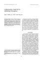
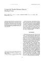
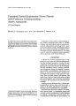
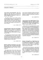
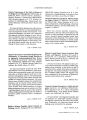
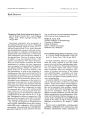
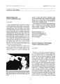
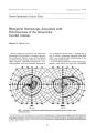
Show JOUT1lQI of CliniCJlI Neuro- ophthalmology 10( 1): 35- 37, 1990. Bilateral Trochlear Nerve Palsies from a Brainstem Hematoma Hisao Tachibana, M. D., Osamu Mimura, M. D., Mitsuo Shiomi, M. D., and Tadatsugu Oono, M. D. © 1990 Raven Press, Ltd., New York We present a case of bilateral superior oblique palsies after a spontaneous brainstem hematoma. A computerized tomographic scan of the brain revealed a highdensity mass lesion consistent with bleeding in the area caudal to the inferior colliculi, where the trochlear nerves decussate and exit the dorsal brainstem. Subsequent studies showed resolution of the density and persistent failure to enhance. Bilateral trochlear nerve palsies due to the nontraumatic brainstem bleeding are extremely rare. Key Words: Bilateral trochlear nerve palsies-- CT scanBrainstem bleeding. From the Fifth Department of Internal Medicine ( H. T.), Department of Ophthalmology ( a. M.), and Fourth D~~ artment ~ f Internal Medicine ( T. O.), Hyogo College of Medlone, Nlshlnorniya, and Department of Internal Medicine, Tanaka HospI-tal, Amagasaki ( M. s.), Japan. . Address correspondence and repnnt requests t? Dr. H. Tachibana at The Fifth Department of Internal Medlcl~ e, Hyogo College of Medicine, 1- 1, Mukogawa- cho, Nlshmomlya 663, Japan. 35 Bilateral superior oblique palsies are usually congenital or the consequence of closed head trauma ( 1- 7). Other causes are extremely rare. We describe a patient with bilateral trochlear nerve palsies after bleeding into the lower midbrain. CASE REPORT A 60- year- old man suddenly collapsed while working outdoors and then lost consciousness. He was taken to an emergency hospital. Computerized tomographic ( CT) scan of the brain without contrast ( Fig. 1) revealed bilateral high- density lesions in the midbrain tegmentum, predominantly on the left side, at the level of the inferior colliculi. The patient was treated conservatively. His level of consciousness gradually improved during 2 hand he began to complain of double vision. Twelve days after onset he was transferred to Tanaka Hospital for detailed examination and treatment. He had neither a previous history of neurological disease nor risk factors for stroke, such as hypertension, diabetes mellitus, or coronary arterial disease. On admission, his blood pressure was 140/ 72 mm Hg and the results of a general examination were unremarkable. Neurological examination revealed mild dysarthria, mild left- sided hemiparesis with normoactive stretch reflexes, left- sided hemiataxia, and an ataxic gait. The patient had a right esotropia and reported torsional and vertical diplopia, which became worse in downgaze. Ductions and versions were not limited in any direction. His ocular motility examination was remarkable for a 24- prism- diopter esotropia and 4prism- diopter left hypertropia in the primary position with an amblyoscope. Excyclotorsion exceeding 30° was noted using vertically linear fusion 36 H. TACHIBANA ET AL. FIG. 1. Brain CT scan demonstrating a high density mass in the midbrain tegmentum at the level of the inferior colliculi with extension to the left ventral side, and ventricular enlargement. targets. The degree of excyclotorsion increased in a downgaze. A Hess screen confirmed a V- pattern esotropia in excess of 25 prism diopters ( Fig. 2). Investigations including erythrocyte sedimentation rate, complete blood count, renal function and electrolytes, plasma glucose and lipid level, tests of bleeding and clotting, urinalysis, occult blood for stool, electrocardiogram, and skull and chest X- ray films showed no abnormalities. Liver function Left Eye tests initially showed moderate abnormalities, although 1 month later liver function was normal. A repeat CT scan on day 12 showed complete resolution of the high- density mass and with contrast showed no contrast enhancement. Subsequent studies showed persistent failure to enhance. With conservative treatment, his double vision and other neurological signs and symptoms grad- Right Eye FIG. 2. Results of Hess screen examination demonstrating a V- pattern esotropia. J I j/ f/ ,"' 1.1':"",. "., BILATERAL TROCHLEAR NERVE PALSIES 37 ually improved and ceased to be evident by 6 months. DISCUSSION The patient suddenly experienced brainstem bleeding although he had neither a history of hypertension nor hemorrhagic tendency. Recently, Mangiardi and Epstein ( 8) classified brainstem bleeding into two forms and stressed the distinction between brainstem " hematoma" and " hemorrhage." Subependymal hematoma is a focal, compressive lesion that displaces rather than destroys brain tissue. It occurs in the younger age group and causes neurological deficits that are often partially reversible. On the other hand, hypertensive brainstem hemorrhage usually causes a diffuse lesion occurring in old age and is most often associated with profound, irreversible neurological deficits that are often fatal. On the basis of the clinical findings, our patient's lesion may have been a brainstern hematoma. These often result from bleeding from a cryptic brainstern vascular malfonnation, usually telangiectasia ( 8). However, the patient was older than cases reported in the literature ( 8). Therefore, the etiology of his bleeding remains unclear. The incidence of trochlear nerve palsy is relatively low. In a retrospective analysis of 1,000 cases of paralysis of cranial nerves III, IV, and VI, there were 172 patients ( 17.2%) with trochlear palsy ( 4). Of these, only 13 ( 7.6%) were bilateral. Furthermore, the incidence of bilateral trochlear nerve palsies in other studies has been noted to be between 7.7 and 17.5% of cases with only trochlear nerve palsy ( 1- 3,6). Most of the causes are head injury, as in traffic accidents. Some authors have reported myasthenia gravis ( 5,7), polyneuropathy ( 7), tumor ( 9), and neurosurgical complications ( 10) as causes. However, there are few reports of bilateral trochlear nerve palsies after nontraumatic brainstem bleeding, although unilateral trochlear nerve palsy from midbrain hemorrhage has been reported ( 11,12). The nucleus of the trochlear nerve is located at the level of the interior colliculus and is ventral to the cerebral aqueduct. The fibers leave the nucleus and pass caudally and superiorly to the anterior medullary decussation just above the fourth ventricle. At this site, a single lesion may impair both nerve functions. The most common cause of bilateral trochlear nerve palsies is a contrecoup injury to the decussation region after frontal head injury ( 2,3). Murray and Ajax ( 9) reported a case with bilateral trochlear nerve palsies due to a metastatic adenocarcinoma localized in the anterior cerebellar vermis. They speculated that the lesion compressed the area caudal to the inferior colliculi where the fourth nerves decussate and exit the dorsal brainstem. In our case, the hematoma was located in the dorsal brainstem, the area of the trochlear nerve nucleus and decussation, and directly caused bilateral trochlear nerve injury. REFERENCES 1. Khawan E, Scott AB, Jampolsky A. Acquired superior oblique palsy. Diagnosis and management. Arch Ophthalmol 1967; 77: 761~. 2. Burger LJ, Kalvin NH, Smith JL. Acquired lesions of the fourth cranial nerve. Brain 1970; 93: 567- 74. 3. Younge BR, Sutula F. Analysis of trochlear nerve palsies. Diagnosis, etiology, and treatment. Mayo Clin Proc 1977; 52: 11~. 4. Ruth JA, Younge BR. Paralysis of cranial nerves III, IV, and VI. Causes and prognosis in 1000 cases. Arch Ophthalmol 1981; 99: 76- 9. 5. Lee 1. Flynn JT. Bilateral superior oblique palsies. Br J Ophthalmol 1985; 69: 50S- 13. 6. von Noorden GK, Murry E, Wong SY. Superior oblique paralysis. A review of 270 cases. Arch Ophthalmol 1986; 104: 1771~. 7. Mori M, Ohira A, Goto K, Ozawa T. Acquired bilateral superior oblique palsies. Jpn J Clin OphthalmoI1988; 42: 65- 8. 8. Mangiardi JR, Epstein FJ. Brainstem hematomas: review of the literature and presentation of five cases. J Neural Neurosurg Psychiatry 1988; 51: 966- 76. 9. Murray RS, Ajax ET. Bilateral trochlear nerve palsies. A clinicoanatomic correlate. J Clin Neuro- ophthalmol 1985; 5: 57~. 10. Yoss RE, Rucker CW, Miller RH. Neurosurgical complications affecting the oculomotor, trochlear, and abducent nerves. Neurology 1968; 18: 59~ 0. 11. Mansour AM, Reinecke RD. Central trochlear palsy. Surg OphthalmoI1986; 30: 279- 97. 12. Kamei T, Uchiyama F, Fukuyama J. Primary tectal mesencephalic hemorrhage with isolated trochlear nerve palsy. A case report. Rinsho Shinkeigaku 1987; 27: 1167- 9. I elin NeurCHJphthalmol, Vol. 10, No. 1, 1990 |