| OCR Text |
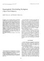
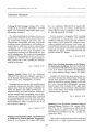
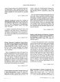
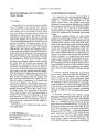
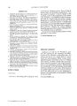
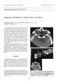
Show lourTlJ2l of Clinical Neuro-ophthalmology 8(4): 215-220, 1988. Cyclosporin A-Induced Reversible Cortical Blindness Steven E. Wilson, M.D., Piet C. de Groen, M.D., Allen J. Aksamit, M.D., Russell H. Wiesner, M.D., James A. Garrity, M.D., and Ruud A. F. Krom, M.D. © 1988 Raven Press, Ltd., New York Despite the occurrence of serious side effects, the use of cyclosporin A after organ transplantation has increased because of its ability to effectively suppress allograft rejection. Its use in the treatment of ophthalmic disease has also recently increased. Central nervous system toxicity due to cyclosporin A is a significant but apparently clinically reversible side effect. A liver transplant patient in whom cortical blindness from profound neurotoxicity was the initial presentation is described. Neurologic abnormalities, including cortical blindness, resolved completely after discontinuation of cyclosporin A. However, pathologic studies performed 8.5 months after the initial transplant revealed residual central nervous system demyelination. Key Words: Cyclosporin A-Cortical blindness-Neurotoxicity. From the Departments of Ophthalmology (S.E.W., ).A.G.), Internal Medicine (P.c. de G., R.H.W.), Surgery (R.A.F.K.), and Neurology (A.).A.), Mayo Clinic Foundation, Rochester, Minnesota. Address correspondence and reprint requests to Dr. J. A. Garrity at Department of Ophthalmology, Mayo Clinic, Rochester, MN 55905, U.S.A. 215 Since the discovery of the immunosuppressive effects of cyclosporin A by Borel (1) in 1976, there has been a remarkable increase in its utilization after organ transplantation and for the treatment of autoimmune diseases. Recently, a review of cyclosporin A immunology, pharmacology, and therapeutic uses was published in the ophthalmic literature (2). Many trials have reported use of cyclosporin A in the treatment of human ocular disease (Table 1) (3--21). Many common side effects and toxicities have been attributed to cyclosporin A (22) (Table 2), some of which may be serious. Recently, severe reversible central nervous system toxicity was noted in liver transplant patients treated with systemic cyclosporin A at our institution (23). A case in which the initial manifestations of the cyclosporin A toxicity were neuroophthalmologic is described below. CASE REPORT A 55-year-old woman underwent liver transplantation because of advanced cryptogenic cirrhosis on July 26, 1986. Four months prior to transplantation, ophthalmologic examination was normal with a best corrected visual acuity of 20/20 in both eyes. A cerebral computed tomographic scan obtained for hepatic encephalopathy prior to transplantation was normal. Immunosuppression was started immediately prior to surgery with 1 g of methylprednisolone and 100 mg cyclophosphamide given intravenously, and an additional 1 g of methylprednisolone was administered immediately postoperatively. One hundred milligrams of cyclophosphamide was given intravenously on days 0, 1, and 2 after transplantation. Use of cyclosporin A, 100 mg intravenously three times a day, was initiated on day 2. The dose was in- 216 S. E. WILSON ET AL. TABLE 1. Cyclosporin A in ophthalmologic disease TABLE 2. Cyclosporin A toxicities and side effects Anemia Elevated sedimentation rate Gingival hyperplasia Hepatotoxicity Hidra-adenitis Hypertrichosis Hypertension Nausea and vomiting Nephrotoxicity NeurotOXicity Opportunistic infections Malignancy creased to 120 mg three times per day on day 4 after a whole blood trough level measured by high performance liquid chromatography of 199 ng/ml was obtained. Cyclosporin A trough levels were carefully monitored throughout the hospitalization and did not exceed 240 ng/ml. The patient was awake and alert, but on the morning of day 5 complained of having had bad dreams. At 8 A.M. on day 5, she complained of pain in the left eye and then in both eyes. At 10 A.M., she was disoriented to time and place and complained of blindness. The patient was seen in consultation by one of the authors (S.E.W.) and was noted to be alert, cooperative, and oriented to time, place, and persons. She denied light perception in each eye. Optokinetic responses were absent. The pUPils were 3.0 mm with brisk response to light. Th'-"'e was no afferent pupillary defect. There was a left gaze preference. Periodic alternating nystagmus was observed with cycles of right beating and left beating phases lasting 45 to 60 s. The eyes were noted to be motionless for 5 to 10 s between phases. Extraocular movements were otherwise normal. Repetitive nonmyokymic twitching of the upper and lower eyelids was noted bilaterally. The remainder of the examination was normal. Flash-induced visual evoked responses were prolonged to 178 ms (right) and 200 ms (left), with normal being 95-115 ms. The progressive generalized neurologic dysfunc-tion that subsequently developed has been detailed elsewhere (23). A cerebral computed t?mographic scan revealed generalized hypodensIty of the white matter most pronounced in the occipital lobes, with the left greater than right (Fig. 1). Cyclosporin A was discontinued. A lu~bar punc~re yielded clear cerebrospinal fluid WIth an openmg pressure of 16 cm of wa:er and normal la~oratory studies including bactenal, fungal, and VIral cultures. The neurologic status steadily improved over the subsequent 10 days. She was ~lert on da~ 9, and visual testing on day 10 was finger counting bilaterally. Vision had improved to 20/200 in .each eye and periodic alternating nystagmus had dIsappeared by day 12. Liver function subsequently deteriorated secondary to rejection, and a second liver transplant was performed on day 17. . Postoperatively on days 22 and 30, two addItional episodes of altered sensorium occurred during cyclosporin A immunosuppression. One occurred during intravenous treatment and the other while the patient was being treated orally. In each instance, discontinuation of cyclosporin A was followed by a rapid improvement in neurologic status (23). Cerebral computed tomography performed on day 31 demonstrated considerable resolution of the low attenuation in the white matter. However, magnetic resonance imaging on day 32 (Fig. 2) still detected a marked white matter disorder with changes being most prominent in the posterior cerebrum. The patient slowly improved and was discharged from the hospital on day 62 with oral cyclosporin A at 50 mg three times per day. During repeat ophthalmologic examination 5 months after discharge, she was noted to be mildly jaundiced with normal sensorium. She had not taken cyclosporin A for approximately 3 months. Best corrected acuity was 20/20 in each eye. There was no nystagmus, and the optokinetic responses were normal. The pupillary responses were normal. She could correctly identify all of the Ishihara color plates in each eye. Ocular rotations were full. There were no abnormalities detected on funduscopic examination. Computer assisted perimetry (program 31) was normal in each eye, Flashinduced visual evoked responses were 100 ms in each eye. ' Eight and one-half months after initial transplantation, the patient died with severe hepatic failure due to rejection. At autopsy, the white matter of the posterior cerebral hemispheres appeared grossly normal. Myelin staining of the sections 34 5, 6 7-10 11 12 13 14 15 16-20 21 Ocular disease References Behcet's disease Corneal peripheral melting syndrome Corneal transplantation Graves' ophthalmopathy Herpes keratitis Mooren's ulcer Myasthenia gravis Optic neuritis Sjogren's syndrome Uveitis Vernal keratoconjunctivitis CYCLOSPORIN A-INDUCED BLINDNESS 217 FIG. 1. Computed tomography performed on day 5 demonstrates extensive areas of decreased attenuation (arrows) most pronounced in the subcortical and white matter regions of the posterior parietal and occipital lobes bilaterally. from the region, however, showed a mild pallor of the white matter (Fig. 3), and microscopically a few scattered macrophages were found among nerve fibers, which frequently showed irregular focal swelling of myelin sheaths. Mild astrocytosis was also evident within the white matter. Additional findings were multiple scattered foci of resolving petechiae in the cerebral white matter and a small (0.8 x 0.5 cm) old hemorrhage in the right cerebellar white matter. A few scattered minute demyelinating lesions characteristic of progressive multifocal encephalopathy were found in the cerebral and cerebellar white matter. In the vicinity of the small hemorrhage mentioned above, they were more numerous and showed early confluence (pathology provided by Harno Okazaki, M.D.). DISCUSSION Cyclosporin A-induced central nervous system toxicity has been reported in several letters and articles in the recent medical literature. Toxicity has also occurred in patients receiving renal and bone marrow transplants and thus is not restricted to liver transplant recipients. This form of toxicity has only been observed with systemic administration, either intravenous or oral. Berden et al. (24) reported prolonged coma in a renal transplant patient treated with oral cyclosporin A. They also described diffuse white matter hypodensity on cerebral computed tomography and electromyographic signs of axonal degeneration and demyelination. The clinical computed tomography and electromyography changes gradually resolved with discontinuation of the cyclosporin A. Wilczek et al. (26) also reported coma with electroencephalographic abnormalities in a renal transplant patient treated with cyclosporin A. The coma resolved when the medication was stopped. Two subsequent attempts to reintroduce cyclosporin A were followed by a deterioration in mental status. There have been many reports of convulsion in patients receiving cyclosporin A following bone marrow (26-31), renal (32-34), and liver (35) transplants. Velu et al. (31) reported that postmortem examination of the brain revealed only diffuse edema after death due to status epilepticus. Atkinson et al. (36) reported five patients who devel- I Clill Ncuro-ophthalmol, Vol. 8. No.4, 1988 218 FIG. 2. Magnetic resonance imaging performed on day 32. Extensive hyperintense signal (arrows) on a T2-weighted image is present, predominantly involving the white matter and most pronounced in the parietal and occipital lobes bilaterally. S. E. WILSON ET AL. oped various combinations of confusion, amnesia, urinary retention, tremor, paresis, and ataxia while being treated with cyclosporin A after bone marrow transplantation. In all cases, the neurologic symptoms and signs resolved when the medication was stopped. In two patients, the neurologic disorder reoccurred when the cyclosporin A was reintroduced. Noll and Kulkarni (37) reported complex visual hallucinations and a "slow, insidious loss of visual acuity" in both eyes to perception of hand motion only while being treated with cyclosporin A after bone marrow transplantation. Both abnormalities resolved when cyclosporin A was discontinued. Recently, Rubin and Kang (38) reported global encephalopathy, seizures, and cortical blindness in a 5-year-old bone marrow transplant recipient during treatment with cyclosporin A. The serum cyclosporin A level was 1704 ng/ml. A cerebral computed tomographic scan demonstrated white matter abnormalities. All abnormalities resolved when cyclosporin A was discontinued. Visual fields and visual evoked responses were not reported. In 48 patients who have received liver transplants at the Mayo Clinic during the past 2 years, ., ,'. ,f 1 '"I S, No.4, 1988 13 were believed to have experienced cyclosporin A induced central nervous system toxicity (23). Of these, three have developed reversible cortical blindness. All three patients had diffuse white matter abnormalities most prominent in the posterior cerebrum on both computed tomography and magnetic resonance imaging. In all three cases, the blindness completely resolved when the cyclosporin A was discontinued. In the case presented, recurrence and resolution of neurological symptoms and signs follow reintroduction and discontinuation, respectively, of cyclosporin A on two occasions. Apparent complete clinical reversability of the cyclosporin A induced central nervous system toxicity was noted with a return of visual acuity to the normal pretransplant level. There was normalization of visual evoked responses, and there were no detectable visual field defects 8.5 months after the cortical blindness occurred. Pathologic examination revealed residual evidence of central nervous system demyelination. Clinically, however, it was apparent that all of the deficits had completely resolved. It appears that a certain degree of subclinical structural damage had taken place in the white matter, where reversible CYCLOSPORIN A-INDUCED BLINDNESS 219 FIG. 3. Whole mount view of the left occipital lobe showing diffuse central demyelination. Compare the pallor of this white matter (large arrows) with the normal myelinated zone at the apex of the gyrus indicated by the small arrow. Lacunae present within the white matter represent blood vessels and perivascular spaces (open arrow). (Luxol fast blue; original magnification, x 1.) computed tomography and magnetic resonance imaging abnormalities were noted. The mechanism of the cyclosporin A-induced neurotoxicity is unknown. In many cases of cyclosporin A neurotoxicity described in the literature, cyclosporin A trough levels were noted to be elevated (24,28,31-33,36,38). In other cases, levels have been normal (23,27,36). Cyclosporin A levels were well documented in the patient under discussion and were never noted to be above the upper limit (350 ng/ml). In a recent retrospective study by de Groen et al., mean total serum cholesterol levels for the first 7 days after liver transplantation were lower in 13 patients who developed cyclosporin A neurotoxicity than in 35 patients who did not (23). In the case discussed, the average cholesterol level in the first postoperative week was 94 mg/dl. In the same study, mean cyclosporin A levels over the same period did not differ between the two groups. The authors hypothesized that since cyclosporin A is a highly lipophilic molecule, the majority of which is bound to serum lipoproteins, a low serum cholesterol level might result in an elevation of unbound cyclosporin A (23). Thus, even patients with what are normally considered to be nontoxic cyclosporin A levels could develop neu-rotoxicity. By whatever mechanism, presumably the end result is an interference with normal neuronal function. Reversible cortical blindness thought to be directly associated with occipital lobe seizures has been noted in two patients treated with cisplatinum (39). Interestingly, in one of these patients, a subtle low density zone localized primarily to the white matter of the occipital lobes was noted. The development of period alternating nystagmus (PAN) in our patient is worthy of mention. Its onset was coincident with the development of cortical blindness and resolved 7 days later when the vision improved. There are many conditions associated with PAN (40). The most likely etiology in the present case would either be profound bilateral loss of vision (41) or a generalized central nervous system demyelination (42). Although it is not possible to determine exactly the cause, the time course suggests the former as the most likely etiology. Ophthalmologists should be aware of cyclosporin A neurotoxicity for two reasons. First, they may be consulted in cases where the initial presentation is decreased vision and have the opportu- , Clill Nellro-ophlhalmol, Vol. 8, No.4, 1988 220 S. E. WILSON ET AL. nity to aid in the recognition and management of the disorder. Secondly, with the increased utilization of cyclosporin A in the treatment of ophthalmologic diseases, the ophthalmologist should be alert to the possible development of this reversible complication. Acknowledgment: This work was supported by Research to Prevent Blindness, New York, NY. REFERENCES 1. Borel JF, Feurer C, Gubler HU, et al. Biological effects of cyclosporin A: A new antilymphocytic agent. Agents Actions 1976;6:468-75. 2. Nussenblatt RB, Palestine A. Cyclosporin: Immunology, pharmacology and therapeutic uses. Surv Ophthalmol1986; 31:159-69. 3. Nussenblatt RB, Palestine AG, Chan CC, Mochizuki M, Yancey K. Effectiveness of cyclosporin therapy for Behcet's disease. Arthritis Rheum 1985;28:671-9. 4. Kruit PJ. Van Balen AT, Stilma JS. Cyclosporin A treatment in two cases of corneal peripheral melting syndrome. Doc OphthalmoI1985;59:33. 5. Hoffmann F, Wiederholt M. Lokale behandlung des hornhaut transplantates beim Menscheu mit cyclosporin A. Klin Monatsbl Augenheilkd 1985;187:92--6. 6. Hill JC The use of systemic cyclosporin A in human corneal transplantation: a preliminary report. Doc Ophthalmol1986; 62:337-44. 7. Witte A, Landgraf R. Markl A, Boergen KP, Hasenfratz G, Pickardt CR. Treatment of Graves' ophthalmopathy with cyclosporin A. Klin Wochenschr 1985;63:1000-4. 8. Utech C, Wulle KG, Biele EU, Pfannenstiel P, Panitz N, Kiefer H. Treatment of severe Graves' ophthalmopathy with cyclosporin A. Acta EndocrinoI1985;11:493--8. 9. Kvetny J, Frandsen NE, Johnsen T, Dieperinck H, Mogensen E. Treatment of Graves' ophthalmopathy with cyclosporin A. Acta Med Scand 1986;220:189-91. 10. Kahaly G, Schrezenmeir J. Schweikert B, Muller W, Krause U, Beyer J. Remission-maintaining effect of cyclosporine in endocrine ophthalmopathy. Transplant Proc 1986;18:844. 11. Colin J. Chastel C, Bonissent JF. Herpetic stromal keratitis. Treatment with cyclosporin eyedrops. Presse Med 1986;15: 1245. 12. Hill JC, Potter P. Treatment of Mooren's ulcer with cyclosporin A. Report of three cases. Br J Ophthalmol 1987;71: 11-4. 13. Tindall RS, Rollins JA, Phillips JT, Greenlee RG, Wells L, Belendiuk G. Preliminary results of a double-blind, randomized, placebo-controlled trial of cyclosporine in myasthenia gravis. N Engl JMed 1987;316:719-24. 14. Nussenblatt RB, Palestine AG, Chan CC, Breen L, Caruso R. Improvement of uveitis and optic nerve disease by cyc1osporine in a patient with multiple sclerosis. Am J OphthlamoI1984; 97:790-1. 15. Drosos AA, Skopouli FN, Costopoulos JS, Papadimitriou CS, Moutsopoulos HM. Cyclosporin A (CyA) in primary Sjogren's syndrome: a double blind study. Ann Rheum Dis 1986;45:732-5. 16. Nussenblatt RB, Palestine AG, Chan CC Cyclosporin A therapy in the treatment of intraocular inflammatory disease resistant to systemic corticosteroids and cytotoxic agents. Am JOphthalmol 1983;96:275-82. 17. Nussenblatt RB, Palestine AG, Rook AH, et al. Treatment of intraocular inflammatory disease with cyclosporin A. Lancet 1983;2:235-8. 18. Graham EM, Sanders MD, James DG, Hamblin A, KaspGrochowska E, Dumonde D. Cyclosporin A in the treat- ' •. " .. ° "f'idl""mol. l'ryl. 8, No.4. 1988 ment of posterior uveitis. Trans Ophthalmol Soc UK 1985; 104:146--51. 19. Nissen C Bendtzen K, Tvede N, Andersen V. The treatment of p~esumed non-infective uveitis with cyclosporin A. Acta Ophthalmol 1985;63(suppl 173):72-3. 20. Fite Kv, Pardue S, Bengston L, Hayden 0, S~yth J~ !r. Effects of cyclosporine in spontaneous, postenor uveitis. Curr Eye Res 1986;5:787-96. . 21. BenEzra 0, Peer J. Brodsky M, Cohen E. Cyclospo~e ey~drops for the treatment of severe vernal keratoconJunCtiVi-tis. Am JOphthalmoI1986;101:278--82. . 22. Palestine AG, Nussenblatt RB, Chan CC Side effects of systemic cyclosporine in patients not undergoing transplantation. Am JMed 1984;77:652-6. 23. De Groen PC, Aksamit AJ. Rakela J, Forbes GS, Krom RAF. Central nervous system toxicity after liver transplantation. The role of cyclosporine and cholesterol. N Engl J Med 1987;317:861-6. 24. Berden JHM, Holtsma AJ, Merx JL, Keyser A. Severe central nervous system toxicity associated with cyclosporin. Lancet 1985;1:219-20. 25. Wilczek H, Ringden 0, Tyden G. Cyclosporine associated central nervous system toxicity after renal transplantation. Transplantation 1985;39:110. 26. Joss DV, Barrett AJ. Kendra JR, Lucas CF, Desai S. Hypertension and convulsions in children receiving cyclosporin A. Lancet 1982;1:906. 27. Durrant S, Chipping PM, Palmer S, Gordon-Smith EC Cyc1osporin A, methylprednisolone, and convulsions. Lancet 1982;2:829-30. 28. Boogaerts MA, Zachee P, VerwiIghen RL. Cyclosporin, methylprednisolone, and convulsions. Lancet 1982;2:1216 -7. 29. Thompson CB, Sullivan KM, June CH, Thomas ED. Association between cyclosporin neurotoxicity and hypomagnesaemia. Lancet 1984;2:1116--20. 30. Lo'pez Messa JB, Gonza'lez Go'mez N, Alonso Alonso P, et al. Convulsiones e hipertensio'n arterial en tres pacientes sometidos a trasplante de medula o'sea y en traramiento con ciclosporina A. Rev Clin Esp 1986;178:186. 31. Velu T, Debusscher L, Stryckmans PA. Cyclosporin associated fatal convulsions. Lancet 1985;1:219. 32. Dilip Shah, Rylance PB, Rogerson ME, Bewick M, Parsons V. Generalized epileptic fits in renal transplant recipients given cyclosporin A. Br Med J 1984;289:1347-8. 33. Beaman M, Parvin S, Veitch PS, Walls J. Convulsions associated with cyclosporin A in renal transplant recipients. Br Med J1985;290:139-40. 34. Nordal KP, Talseth T, Dahl E, et al. Aluminum overload, a predisposing condition for epileptic seizures in renaltransplant patients treated with cyclosporin? Lancet 1985;2: 153-4. 35. Powell-Jackson PR, Carmichael FJL, CaIne RY, Williams R. Adult respiratory distress syndrome and convulsions associated with administration of cyclosporine in liver transplant recipients. Transplantation 1984;38:341-3. 36. Atkinson K, Biggs J. Darveniza P, Boland J, Concannon A, Dodds A. Cyclosporine associated central nervous system toxicity after allogenic bone marrow transplantation. Transplantation 1984;38:34. 37. Noll RB, Kulkarni R. Complex visual hallucinations and cyclosporine. Arch Neural 1984;41:329. 38. Rubin AM, Kang H. Cerebral blindness and encephalopathy with cyclosporin A toxicity. Neurology 1987;37:1072-6. 39. Kattah JC, Potolicchio SJ. Kotz HL, Kolsky MP, Thomas D. Cortical blindness and occipital lobe seizures induced by cis-platinum. Neuro-Ophthalmology 1987;7:99-104. 40. Baloh RW, Honrubia V, Kenrad HR. Periodic alternating nystagmus. Brain 1976;99:11-26. 41. Cross SA, Smith JL, Norton EWD. Periodic alternating nystagmus clearing after vitrectomy. JClin Neuro-ophthalmol 1982;2:5--11. 42. Keane JR. Periodic alternating nystagmus with downward beating nystagmus. Arch Neural 1974;30:399-402. |