| OCR Text |
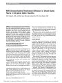
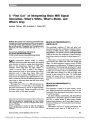
Show Clinical and Imaging Features of Fludarabine Neurotoxicity Michael S. Lee, MD, Alexander M. McKinney, MD, Jeffrey R. Brace, MD, Karen SantaCruz, MD Abstract: Neurotoxicity from intravenous fludarabine is a rare but recognized clinical entity. Its brain imaging features have not been extensively described. Three patients received 38.5 mg or 40 mg/m2 per day fludar-abine in a 5-day intravenous infusion before bone marrow transplantation in treatment of hematopoietic malignan-cies. Several weeks later, each patient developed pro-gressive neurologic decline, including retrogeniculate blindness, leading to coma and death. Brain MRI showed progressively enlarging but mild T2/FLAIR hyper-intensities in the periventricular white matter. The lesions demonstrated restricted diffusion but did not enhance. Because the neurotoxicity of fludarabine appears long after exposure, neurologic decline in this setting is likely to be attributed to opportunistic disease. However, the imaging features are distinctive in their latency and in being mild relative to the profound clinical features. The safe dose of fludarabine in this context remains controversial. Journal of Neuro-Ophthalmology 2010;30:37-41 doi: 10.1097/WNO.0b013e3181ce8087 2010 by North American Neuro-Ophthalmology Society Fludarabine, a purine analog that inhibits DNA syn-thesis, acts against both dividing and resting cells. Other potential mechanisms of action include inhibition of RNA synthesis and activation of apoptosis. Treatment indications for fludarabine include myeloproliferative malignancies and immunosuppression before a bone marrow transplant (BMT) (1,2). Fludarabine is typically infused once daily for 5 days. In phase I and phase II clinical trials, several patients developed severe neurotoxicity at higher doses (2). We report the clinical and neuroimaging findings of 3 patients who experienced fludarabine-induced neurotoxicity that prominently involved loss of vision. CASE REPORTS Case 1 A 48-year-old man had acute myelogenous leukemia. Eighteen days before receiving a BMT, he received a 5-day infusion of 38.5 mg/m2 fludarabine per day, 1965 mg/m2 cyclophosphamide per day, and total body irradiation of 400 cGy in 2 fractions. Renal function was normal. Two weeks after receiving the BMT, he developed bilateral upper extremity weakness and declining vision in both eyes. Neuro-ophthalmologic examination revealed an alert, oriented, and cooperative man. Visual acuities were light perception in the right eye and hand motions in the left eye. Results of pupillary, extraocular motility, and slit lamp examinations were normal. Ophthalmoscopy showed Roth spots bilaterally. The optic discs appeared normal. He had weakness of all 4 extremities, which was greatest in the proximal lower extremities. He was generally hyperreflexive with extensor plantar reflexes. There was numbness to all modalities below the T4 dermatome. Brain MRI showed mild diffuse T2 and FLAIR hyper-intense lesions in the periventricular white matter with restricted diffusion and corresponding low signal on appa-rent diffusion coefficient (ADC) maps, confirming cyto-toxic effects (Fig. 1A). The lesions did not enhance. Spine MRI demonstrated no abnormalities. Lumbar puncture showed normal cellularity (1 white blood cell and 0 red blood cells), 34 mg/dL protein, 66 mg/dL glucose, and negative results for VDRL, cryptococcal antigen, group B streptococcus, fungal stain, Gram stain, oligoclonal bands, cytology, flow cytometry, and Departments of Ophthalmology (MSL), Neurology (MSL), Neuro-surgery (MSL), Neuroradiology (AMM, JRB), and Neuropathology (KS), University of Minnesota, Minneapolis, Minnesota. This work was presented in part at the annual meeting of the Frank B. Walsh Society, Lake Tahoe, NV, February 2009. This study was supported by an unrestricted grant from Research to Prevent Blindness (New York, NY) and the Minnesota Lions (to MSL). Address correspondence to Michael S. Lee, MD, 420 Delaware Street SE, MMC 493, Minneapolis, MN 55455; E-mail: mikelee@umn.edu. Lee et al: J Neuro-Ophthalmol 2010; 30: 37-41 37 Original Contribution FIG. 1. Case 1. A. Brain MRI performed 1 month after the patient had received 38.5 mg/m2 fludarabine per day intravenously for 5 days. Axial FLAIR (left) shows subtle white matter changes (arrows). Diffusion-weighted imaging (center) shows high signal in the lesions. Apparent diffusion coefficient map (right) shows that the corresponding areas are of low signal, confirming that the lesions contain restricted diffusion. B. Brain MRI performed 2 weeks later shows signal abnormalities of greater intensity and size. C. Autopsy histopathology of the brain shows periventricular white matter axonal and myelin damage. Left panel. Hematoxylin and eosin (original magnification: 320) stain shows abundant axonal spheroids (arrows). Right panel. Luxol fast blue/periodic acid-Schiff (PAS) (original magnification: 310) stain shows white matter vacuolization and abundant macrophages with lightly PAS-positive macrophages (arrows). Original Contribution 38 Lee et al: J Neuro-Ophthalmol 2010; 30: 37-41 polymerase chain reaction (PCR) for human herpesvirus-6 (HHV6), vesicular stomatitis virus (VZV), Epstein-Barr virus (EBV), JC virus, and cytomegalovirus (CMV). The myelin basic protein level was elevated 80-fold. The patient became increasingly encephalopathic over the next week. Repeat brain MRI showed increasing size and intensity of the periventricular white matter lesions (Fig. 1B). Results of a repeat spine MRI were unremarkable. Electroencephalography showed theta and delta slowing consistent with diffuse cerebral dysfunction. The patient died 9 weeks after fludarabine exposure. Autopsy showed axonal and myelin damage in the periventricular white matter. In the periventricular white matter of the temporal and occipital lobes, prominent white matter vacuolization, axonal spheroids, and abundant macrophages were seen (Fig. 1C), consistent with toxic leukoencephalopathy and previous descriptions of fludar-abine neurotoxicity (1-7). The medulla and spinal cord also showed white matter vacuolization, and atypical in-travascular cells consistent with leukemia were present. Case 2 A 58-year-old woman received a diagnosis of multiple mye-loma. Four years later, she received at a dose of 40 mg/m2 fludarabine per day intravenously for 5 days, a single dose of 1950 mg/m2 cyclophosphamide on the first day, and a single 200-cGy fraction of whole body radiation. Renal function was normal. One week later she underwent a nonmyeloablative double umbilical cord BMT. Five weeks after fludarabine infusion, the patient de-veloped progressive confusion and lethargy. Nine days later she became nonverbal but responded appropriately with head nodding. The following day she denied seeing light. Brain MRI showed periventricular white matter T2 hyperintensities with restricted diffusion. The lesions did not enhance (Fig. 2A). Two days later, she did not respond to oral commands. Pupils were poorly reactive to light. She showed no spontaneous movement of her extremities, but she would withdraw to painful stimuli. Her extremities were spastic, her reflexes were brisk, and plantar reflexes were extensor. A repeat brain MRI showed that the lesions had expanded in size and intensity (Fig. 2B). Spine MRI dis-closed no contributory abnormalities. Lumbar puncture demonstrated no abnormalities. She was intubated electively 2 weeks later, and support was withdrawn after 1 week. No postmortem examination was performed. Case 3 A 35-year-old woman received a diagnosis of acute mye-logenous leukemia for which she underwent a non-myeloablative allogenic peripheral blood stem cell BMT 6 months later. One week before the BMT, she had received 38.5 mg/m2 fludarabine per day for 5 days and a single dose of 2010 mg/m2 cyclophosphamide for 1 day. Renal func-tion was normal. Her post-BMT course was complicated by Aspergillus pneumonia leading to sepsis and acute renal failure. Twelve days after the BMT, she was intubated for progressive dyspnea. Before intubation she was interactive and capable of transferring herself; however, the patient did not regain consciousness after sedation was discontinued 8 days later. Four weeks after fludarabine infusion, she could not follow verbal commands or react to painful stimuli, but she opened her eyes spontaneously. Pupils were normal. Ocu-locephalic testing revealed normal results. Deep tendon reflexes were reduced in the upper extremities and absent in the lower extremities. Brain MRI showed subtle T2 hyperintensity in the splenium of the corpus callosum. There was no restricted diffusion or enhancement (Fig. 3A). An electroencephalo-gram demonstrated diffuse theta and delta slowing. Results of lumbar puncture was negative, including PCR testing for the JC virus. Repeat brain MRI over the next 2 weeks demonstrated increasing size and intensity to the white matter changes with mild restricted diffusion (Fig. 3B). She failed to improve and died 2 months after the BMT. There was no postmortem examination. DISCUSSION Previous reports of fludarabine neurotoxicity have relied on brain CT or have described MRI abnormalities without detail (1-7). Our patients showed variable, ill-defined, mildly hyperintense lesions in the periventricular and per-iatrial cerebral white matter on T2 and FLAIR sequences with restricted diffusion but no enhancement. The brain MRI abnormalities were mild in relation to the profound clinical deficits and histopathology. The imaging abnor-malities increased in size and intensity in repeated studies. Although clinical and postmortem examination demon-strated spinal cord involvement, spinal MRI did not show any abnormalities. Other toxic leukoencephalopathies show similar white matter changes, but they occur concomitantly with drug administration and later stabilize or improve with de-challenge (8). Fludarabine neurotoxicity appears unique in the delayed and progressive lesion appearance many weeks after drug cessation. The progressive neurologic decline may be confused with progressive multifocal leukoencephalopathy (PML), but imaging should differentiate these entities. In contrast to fludarabine toxicity, PML lesions involve the subcortical white matter and do not show restricted diffusion, and lesion size correlates with clinic clinical findings. Similarities include the absence of enhancement or mass effect (9). Initial animal studies of fludarabine in dogs failed to show neurotoxicity after a single dose or 5 daily doses (3). In phase I clinical trials, neurotoxicity occurred in up to 18% Original Contribution Lee et al: J Neuro-Ophthalmol 2010; 30: 37-41 39 FIG. 2. Case 2. A. Brain MRI performed 7 weeks after the patient received 40 mg/m2 fludarabine per day intravenously for 5 days. Axial FLAIR imaging (left) shows periventricular high signal areas. Diffusion-weighted imaging (DWI, center) shows that the lesions have high signal and an apparent diffusion coefficient (ADC) map (right) shows corresponding low signal, indicating restricted diffusion. B. Brain imaging performed 1 week later shows increasing size and intensity of the lesions on FLAIR (left), DWI (center), and ADC map (right). FIG. 3. Case 3. Sequential axial FLAIR imaging studies at 4 weeks (A), 5 weeks (B), 6 weeks (C), and 8 weeks (D) after the patient had received 38.5 mg/m2 fludarabine per day intravenously for 5 days. The lesions show progressive increase in size and intensity. Original Contribution 40 Lee et al: J Neuro-Ophthalmol 2010; 30: 37-41 of patients treated with high-dose fludarabine (60-150 mg/m2 per day for 5 days) (1,2,4,5). Further study revealed that the toxicity appeared to be dose dependent. Among 443 patients receiving fludarabine, 36% receiving high doses (>450 mg/m2 total dose) developed severe central nervous system toxicity compared with only 0.2% receiving low doses (,125 mg/ m2 total dose) (6). Several large BMT centers use daily doses of 40 mg/m2 (4,10-15) or 50 mg/m2 in a 5-day infusion (16,17). Others have advocated lower doses-18-25 mg/m2 per day for a 5-day infusion (1,7). At the University of Minnesota, 245 patients received fludar-abine from September 2005 to July 2007 at doses of 38.5 or 40 mg/m2 per day for 5 days before BMT. The neurotoxicity of fludarabine may result in mild symptoms including peripheral neuropathy, altered mental status with hallucinations, motor weakness, paralysis, or seizure. Severe neurotoxicity may produce progressive worsening to death over the course of weeks to months. Visual disturbances are the most commonly reported symptom and result from cortical blindness (4-6), visual pathway demyelination (4-6), and/or retinal bipolar cell loss (7) based on neuroimaging (4-7), clinical examination (4-7), and histopathologic evaluation (4-7). Resolution of neurotoxicity rarely occurs, with most patients suffering irreversible and severe dysfunction. The risk factors for toxicity at recommended doses have not been identified. Fludarabine is distinctive among agents causing central nervous system toxicity in that its clinical effects do not manifest until weeks to months after exposure (1,2,4-7). This phenomenon can be diagnostically confusing as the offending agent no longer appears on the patient's medi-cation list at the time of symptom onset. Moreover, in the immunosuppressed patient, the inclination will be to attribute the clinical and imaging abnormalities to an opportunistic infection. REFERENCES 1. Von Hoff DD. Phase I clinical trials with fludarabine phosphate. Semin Oncol 1990;17:33-8. 2. Cheson BD, Vena DA, Foss FM, et al. Neurotoxicity of purine analogs: a review. J Clin Oncol 1994;12:2216-28. 3. Noker PE, Duncan GF, El Dareer SM, et al. Disposition of 9-d-arabinofuranosyl-2-fluoroadenine 5'-phosphate in mice and dogs. Cancer Treat Rep 1983;67:445-56. 4. Spriggs DR, Stopa E, Mayer RJ et al. Fludarabine phosphate (NSC 312878) infusions for the treatment of acute leukemia: phase I and neuropathological study. Can Res 1986;46:5953-8. 5. Warrell RP Jr, Berman E. Phase I and II study of fludarabine phosphate in leukemia: therapeutic efficacy with delayed central nervous system toxicity. J Clin Oncol 1986;4:74-9. 6. Chun HG, Leyland-Jones BR, Caryk SM, et al. Central nervous system toxicity of fludarabine phosphate. Cancer Treat Rep 1986;70:1225-8. 7. Ding X, Herzlich AA, Bishop R, et al. Ocular toxicity of fludarabine: a purine analog. Expert Rev Ophthalmol 2008; 3:97-109. 8. McKinney AM, Kieffer SA, Paylor RT, et al. Acute toxic leukoencephalopathy: potential for reversibility clinically and on MRI with diffusion-weighted and FLAIR imaging. AJR Am J Roentgenol 2009;193:192-206. 9. Smith AB, Smirniotopoulos JG, Rushing EJ. From the archives of the AFIP: central nervous system infections associated with human immunodeficiency virus infection: radiologic-pathologic correlation. Radiographics 2008;28: 2033-58. 10. Bachanova V, Verneris MR, DeFor T, et al. Prolonged survival in adults with acute lymphoblastic leukemia after reduced-intensity conditioning with cord blood or sibling donor transplantation. Blood 2009;113:2902-5. 11. Brunstein CG, Cantero S, Cao Q, et al. Promising progression-free survival for patients low and intermediate grade lymphoid malignancies after nonmyeloablative umbilical cord blood transplantation. Biol Blood Marrow Transplant 2009;15:214-22. 12. Majhail NS, Brunstein CG, Tomblyn M, et al. Reduced-intensity allogenic transplant in patients older than 55 years: unrelated umbilical cord blood is safe and effective for patients without a matched related donor. Biol Blood Marrow Transplant 2008;14:282-9. 13. Brunstein CG, Barker JN, Weisdorf DJ, et al. Umbilical cord blood transplantation after nonmyeloablative conditioning: impact on transplantation outcomes in 110 adults with hematologic disease. Blood 2007;110:3064-70. 14. Markova M, Barker JN, Miller JS, et al. Fludarabine vs cladribine plus busulfan and low-dose TBI as reduced intensity conditioning for allogeneic hematopoietic stem cell transplantation: a prospective randomized trial. Bone Marrow Transplant 2007;39:193-99. 15. Barker JN, Weisdorf DJ, DeFor TE, et al. Rapid and complete donor chimerism in adult recipients of unrelated donor umbilical cord blood transplantation after reduced-intensity conditioning. Blood 2003;102:1915-9. 16. Russell JA, Duan Q, Chaudhry MA, et al. Transplantation from matched siblings using once-daily intravenous busulfan/fludarabine with thymoglobulin: a myeloablative regimen with low nonrelapse mortality in all but older patients with high-risk disease. Biol Blood Marrow Transplant 2008;14:888-95. 17. Russell JA, Tran HT, Quinlan D, et al. Once daily intravenous busulfan given with fludarabine as conditioning for allogeneic stem cell transplantation: study of pharmacokinetics and early clinical outcomes. Biol Blood Marrow Transplant 2002;8:468-76. Original Contribution Lee et al: J Neuro-Ophthalmol 2010; 30: 37-41 41 |