| OCR Text |
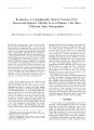
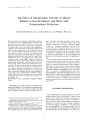
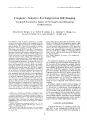
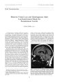
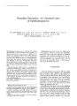
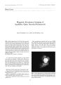
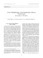
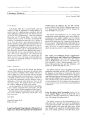
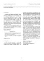
Show Journal of Neuro- Opiiihahuology 17( 1): 33- 35, 1997. © 1997 Lippineoil- Raven Publishers, Philadelphia Cataract in Tuberous Sclerosis Judith E. A. Warner, M. D., and Simmons Lessell, M. D. We evaluated two patients with tuberous sclerosis ( TS) and cataracts. In one, the cataract had been noted at an early age. In the second, it was noted at her first ophthalmic examination, age 31. Review of the literature indicates that although in some cases, cataract is attributable to local ocular disease, it may be a rare manifestation of TS. Key Words: Cataract- Tuberous sclerosis. We evaluated two patients with tuberous sclerosis ( TS) and unilateral cataracts. In one of them, the cataract had been detected in childhood. The other patient's cataract was discovered at age 31, at her first eye examination. A review of the literature showed few similar cases. Case Reports Case One This patient, the product of a normal pregnancy, labor, and delivery, developed infantile spasms in the first year of her life. At age 2, recognition of typical cutaneous stigmata led to the diagnosis of TS. Her family history was negative for neurological disorders, and examination of the parents failed to show the skin lesions of the disease. A routine eye examination at age 6 disclosed a cataract of the right eye, with vision of 20/ 30. Her examination was otherwise unremarkable. Over the succeeding years, vision declined somewhat in the right eye. When she was Manuscript received May 2, 1996; accepted June 5, 1996. From the Departments of Ophthalmology and Neurology, University of Utah, Salt Lake City, Utah; and Department of Ophthalmology ( S. L.), Massachusetts Eye and Ear Infirmary, Harvard Medical School, Boston, Massachusetts, U. S. A. Address correspondenee and reprint requests lo Dr. J. E. A. Warner at Department of Ophthalmology, 50 North Medical Drive, University of Utah, Sail Lake City, UT 84132, U. S. A. 14, a subependymal giant cell astrocytoma of the third ventricle causing hydrocephalus was resected. Although she was " mainstreamed" in school, she was only able to obtain " low- average" grades. Examination at age 26 showed adenoma sebaceum. Her best- corrected visual acuities were 20/ 70 right eye and 20/ 20 left eye. She could not identify any Ishihara color test plates with her right eye, and made many errors with the left. Goldmann visual fields were full. There were no defects in lid and pupillary function. She had a small right exotropia. There was a cortical cataract of the right eye sparing only the nasal side ( Fig. 1). Both discs were pale, but there were no retinal lesions. Case Two A 31- year- old woman was referred because she complained of inability to see things to her right. Her family history was allegedly noncontributory, but close relatives were never examined. Her only eye history was of refractive error. The patient's intelligence was always subnormal. In her early teens, she began having seizures. At age 19, she developed obstructive hydrocephalus from a subependymal giant cell astrocytoma. There were signs of TS including adenoma sebacea, shagreen patches, ash leaf spots, and subungual fibromas. She had subtotal resection of the tumor, radiation, and shunting, but the tumor recurred 3 years later. After another resection, she incurred a persistent right hemianopia. Examination showed that her best- corrected visual acuities were 20/ 25 each eye. Color vision ( Ishihara) was normal. There was an absolute, macula- splitting right homonymous hemianopia. There were two opacified areas in the fetal nucleus of the left eye ( Fig. 2). Her fundi had areas of peripheral hyperpigmentation, but no retinal hamartoma. The findings on ophthalmic examination were unchanged 6 years later. Comment Eye manifestations are common in TS. Robertson ( 1) evaluated 139 patients and found that 49% had retinal 33 34 J. E. A. WARNER AND S. LESSELL FIG. 1. Patient 1. Slit lamp images of the lens OD with ( A) direct and ( B) retroillumination showing the dense cataract sparing the nasal region. hamartomas. Foci of fundus and iris depigmentation and pedunculated conjunctiva] lesions have also been described. Papilledema is by no means uncommon because, as in our cases, expanding brain tumors may elevate the intracranial pressure. There have only been a few reported instances of cataract in TS. Only one of Robertson's patients had cataract, and no details of the case are available ( personal communication, May 1995). Three reports of single cases of TS note band keratopathy and cataract ( 2- 4). In these cases, the cataract might have been secondary to some other local eye disease and not a primary manifestation of TS. Yakovlev ( 5) described a case of bilateral congenital cataracts in the disease. In 1940 and 1946, Hall encountered a 17- year- old boy with TS who had " peripheral degeneration" of both lenses and a " small nodular mass" in the center of one lens ( 6,7). These abnormalities did not change during 4.5 years of observation. Histopathologic examination of the mass at autopsy showed an almost structureless matrix with faint calcification and some retained nuclei. It was interpreted as a " heterotopous formation." How- FIG. 2. Patient 2. Slit lamp images of the lens OS with retroillumination showing the fetal nuclear cataract. ever, the lesion could simply have been a congenital cataract. Ross and Dickerson ( 8) described a unilateral posterior polar cataract in a 19 year old with TS and bilateral keratoconus. Walsh ( 9) observed opaque nodules in the subcapsular cortex in a 16- year- old boy with TS. Our first patient's cataract was present from at least age 6. The location of the opacities in our cases, like the four described previously, is compatible with a congenital cataract. Although the association of cataract with TS might be fortuitous, and in some cases seems ascribable to other local eye, it appears most likely that congenital cataract is a valid if uncommon ocular manifestation of the other local eye disease. Acknowledgment: This work was supported in part by a grant from Research to Prevent Blindness, Inc., New York, NY, to the Department of Ophthalmology, University of Utah. References 1. Robertson DM. Ophthalmic findings. In: Gomez M, ed. Tuberous sclerosis. New York: Raven Press, 1979: 12 l^ t2. 2. Thomas C, Cordier J, Algan B. Keratite en bandelette et sclerose tubereuse de Bourneville. Bull Soc Ophthalmol ( Paris) 1948: 7: 465- 7. 3. Ardouin M, Feuvrier YM, Urvoy M. Sclerose tubereuse de Bourneville avec manifestations oculaires peu habituelle: Oedeme ./ Newo- Ophtlialmol. Vol. 17. No. I, 1997 CATARACT IN TUBEROUS SCLEROSIS 35 papillaire el kcratite en bandelette. Rev Olo Nemo Oplital 1961; 32: 499- 503. 4. Busch KT, Busch G. Neuro- ophthalmological and neuropatholog-ical findings in tuberous sclerosis ( Bourneville's disease). Klin Monatsble Augenheilk 1962; 141: 388. 5. Yakovlev PI, Guthrie RH. Congenital ectodermoses ( neurocutaneous syndromes) in epileptic patients. Arch Neurol Psychial 1931; 26: 1145- 94. 6. Hall GS. Tuberose sclerosis, rheoslosi. s and neurofibromatosis. Q J Med 1940; 9: 1- 10. 7. Hall GS. The ocular manifestations of tuberose sclerosis. Q J Med I946; 15: 209. 8. Ross AT, Diekerson WW. Tuberous sclerosis. Arch Neurol P. vy-chial 1943; 50: 233- 57. 9. Walsh FB, Hoyt WF. Clinical neuro- opluhcilmologv; vol 3. Baltimore: Williams and Wilkins, 1969: 1962. ./ Newo- Ophllmlmol, Vol. 17. No. I. 1997 |