| OCR Text |
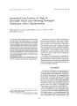
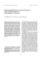
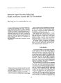
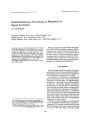
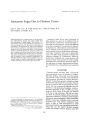
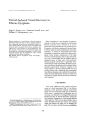
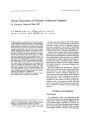
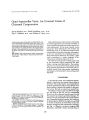
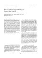
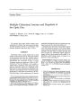
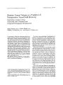
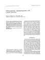
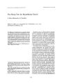
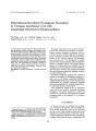
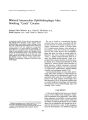
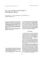
Show Journal of Clinical Neuro- ophthalmology 11( 4): 293- 296, 1991. © 1991 Raven Press, Ltd., New York Intermittent Downbeat Nystagmus Secondary to Vermian Arachnoid Cyst with Associated Obstructive Hydrocephalus Tin Chan, M. B., B. S., Patricia Logan, F. R. C. S. I., and Peter Eustace M. Ch., F. R. C. S., F. R. C. S. I., F. C. O. P. H. T. H. A 27- year- old man presented with a history of dizziness and intermittent vertical oscillopsia after oblique extension of the head and neck, following a neck injury 2 years previously. He had no other symptoms of cerebellar or brainstem dysfunction. On examination, an intermittent downbeat nystagmus lasting about fifty seconds was elicited by head extension and rotation. Radiologic examination showed slight lateral instability of the odontoid on lateral rotation, and computed tomography brain scan with Iopamidol 300 ( Niopam) contrast revealed a 5- em vermian arachnoid cyst in the posterior fossa with obstructive hydrocephalus. Removal of the cyst cured the nystagmus. Key Words: Downbeat nystagmus- Vermian arachnoid cyst. From the Department of Ophtha. lmology, I! niversity College Dublin Mater Misericordiae Hospital, and Richmond Institute of Neu~ oscience, Beaumont Hospital, Dublin, Ireland. Address correspondence and reprint requests to Professor P. Eustace at Department of Ophtha~ mology, University College Dublin, Mater Misericordiae Hospital, Eccles Street, Dublm 7, Ireland. 293 This paper reports the case of a patient who presented with intermittent downbeat nystagmus. Primary position downbeat nystagmus was described in detail in 1968 by Cogan ( 1). He reported that it is typically associated with structural abnormalities at the foramen magnum ( cervicomedullary junction) area. The nystagmus is usually present in the primary position, with upward slow phases of constant velocity. The upward drift is greatest on looking down and laterally, and the nystagmus is most prominent in these positions. The slow- phase velocity may increase during and after neck extension of the neck, probably owing to an otolith influence ( 2). Downbeat nystagmus suggests that a lesion is present in the region of the foramen magnum. It may occur as an benign congenital hereditary childhood phenomenon ( 3), or with Arnold- Chiari malformation ( 4), multiple sclerosis ( 5), herpetic brainstem encephalitis ( 6), cerebellar degeneration ( 7), and other posterior fossa lesions. Toxic substances, drugs ( 8- 12), or deficiency states ( 13,14) can produce reversible downbeat nystagmus. We present a patient in whom the presence of a previously unrecognised downbeat nystagmus lead to the diagnosis of a posterior fossa lesion. CASE REPORT A 27- year- old male was admitted for investigation of symptoms of dizziness. He was healthy until 20 months previously, when he was knocked over by a wave while surfing on Bondi beach and sustained an extension injury of his neck. He developed symptoms of dizziness and unsteadiness for about 3 days after the injury. Subsequently, he 294 T. CHAN ET AL. o~ lnUSA AMPL DIV.•. 031 I I '. . : II .: J:'" I::: .:::, ii:: :: ::::: i:::" . ..' ~ ::: ,.:: . ... 1i :: .. . I " ' 1\ .. :': ::. . :': 1 i':: I . ' f IIi. ' I I 1:: lJ '" .::: :: '. : I I:' ':. ::..... ::: . . .: : : I"'" "" ': If: iii: i: ': .... ,: .1 ::. :' FIG. 1. An infrared oculographic recording of vertical eye movements shows downbeat nystagmus after oblique head and neck extension. Upper tracing represents the right eye and lower tracing for the left eye. ( Paper speed 25 mm/ sec) developed acute vertigo, ataxia, vertical oscillopsia, and nausea for 1- 2 minutes on turning his head; this lasted for approximately 1 week. Six months later, he developed neck pain, which was low- grade but came on if he was tired and after alcohol ingestion. It radiated across to the base of the occipital area. There was no history of alcohol abuse or any other exposure to toxins. Family history was normal. Physical examination was also normal, in particular, there was no evidence of any neurological deficit. Ophthalmological examination showed normal visual acuity, visual field, pupillary reaction, saccades and pursuit eye movements, vestibulo- ocular reflex, and convergence. Intermittent downbeat nystagmus developed after oblique head and neck extension laterally and was sustained for about 50 seconds, then gradually resolved ( Fig. 1). No nystagmus was present while his head was held in the primary pOSition. Fundoscopy was normal. Laboratory findings, including full blood count, urea and electrolytes analysis were normal. Radiological examination included plain radiograph views of the odontoid process that showed slight lateral instability of the atlanto- axial joint, especially on lateral rotation to the left. A computed tomography ( CT) brain scan revealed a vermian cyst in the midline posterior fossa 5 cm in diameter. It extended from the level of the foramen magnum to the level of the tentorium, causing anterior displacement of the fourth ventricle and dilatation of the third and lateral ventricles ( Fig. 2). FIG. 2. CT brain scan reveals a vermian cyst in the midline posterior fossa and hydrocephalus. J Clin Neuro- ophthalmol. Vol. 11. NiJ 4. 199] DOWNBEAT NYSTAGMUS WITH VERMIAN ARACHNOID CYST 295 FIG. 3. CT brain scan shows residual widening of the valleculla and resolution of the hydrocephalus. At surgery 2 weeks later, a posterior fossa craniectomy and biopsy and drainage of an arachnoid cyst were performed. He made an uneventful recovery, and a CT brain scan taken one week later showed some residual widening of the valleculla. The cystic lesion was removed, and there was resolution of the hydrocephalus ( Fig. 3). Histological examination of the biopsy specimens showed a thin strip of dense fibrocollagenous connective tissue with dense arachnoid cells at the central segment. Occasional psammoma bodies were seen. These findings are commonly seen in arachnoid cyst ( Fig. 4). Postoperatively there has been complete resolution of his symptoms and of the downbeating nystagmus. A small residual skew deviation ( Fig. 5) is corrected by a two- diopter prism. DISCUSSION The etiology of downbeat nystagmus was extensively considered by various authors. Berger et al. ( 11) reviewed the literature in 1982 and described the three main causes: structural lesions at the craniocervical junction; diseases of the cerebellum or lower brain stem; and metabolic disorders. Intermittent downbeat nystagmus is very uncommon. Other cases reported were due to Arnold- Chiari malformations ( 15,16). The pathogenesis of downbeat nystagmus is not clear, but various mechanisms are proposed. First, upward drifts of the eyes may be caused by unopposed tonic inputs from anterior semicircular canals ( 17). Central projections from posterior semicircular canals cross in the fourth ventricle's floor. Thus, midline medullary lesions could be expected to cause downbeat nystagmus. A loss of inhibition of central vestibular connection of the anterior semicircular canals could also cause upward drift of the eyes. Also, a vertical pursuit imbalance could lead to upward drift ( 18). The presence of a skew deviation, as in this case, may reflect imbalance of the otolith inputs that cross in the medulla and ascend in the medial longitudinal fasiculus. The obstructive effect from the FIG. 4. Histopathological appearance of the biopsy, showing the psammoma bodies ( arrow), fibro- collagenous connective tissue, and arachnoid cells. I Gill Neuro- ophthalmol, Vol. 11, No. 4. 1991 296 T. CHAN ET AL. - ". 1.- 1_ FIG. 5. Hess screen chart shows a skew deviation with left over right hypertropia. arachnoid cyst may have compressed the brainstem and cerebellum. Acknowledgment: We would like to thank Dr. Michael A. Farrell for his help on the histopathology report. REFERENCES 1. Cogan DG. Downbeat nystagmus. Arch Ophthalmol 1968; 80: 757. 2. Leigh RJ, Zee OS. The neurology of eye movement. Philadelphia: FA Davis, 1983: 197. 3. Bixenman WW. Congenital hereditary downbeat nystagmus. Can JOphthalmol 1983; 18: 344- 8. 4. Hostovsky M, Tubman DE, Wirtchafter JD. Intrathecal metrizamide computed tomography: diagnosis of downbeat nystagmus in Amold- Chiari I malformation. Sun' Ophthalmol 1982; 27: 123- 5. 5. Masucci EF, Kurtzke JF. Downbeat nystagmus secondary to multiple sclerosis. Ann Ophthalmo/ 1988; 20: 347-- 8. 6. Hirst LW, Clark AW, Wolinsky JS, et al. Downbeat nystagmus- a case report of herpetic brain stem encephalitis. , Clin Neuro- ophthalmo/ 1983; 3: 245- 9. 7. Zasorin NL, Baloh RW. Downbeat nystagmus with alcoholic cerebellar degeneration. Arch Neuro/ 1984; 4l: 1301- 2. 1Cli" Neuro- ophllu1l11101, VOl. 11. i, · u. - 1. j9~ 1 8. Wheeler SO, Ramsay RE, Weiss J. Drug- induced downbeat nystagmus. Ann NeuroI1982; 12: 227-- 8. 9. Palakurthy PR, Iyer V, Meckler RJ. Unusual neurotoxicity associated with Amiodarone therapy. Arch Intern Med 1987; 147( 5): 881- 4. 10. Rosenberg ML. Reversible downbeat nystagmus secondary to excessive alcohol intake. J Clin Neuro- ophthalmol 1987; 7: 1: 23- 5. 11. Berger JR, Kovacs AG. Downbeat nystagmus with phenytoin. JClin Neuro- ophthalmoI1982; 2: 209- 11. 12. Williams DP, Troost T, Rogers J. Lithium- induced downbeat nystagmus. Arch Neurol 1988; 45: 1022- 3. 13. Saul RF, Selhorst JB. Downbeat nystagmus with magnesium depletion. Arch NeuroI1981; 38: 650- 2. 14. Mayfrank L, Thoden U. Downbeat nystagmus indicates cerebellar or brain- stem lesions in vitamin B 12 deficiency. J NeuroI1986; 233: 14S-- 8. 15. Yee 0, Baloh RW. Honrubia V. Episodic vertical oscillopsia and downbeat nystagmus in a Chiari malformation. Arch OphthalmoI1984; 102: 723- 5. 16. Pedersen RA, Troost T, Abel L, Zorub O. Intermittent downbeat nystagmus and oscillopsia reversed by suboccipital craniectomy. Neurology 1980; 30: 1237- 42. 17. Baloh RW, Spooner JW. Downbeat nystagmus: a type of central vestibular nystagmus. Neurology 1981; 31: 304- 10. 18. Zee OS, Freeman JM, Robinson OA. The mechanism of downbeat nystagmus. Arch NeuroI1974; 30: 277. |