| OCR Text |
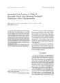
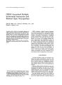
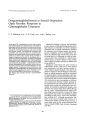
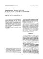
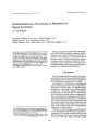
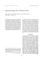
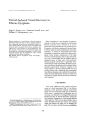
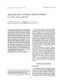
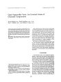
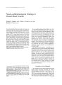
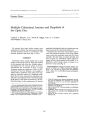
Show ! oUTnQI of Clinical NeurD- ophthalmology 11( 4): 233- 240, 1991. CREST- Associated Multiple Intracranial Aneurysms and Bilateral Optic Neuropathies Julio R. Ortiz, M. D., Nancy J. Newman, M. D., and Daniel L. Barrow, M. D. © 1991 Raven Press, Ltd., New York A patient with a clinical and serological diagnosis of CREST syndrome ( calcinosis, Raynaud's phenomenon, esophageal motility abnormalities, sclerodactyly, telangiectatic lesions), a variant of PSS ( progressive systemic sclerosis), was found to have multiple intracranial aneurysms. She subsequently developed bilateral optic neuropathies. Ischemic and compressive etiologies are proposed and related to the histopathology of the patient's underlying systemic disease. Key Words: CREST syndrome- Optic neuropathyProgressive systemic sclerosis ( PSS)- Intracranial aneurysms. From the Departments of Ophthalmology a · R. O., N. J. N.), Neurology ( N. J. N.), and Surgery ( Neurosurgery) ( D. L. B.), Emory University School of Medicine, Atlanta, Georgta, U. S. A. Address correspondence and reprint requests to Dr. Nancy J. Newman at Neuro- ophthalmology Unit, Emory Eye Center, 1327 Clifton Rd., N. E., Atlanta, GA 30322, U. S. A. 233 CREST syndrome, a clinical variant of progressive systemic sclerosis ( PSS), is typically characterized by the development of subcutaneous calcific nodules, Raynaud's phenomenon, esophageal motility abnormalities, sclerodactyly, and telangiectatic lesions of the skin and mucous membranes. As in many of the collagen- vascular diseases, degenerative and inflammatory lesions of the vascular system commonly occur. Because of the unique morphology and lack of perivascular support of the intracranial cerebral vessels, vasculitic involvement may lead to vascular wall ischemia, weakening, and aneurysmal dilatation. We present the first case ( to our knowledge), of CREST- associated multiple intracranial aneurysms. Aneurysmal compression and CRESTrelated vasculopathy may have combined to produce bilateral optic neuropathies in this patient. CASE HISTORY A 66- year- old black woman was referred to the Emory Eye Center with bilateral visual loss. The patient complained of slowly progressive, painless loss of vision in both eyes. Ophthalmic examination revealed visual acuities of 20/ 60, bilateral nuclear sclerotic cataracts, and normal- appearing optic discs. Her past medical history included an acute subarachnoid hemorrhage at the age of 40. Cerebral angiography at that time revealed multiple intracranial aneurysms. She was managed at another hospital where surgical intervention was considered too hazardous. No surgery was performed. Her past medical history was also notable for Raynaud's phenomenon, arthritic pain and swelling of her hands and knees, recurrent bouts of 234 J. R. ORTIZ ET AI. pneumonia with baseline dyspnea on exertion, dysphagia, a recurrent urticarial rash of the face and upper body, a single subcutaneous calcific nodule, and recurrent episodes of lower extremity thrombophlebitis. She had elevated serum globulins ( 3.5 g/ dl) with an electrophoretic pattern consistent with the presence of circulating immune complexes. Rheumatoid factor was positive; antinuclear antibodies were detected in a high titered homogeneous pattern; and anticentromere antibodies were elevated. Based on her clinical and serologic profile, the diagnosis of CREST syndrome was made. One month after presentation, the patient had a left extracapsular cataract extraction with a posterior chamber intraocular lens implant. Immediately after surgery, the patient complained of severe pain and visual loss in the left eye. Vision in that eye was limited to light perception; there was an afferent pupillary defect; and the intraocular pressure was elevated at 48 mm Hg. Two and onehalf hours of aggressive therapy brought the intraocular pressure under control, but visual acuity did not improve to better than recognition of hand motions. Ischemic optic neuropathy was suspected, although disc edema was not appreciated. During the next 3 weeks, the patient noted a gradual decline in the vision of her right eye, and she was referred to the Neuro- ophthalmology Unit. Visual acuity was 20/ 300 in the right eye and hand motions in the left. There was a left relative afferent pupillary defect. Motility was full. Intraocular pressures were normal, and slit lamp examination unremarkable. Ophthalmoscopic examination revealed a normal- appearing disc in the right eye and temporal pallor in the left. Visual fields showed a superior altitudinal defect involving fixation on the right and an inferotemporal island of remaining vision on the left ( Fig. 1). Findings were diagnostic of bilateral optic neuropathies. A contrast- enhanced computed tomography ( CT) scan of the brain and orbits showed multiple enhancing soft tissue masses with rim calcifications in the region of the circle of Willis, abutting the anterior visual pathways bilaterally ( Fig. 2). Four- vessel cerebral arteriogram revealed multiple aneurysms ( Figs. 3 and 4): ( a) an aneurysm of the right supraclinoid carotid at the origin of the superior hypophyseal artery ( 0.3 cm); ( b) a bilobed, intracavernous aneurysm of the right internal carotid artery ( 1.2 cm); ( c) a bilobed aneurysm of the left ophthalmic artery at its origin ( 0.8 cm); ( d) an aneurysm at the bifurcation of the right middle cerebral artery ( 1.0 cm); ( e) an aneurysm of the left superior hypophyseal artery ( 0.8 cm); ( f) an aneurysm of the left anterior choroidal artery ( 1.6 cm). Given the progressive visual loss in the right eye and a normal- appearing optic nerve head, recent aneurysmal compression of the right optic nerve was suspected. The patient was referred for neurosurgical intervention and underwent a right frontotemporal craniotomy. The right side was 210 LEFT RIGHT ~ IG. 1. Pre- operative Goldmann visual fields. Inferotemporal island of vision, left eye. "' y'r- lr! or Clltltudlnal defect involving fixation, right eye. 1elin Neuro- ophthalmol. VA/. 11, ,,",, CREST- ASSOCIATED OPTIC NEUROPATHIES 235 A a FIG. 2. Contrast- enhanced coronal ( A) and axial ( B) CT scan demonstrates multiple soft tissue masses in the region of the anterior circle of Willis, bilaterally. A chosen to provide the best exposure of the right optic nerve, which had the most preserved vision. The right middle cerebral artery aneurysm was clipped without difficulty. The right supraclinoid carotid artery aneurysm arising from the region of the superior hypophyseal artery was pointing medially, indenting the right optic nerve, and was also clip- ligated. A bilobed aneurysm of the distal right intracavernous carotid artery was not compressing the visual apparatus and was reinforced with muslin. The left optic nerve was markedly deviated to the right and lay beneath the right optic nerve. This deviation was caused by a medially directed B FIG. 3. A: Right internal carotid arteriogram, anteroposterior view, shows right middle cerebral artery ( arrowhead) intracavernous ( large arrow), and superior hypophyseal ( small arrow) artery aneurysms. B: Left internal carotid injection shows left anterior choroidal ( large arrow) and ophthalmic artery ( small arrow) aneurysms. J Clin Neuro- ophthalmol, Vol, II, No, 4, 1991 236 J. R. ORTIZ ET AL. A B FIG. 4. Right internal carotid arteriogram, lateral views. A: Right middle cerebral ( large arrowhead) and intracavernous ( large arrow) artery aneurysms are shown. B: Left anterior choroidal ( large arrow) and ophthalmic ( small arrow) artery aneurysms are seen. aneurysm arising from the region of the left superior hypophyseal artery. It was felt that this lesion and the other two left- sided aneurysms could most safely be addressed from a left frontotemporal approach. Postoperatively, the patient had a transient superior division third nerve paralysis. Vision in both eyes was limited to counting fingers. The visual field on the right was notable for restoration of some peripheral vision temporally and superiorly, with further field loss centrally ( Fig. 5). At 5- month follow- up, her vision was 20/ 40 in the right eye and 20/ 100 in the left. Visual fields were remarkably improved, with only a central defect nasally in the right eye and some expansion of isopters in the left eye, which involved some fixation inferiorly ( Fig. 6). An elective left- sided neurosurgical procedure to address the remaining aneurysms is planned. DISCUSSION PSS and CREST Progressive systemic sclerosis is a generalized disorder of connective tissue characterized by de(', · n"' r~~;' · · -"-,.- 1 _,. 1 ' m'lnJurv ch, lngt" s. Eventually "' li\.. rial leads / Gin NeuTo- ophthalmol. Vol. 11. No I. 1~~" 1 to severe fibrosis. Progressive systemic sclerosis manifests clinically by diffuse induration of the skin and dysfunction of visceral structures, particularly the gastrointestinal tract, kidneys, heart, and lungs. Manifestations of this disorder are often based on the functional and structural vascular compromises that occur after repeated vascular insults, subsequent healing, and proliferative vascular response ( 1). The cause of vascular injury in PSS remains unknown. The leading hypothesis suggests that there exists an underlying systemic abnormality of collagen metabolism that specifically affects the small vessels and capillaries ( 2). Histopathology Histopathologically, the vascular lesions are characterized by subintimal proliferation of connective tissue, subendothelial fibrin deposits, basement membrane thickening, and fragmentation of elastic tissue ( 3,4). Devascularization of tissues is a generalized phenomenon in PSS, and it has been proposed that arteriolar and capillary endothelia may be the primary target cell population. Kaheleh and colleagues noted that the sera of 31 of 52 patients with PSS contained cytotoxic activity specific for endothelial cells ( 1). In addition, immune complex de- CREST- ASSOCIATED OPTIC NEUROPATHIES 210 LEfT RIGHT FIG. 5. Postoperative visual fields. Visual field unchanged, left eye. Restoration of some field temporally and superiorly, with further loss centrally, right eye. 237 position has been known to induce endothelial damage in this disease ( 5). The coagulation cascade may be triggered by the intimal lesion, leading to fibrin deposition, decreased blood flow, and local ischemia. Exposed subendothelium will also induce platelet adherence and aggregation at the site of injury. If this process involves vasa vaso-rum, it may theoretically lead to weakening and generalized dilatation of larger vascular structures. Although extremely rare, large blood vessel involvement occasionally gives rise to major symptoms. Chaithiraphan and associates have described the devascularization of a coronary arterial wall leading to the development of three aneurys- 210 LEfT RIGHT FIG. 6. Visual fields at 5- month follow- up. Expansion of the visual field involves some fixation inferiorly, left eye. Normalization of the field with the exception of a central defect nasally, right eye. I Clin Neuro- ophthalmoi, Vol. 11, No. 4, 1991 238 ]. R. ORTIZ ET AL. mal bulges and anginal chest pain in a young patient with PSS ( 6). Clinical Manifestations CREST syndrome, a clinical variant of PSS, is characterized by at least three of the following features: calcinosis, Raynaud's phenomenon, esophageal motility abnormalities, sclerodactyly, and telangiectatic lesions of the skin and mucosal surfaces. The majority of patients (> 85%) present with Raynaud's phenomenon, which may antedate further manifestations of the disease by several years. The interval from the onset of symptoms to a final diagnosis often exceeds 1~ 15 years ( 7). Patients with PSS and CREST have remarkably normal results on routine laboratory tests ( 8). The erythrocyte sedimentation rate tends to be normal or only slightly elevated. However, various autoantibodies have been found in these syndromes. Approximately 30% of patients are rheumatoid factor positive. Antibodies to collagen ( 9) and antinuclear antibodies are very common. Anticentromere antibodies are found in 5~ 98% of patients with CREST and 3- 18% of patients with PSS, yet are rarely found in systemic lupus erythematosus ( 2%) and mixed connective tissue disease ( 6%) ( 7, 1~ 15). Anticentromere antibodies may be detected in a patient's serum prior to the development of the full CREST clinical syndrome. Autoantibodies found in other connective tissue diseases ( e. g., anti- DNA, anti- RNP, anti- Sjogren's syndrome antigen B) are not found in CREST patients. Neurologic and cerebrovascular abnormalities are uncommon in PSS and CREST syndrome, unlike related diseases such as systemic lupus erythematosus and polyarteritis nodosa. Gordon and Silverstein suggested that neurological complications of PSS are a result of concurrent drug therapy, involvement of other internal organs with secondary neurologic sequelae ( i. e., hypertensive cerebrovascular disease from renal involvement, tetany from gastrointestinal malabsorption syndromes, or confusion from pulmonary damage and hypoxia), or simply coincidental ( 16). The infrequency of primary neurologic disease may reflect the lack of collagenous connective tissue in the central nervous system. Cerebral blood vessels are only rarely involved by the disease process ( 16). Lee and Haynes described an unusual patient with PSS who died of a massive left cerebral inLn.~ ti(> T-' "~' 1JI",,~ to C\ l... Ct internal carotid arteritis ( 5). . .!;,- kening of the 1 Clin Neuro- ophlhalmol. Vol. 11. Nu. 4. lY~; carotid vessel wall and vasculitis of the vasa vasorum surrounding the internal carotid and anterior and middle cerebral arteries. No sclerodermatous fibrous thickening was seen in the anterior and middle cerebral arteries. Ophthalmologic manifestations of PSS are rare, but include keratitis ( 17,18) and keratoconus ( 19), shallow conjunctival fornices ( 20), keratoconjunctivitis sicca ( 21), eyelid tightness ( 19), and lid telangiectasia ( 20). Arnett and Michaels described a single case of inflammatory ocular myopathy ( 22). Gass presented a patient with multiple focal exudative and hemorrhagic retinal detachments ( 23). Multiple retinal vascular abnormalities have been described in association with PSS, including branch retinal vein occlusion ( 24), central vein occlusions ( 25), and branch ( 26) and central retinal arterial occlusions ( 27). Cases with optic nerve head edema ( 17), cotton wool spots ( 28), intraretinaI edema, exudates and hemorrhages ( 17,26,29), and fibrinoid necrosis of choroidal vasculature ( 27) have been reported. We could find no reports of ischemic or compressive optic neuropathies documented in the literature. Intracranial Aneurysms Intracranial aneurysms are discovered predominantly in patients between 40 and 70 years of age, often presenting with catastrophic subarachnoid hemorrhage. Rarely do they present with an insidious, painless, and slowly progressive visual loss secondary to retrobulbar optic nerve compression. Optic disc swelling or associated neurologic signs are typically absent. Central, cecocentral, paracentral, arcuate, altitudinal, nasal, and temporal hemianopic visual field defects have been described ( 3~ 32). Visual loss associated with intracranial aneurysms has been ascribed to vascular insufficiency and/ or mechanical blockage of axonal transport ( 33,34). Acute monocular visual loss can occur, presumably on the basis of sudden interruption of the vascular supply to the optic nerve ( 35). The reported frequency of multiple aneurysms varies widely. Bigelow found more than one aneurysm in 10.2% of 2,237 cases of intracranial aneurysms collected from the literature ( 36). Stehbens reported multiplicity in 30.3% of 122 brains found to harbor aneurysms on autopsy ( 37). Wilson and associates found that 45% of 254 aneurysm patients studied had multiple aneurysms, and of these 68% had 2 aneurysms, 24% had 3 aneurysms, 8% had 4 aneurysms, and only 1 case had 5 CREST- ASSOCIATED OPTIC NEUROPATHIES 239 aneurysms ( 38). Rarely are more than three aneurysms found in anyone brain. When large numbers of aneurysms are discovered, an arteritic process or connective tissue disease should be suspected ( 39). Intracrania~ aneurysms of inflammatory origin occur sporadIcally and may be infectious in etiology or noninfectious arteritis- related. Although ~ are, no~ infectiousaneurysms have been reported m a vanety of diseases. Polyarteritis nodosa has often been associated with splanchnic- mesenteric aneurysms. Cerebral vessels can be involved in as many as 20% of patients with this disease ( 40). An aneurysm ? n a s~ all branch of a vertebral artery wa~ found m a patient with Wegener's granulomatOSIS ( 41). A posterior communicating artery aneurysm pr: senting with subarachnoid hemorrhage was attributed to systemic lupus erythematosis in a 29- year- old woman ( 42). Multiple internal carotid artery aneurysms developed in a 34- month- old child with widespread giant cell arteritis of childhood ( 43). Similarly, a small aneurysm of a cerebellar pial artery was attributed to granulomatous angiitis in a 25- year- old man ( 44). Histologically, the intracranial cerebral arteries are muscular in type and have considerably thinner media and adventitia than extracranial arteries of similar size. They have a prominent internal elastic lamina and a few adventitial elastic fibrils. The media lacks elastica, and the external elastic lamina is notably absent. There are no vasa vasorum in the arterial walls of children and many young adults. As arterial walls age and develop atherosclerotic thickening, the vasa vasorum may become more prominent. Although intracranial vessel wall injury and aneurysm formation has been attributed to involvement of vasa vasorum, anatomical studies have shown a generalized absence of vasa vasorum from the affected portions of the cerebral vasculature ( 45- 48). Rather, endothelial injury, followed by the adhesion of platelets and leukocytes and their activation, playa primary role in aneurysm formation. These events could lead to complement activation, release of lysosomal enzymes and prostaglandins, with further mechanical disruption of the vessel wall ( 49). Chronic hemodynamic stress at sites of vessel wall injury, with fragmentation of the elastic lamina and atrophy of the media, may result in focal wall dilatation and frank aneurysm formation. Because of the architectural peculiarities and the lack of strong perivascular support of the cerebral arterial walls, chronic inflammation may lead to degenerative changes, ischemia, weakening of the arterial wall, and aneurysmal dilatation. Conclusions . Our patient had a clinical and serologic diagnoSIS of PSS and CREST syndrome, characterized by the presence of calcinosis, a long history of Raynaud's phenomenon, dysphagia, arthritis, recurrent ~ hrombophlebitis, skin rashes, mild pulmonary mvolvement, positive rheumatoid factor, and elevated antinuclear and anticentromere antibodies. The sudden and profound visual loss in her left eye after cataract extraction may have been secondary to ischemia to the optic nerve ( 50). A short period of raised intraocular pressure may have changed ocular hemodynamics in an already compromised arteritic circulation. Furthermore, it is likely that both optic nerves were already comp. romised by the effects of aneurysmal compresSIOn. The progressive visual loss in the right eye ~ ay have ~ een related to even further compresSIon of the nght optic nerve by adjacent aneurysms and by a swollen, ischemic left optic nerve, which had come to lie beneath the right nerve due to the deviating effect of a left ophthalmic artery aneurysm. We propose that the patient's multiple intracranial aneurysms may have been related to the chronic degenerative and inflammatory vascular changes typical of PSS and CREST syndrome. We believe this to be the first report of CRESTass. ociate~ intracranial aneurysms. This patient's umque bIlateral optic neuropathy represents another possible ocular manifestation of CREST and PSS in particular, and of the collagen- vascular diseases in general. Acknowledgment: This study was supported in part by a grant from Research to Prevent Blindness, Inc. REFERENCES 1. Kahaleh MB, Sherer GK, LeRoy EC. Endothelial injury in scleroderma. / Exp Med 1979; 149: 1326- 35. 2. Norton WL, Nardo JM. Vascular disease in progressive sys~~~ c sclerosis ( scleroderma). Ann Intern Med 1970; 73: 317- 3. Kinder RR, Fleischmajer R. Systemic scleroderma: a review of organ systems. Int / DermatoI1974; 13: 382- 95. 4. Hupp LH. Giant cell arteritis associated with progressive systemic sclerosis. / Clin Neuro- ophthalmoI1989; 9: 126- 130. 5. Lee JE, Haynes JM. Carotid arteritis and cerebral infarction due . to . scleroderma. Neurology 1967; 17: 18-- 22. 6. Chalthiraphan S, Goldberg E, O'Reilly M, Jootar P. Multiple aneu~ sms of coronary artery in sclerodermal heart disease. Angiology 1973; 24: 86. 7. Frit~ l~ r MJ, Kinse! la T~, Garbutt E. The CREST syndrome: a distinct serolOgIC entity With anticentromere antib d' Am / Med 1980; 69: 520- 6. 0 les. 8. Medsger TA. Systemic sclerosis and localized scleroderma In: Schumacher HR, Klippel JH, Robinson DR, eds. Prime; on the r~ eumatlc diseases. 9th ed. Atlanta, GA: Arth ' ti Foundation, 1988: 111- 7. n s J Clin Neuro- ophthalmol, Vol. 11, No. 4, 1991 240 J. R. ORTIZ ET AL. 9. Mackel AM, Oelustro F, Harper FE, LeRoy EC. Antibodies to coHagen in scleroderma. Arthritis Rheum 1982; 25: 522- 31. 10. Bernstein RM, Steigerwald ) C, Tan EM. Association of antinuclear and antinucleolar antibodies in progressive systemic sclerosis. Clin Exp Immun 1982; 48: 43-- 51. 11. Kallenberg CGM, Pastoor GW, Wouda AA, The TH. Antinuclear antibodies in patients with Raynaud's phenomenon: clinical significance of anticentromere antibodies. Ann Rheum Dis 1982; 41: 382- 7. 12. Moroi Y, Peebles C, Fritzler M), Steigerwald J, Tan EM. Autoantibody to centromere ( kinetochore) in scleroderma sera. Proc Nat Acad Sci 1980; 77: 1627- 31. 13. Tan EM, Rodnan GP, Garcia I, Moroi Y, Fritzler MJ, Peebles C. Diversity of antinuclear antibodies in progressive systemic sclerosis: anti- centromere antibody and its relationship to CREST syndrome. Arthritis Rheum 1980; 23: 617- 25. 14. Tramposch HO, Smith CO, Senecal JL, Rothfield N. A longterm longitudinal study of anticentromere antibodies. Arthritis Rheum 1984; 27: 121- 4. 15. Steen VD, Ziegler GL, Rodnan GP, Medsger TA. Clinical and laboratory associations of anticentromere antibody in patients with progressive systemic sclerosis. Arthritis Rheum 1984; 27: 125-- 31. 16. Gordon RM, Silverstein A. Neurologic manifestations in progressive systemic sclerosis. Arch NeuroI1970; 22: 126-- 34. 17. Manschot WA. Generalized scleroderma with ocular symptoms. Ophthalmologia 1965; 149: 131- 7. 18. Goyle EF. Scleroderma of the cornea. Br JOphthalmol1956; 40: 239. 19. Agatston H). Scleroderma with retinopathy. Am JOphthalmol 1953; 36: 120. 20. Horan EC. Ophthalmic manifestations of progressive systemic sclerosis. Br JOphthalmoI1969; 53: 388. 21. Ramage JH, Kinnear WP. Keratoconjunctivitis sicca and the collagen disease. Br J Ophthalmol 1956; 40: 416. 22. Arnett FC, Michels RG. Inflammatory ocular myopathy in systemic sclerosis ( scleroderma). Arch Intern Med 1973; 132: 740. 23. Gass JOM. A fluoroscein angiographic study of macular dysfunction secondary to retinal vascular disease. VI. X- ray irradiation, carotid artery occlusion, coHagen- vascular disease, and vitTitis. Arch Ophthal 1968; 80: 606. 24. West RH, Barnett AJ. Ocular involvement in scleroderma. Br JOphthalmoI1979; 63: 845. 25. Saari KM, Rudenberg HA, Laitinen O. Bilateral central retinal vein occlusion in a patient with scleroderma. Ophthalmologica 1981; 182: 7. 26. Goder G. Vaskulare augenveranderungen bei der generalisierten sklerodermie. Klin Monatsbl Augenheilkd 1964; 144: 370. 27. Berndt K, Hoffman A. Zentralarterienverschlub der retina bei progressiver sklerodermia. Klin Monatsbl Augenheilkd 1977; 171: 597. 28. PoHack ID, Becker B. Cytoid bodies of the retina in a patient with scleroderma. Am J OphthalmoI1962; 54: 655. 29. MacLean H, Guthrie W. Retinopathy in scleroderma. Trans Ophthal Soc UK 1969; 89: 209. I Clin Neurcrophlhalmol, Vol. 11, NO. 4, 1991 30. CuHen JF, Haining WM, Crombie AL. Cerebral aneurysms presenting with visual field defects. Br J Ophthalmol 1966; 50: 251-- 6. 31. Peiris JB, Ross RusseH RW. Giant aneurysms of the carotid system presenting as visual field defects. JNeurol Neurosurg Psychiatry 1980; 43: 1053-- 64. 32. Fox) L. Intracranial aneurysms. Vol 1. New York: SpringerVerlag, 1983: 171. 33. Hughes EBC. Blood supply of the optic nerves and chiasm and its clinical significance. Br JOphthalmol 1958; 42: 106. 34. Kayan A, Earl q. Compressive lesions of the optic nerves and chiasm. Brain 1975; 98: 13-- 28. 35. Stem WE, Ernest JT. Intracranial ophthalmic artery aneurysm. Am JOphthalmoI1975; 80: 203- 6. 36. Bigelow NH. Multiple intracranial arterial aneurysms: Analysis of their significance. Arch Neurol Psychiatry 1955; 73: 76-- 99. 37. Stehbens WE. Aneurysms and anatomical variation of cerebral arteries. Arch PathoI1963; 75: 45-- 64. 38. Wilson FMA, Jaspan T, Holland 1M. Multiple cerebral aneurysms- a reappraisal. Neuroradiology 1989; 31: 232-- 6. 39. Fox) L. Intracranial aneurysms. Vol 1. New York: SpringerVerlag, 1983: 291. 40. Miller H. Clinical consideration of cerebrovascular disorders occurring during the course of general diseases of an inflammatory or allergic nature. Proc Roy Soc Med 1951; 44: 852. 41. McCormick WF. Problems and pathogenesis of intracranial arterial aneurysms. In: Toole JF, Moosy J, Janeway R, eds. Cerebral vascular diseases, seventh princeton conference. New York: Grune & Stratton, 1971: 219- 31. 42. Kelley RE, Stokes N, Reyes P, Harik 51. Cerebral transmural angiitis and ruptured aneurysm. Arch Neurol 1980; 37: 526. 43. Wagenvoort CA, Harris lE, Brown AL, Veeneklaas GMH. Giant cell arteritis with aneurysm formation in children. Pediatrics 1963; 32: 861- 7. 44. Sandhu R, Alexander 5, Hornabrook RW, Stehbens WE. Granulomatous angiitis of the CNS. Arch Neurol 1979; 36: 433-- 5. 45. Clarke JA. An x- ray microscopic study of vasa vasorum of the intracranial arteries. Z Anat EntwickIungsgesch 1965; 124: 396- 400. 46. Clomer BR, SuHivan OM, Smith RR. Intracranial vessels lack vasa vasorum, JNeurosurg 1984; 61: 44- 8. 47. Hughes JT, Schianchi PM. Cerebral artery spasm: a histolOgIcal study at necropsy of the blood vessels in cases of subarachnoid hemorrhage. J Neurosurg 1978; 48: 515-- 25. 48. Zervas NT, liszczak TM, Mayberg MR. Cerebrospinal fluid may nourish cerebral vessels through pathways in the adventitia that may be analogous to systemic vasa vasorum. , Neurosurg 1982; 56: 475-- 81. 49. Engelberg H. Endothelium in health and disease. Semin Thromb Hemos 1989; 15: 178- 83. 50. Hayreh 55. Anterior ischemic optic neuropathy IV. Occurrence after cataract extraction. Arch Ophthalmol 1980; 98: 141~. |