| OCR Text |
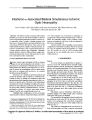
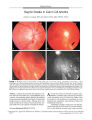
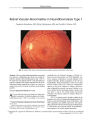
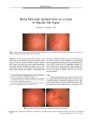
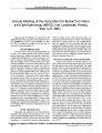
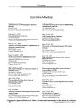
Show PHOTO ESSAY Siegrist Streaks in Giant Cell Arteritis DustinJ. Coupal, MD, and Anil D. Patel, MD, FRCSC, FACS FIGURE 1. A: Right fundus at presentation reveals pallid optic nerve head edema, peripapillary hemorrhages, retinal whitening, and vascular fragmentation consistent with ophthalmic artery occlusion. B: One month later, there is marked optic disc pallor and sclerosis of the retinal vasculature; a portion of a Siegrist streak is visible inferiorly { arrow). C: Peripheral retina shows the typical findings of Siegrist streaks: linear bands of chorioretinal atrophy with hyperpigmented margins. The findings were most prominent in the inferior and nasal mid- peripheral retina and followed the underlying choroidal vessels. D: Fluorescein angiogram ( late phase) shows transmission hyperfluorescence compatible with chorioretinal atrophy. Abstract: A patient who presented with symptoms of gi- A 78- year- old woman was referred for urgent neuro-ant cell arteritis was found to have a right ophthalmic artery /" Aophthalmic evaluation with a 4- day history of transient occlusion. One month after initial evaluation, the peripheral visual obscurations in the OD followed by sudden vision retina demonstrated multiple linear bands of chorioretinal loss in that eye. She described a 6- week history of severe atrophy known as Siegrist streaks. Although most com- headaches, jaw claudication, scalp tenderness, fatigue, and monly described in the setting of acute hypertension, Sie- unintentional weight loss. grist streaks also occur in patients with giant cell arteritis. Examination revealed a visual acuity of no light perception in the OD and 20/ 30 in the OS. External examina- { JNeuro- Ophthalmol 2003; 23: 272- 273) tion revealed bilateral ulcerated scalp lesions consistent Copyright © Lippincott Williams & Wilkins. Unauthorized reproduction of this article is prohibited. 272 J Neuro- Ophthalmol, Vol. 23, No. 4, 2003 Siegrist Streaks in Giant Cell Arteritis JNeuro- Ophthalmol, Vol. 23, No. 4, 2003 with scalp necrosis. Pupillary examination showed a large right afferent pupillary defect. The visual field in the OS was full. The intraocular pressures were within normal limits. Ophthalmoscopy revealed pallid swelling of the optic disc and cloudy swelling of the retina, findings consistent with a right ophthalmic artery occlusion ( Fig. 1 A). The OS was unremarkable. Results of blood work revealed a Westergren erythrocyte sedimentation rate of 26mm/ h and an elevated C-reactive protein of 29mg/ l ( normal = 0- 8mg/ l). The patient was tentatively diagnosed with giant cell arteritis ( GCA) and was admitted to the hospital for intravenous corticosteroid therapy. A temporal artery biopsy confirmed the clinical suspicion of GCA. Reevaluation 1 month later showed marked right optic disc pallor and significant sclerosis of all branches of the right central retinal artery ( Fig. IB). Identified within the mid- peripheral retina were a number of linear and wedge-shaped retinal lesions that are known as Siegrist streaks ( Fig. 1C). Fluorescein angiography vividly demonstrated well- defined radiating bands of retinal atrophy ( Fig. ID). Siegrist streaks initially were identified and documented in 1899 by Siegrist ( 1), who had recognized a peculiar pattern of pigmented retinal lesions in two patients, one with GCA and the other with malignant hypertension. Since that time, other reports of Siegrist streaks have been published, most often in patients with severe arterial hypertension and more rarely in patients having GCA ( 2- 4). Hayreh ( 5) reported that in 123 eyes with ocular involvement in GCA, 10 eyes were found to have chorioretinal ischemic lesions, which we believe are Siegrist streaks. Siegrist streaks develop as a late result of non-perfusion of choroidal vessels causing severe outer retinal From the University of Saskatchewan, Saskatoon, Saskatchewan, Canada. Address correspondence to Anil D. Patel, MD, FRCSC, FACS, 701 Queen Street, Saskatoon, SK, Canada, S7K 0M7; E- mail: anil. patel@ saskatoonhealthregion. ca ischemia ( 2,4). McLeod has suggested that outer retinal infarcts occur more frequently in patients with GCA when there is combined involvement of the central retinal and posterior ciliary circulations ( 6). Histopathologically, Siegrist streaks have been shown to consist of sclerosed choroidal vessels with overlying obliteration of the choriocap-illaris and secondary retinal pigment epithelial atrophy ( 7). A reactive retinal pigment epithelial hypertrophy and hyperplasia occurs along the margins of the streaks 2 or 3 weeks after the onset of choroidal ischemia ( 5). Related to Siegrist streaks are similar manifestations of impaired choroidal perfusion known as Elschnig spots. These spots are small isolated circular areas having central retinal pigment, epithelial pigment clumping, and a surrounding halo of depigmentation ( 8). Whereas Siegrist streaks tend to occur along the course of sclerosed choroidal vessels, Elschnig spots occur in isolation secondary to focal obliteration of the choriocapillaris. Siegrist streaks and Elschnig spots may be found together in the same patient or may occur separately. The significance of Siegrist streaks in patients diagnosed with GCA is not well documented, but when found in the setting of acute hypertension, they are considered to reflect a relatively poor general health prognosis ( 2,3). References 1. Siegrist A. Beitrag zur Kenntnis der arterosklerose der Augenge-fasse. In: IX Int Cong Ophthalmol 1899: 131- 9. 2. Uveal manifestations of systemic disease. In: Duke Elder S, ed.. System of Ophthalmology, Vol. 9. Diseases of the Uveal Tract. St Louis: CV Mosby, 1966: 629- 34. 3. Puri P, Watson AP. Siegrist's streaks: a rare manifestation of hypertensive choroidopathy. Eye. 2001; 15: 233- 4. 4. Hayreh SS, Servais GE, Virdi PS. Fundus lesions in malignant hypertension. VI. Hypertensive choroidopathy. Ophthalmology. 1986; 93: 1383^ 100. 5. Hayreh SS, Podhajsky PA, Zimmerman B. Ocular manifestations of giant cell arteritis. Am J Ophthalmol. 1998; 125: 509- 20. 6. McLeod D, Oji EO, Kohner EM, et al. Fundus signs in temporal arteritis. Br J Ophthalmol. 1978; 62: 591- 4. 7. Green WR. Systemic diseases with retinal involvement. In: Spencer WH, ed. Ophthalmic Pathology: An Atlas and Textbook, v. 2. Philadelphia: WB Saunders, 1985: 1034- 7. 8. Morse PH. Elschnig's spots and hypertensive choroidopathy. Am J Ophthalmol. 1968; 66: 844- 52. Copyright © Lippincott Williams & Wilkins. Unauthorized reproduction of this article is prohibited. 273 |