| OCR Text |
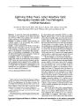
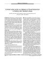
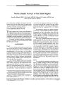
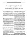
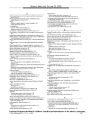
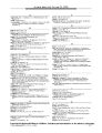
Show ORIGINAL CONTRIBUTION Nerve Sheath Tumors of the Sellar Region Sumedha Bhagat, MBBS, Colin Smith, MBChB, Graham M. Teasdale, MBChB, and Robert M. McFadzean, MBChB Nerve sheath tumors, including schwannomas and neurofibromas, rarely affect the sellar region. The authors report two such cases that were mistaken for pituitary adenomas on the basis of clinical and imaging features. ( JNeuvo- Ophthalmol 2002; 22: 275- 278) The most common cause of a mass in the sellar region is a pituitary adenoma, either secretory or nonsecretory. A cause other than a pituitary adenoma is identified pathologically in about 9% of cases ( 1). There is a wide range of other possibilities, including inflammatory, granulomatous, and vascular lesions, as well as primary and metastatic tumors. We present the clinical, radiologic, and pathologic features of two patients in whom an unusual tumor within the sellar region was found. CASE REPORTS Case 1 A 51 - year- old Caucasian man presented with a 5- year history of erectile impotence, fatigue, and lethargy. He had no visual complaints and did not have a family history of neurofibromatosis, cafe- au- lait spots, or Lisch nodules. Clinical examination showed secondary hypogonadism. He did not have cutaneous neurofibromas, cafe- au- lait spots, or Lisch nodules. Visual acuity was 20/ 15 OU, and Goldmann perimetry showed a superior bitemporal quadrantanopia. The corneal reflexes, pupillary reflexes, extraocular movements, and optic discs were normal. Tests of anterior pituitary function showed panhypopituitarism, as evidenced by deficiencies of growth hormone, cortisone, and thyroxin. The serum prolactin concentration was normal ( 400 mU/ L). Magnetic resonance imaging ( MRI) demonstrated pituitary fossa expansion by a large intrasellar mass that extended into the suprasellar area, producing a " cottage loaf appearance at the point of Departments of Neuro- Ophthalmology ( SB, RMM), Neuropathology ( CS), and Neurosurgery ( GMT), Institute of Neurological Sciences, South Glasgow University Hospitals, NHS Trust, Scotland. Address correspondence to Sumedha Bhagat, MD, Department of Ophthalmology, Ayr Hospital, Dalnellington Road, Ayr KA6 6DX, UK; E- mail: sumedhal@ btinternet. com constriction by the diaphragma sellae ( Fig. 1A). This superior extension elevated and compressed the optic chiasm against the hypothalamus. Intravenous gadolinium produced uniform increase in signal intensity on Tl- weighted MRI. The preoperative diagnosis was pituitary adenoma. At operation, a standard sublabial paraseptal transsphenoidal approach was made to the pituitary fossa, which was thinned but intact. On opening the dura, a fleshy lesion with a distinct capsule was identified. The pituitary fossa was emptied of this tumor, except for a small fragment attached to the base of the pituitary stalk. Microscopic examination demonstrated a tumor with interlacing spindle cells ( Fig. IB). There was mild nuclear pleomorphism but no mitotic figures. Nuclei with a wavy morphology were present within some tumor cells. The tumor was moderately vascular; hyalinization of the vessel walls was not apparent. Immunocytochemistry of the tumor cells was positive with S- 100, a marker of neuroectodermal differentiation. A neurofilament stain showed occasional axons within the tumor. Stains for epithelial membrane antigen, glial fibrillary acidic protein, and anterior pituitary gland hormone production ( prolactin, growth hormone, adrenocorticotropic hormone, luteinizing hormone, follicular stimulating hormone, and thyroid stimulating hormone) markers were negative. Tissue was not available for electron microscopy. The pathologic diagnosis was neurofibroma. Case 2 A 68- year- old Caucasian man presented with a 2- year history of gradual worsening of vision ( OS more than the OD). He experienced sudden loss of vision involving the upper half of his left visual field, which recovered spontaneously over 2 weeks. At presentation, his best- corrected visual acuity was 20/ 20 OU. Pupillary reactions, extraocular movements, optic discs, and other cranial nerve functions were normal. Over the following months, his visual acuity gradually deteriorated to 20/ 40 OD and 20/ 60 OS. Humphrey visual field perimetry revealed bitemporal hemianopia. Direct coronal dynamic computed tomography of the pituitary region demonstrated an intrasellar mass with extension into the suprasellar cistern ( Fig. 2A). The Copyright © Lippincott Williams & Wilkins. Unauthorized reproduction of this article is prohibited. JNeuro- Ophthalmol, Vol. 22, No. 4, 2002 275 JNeuro- Ophthalmol, Vol. 22, No. 4, 2002 Bhagat et al. FIG. 1. ( A) Case 1: preoperative enhanced sagittal T1- weighted magnetic resonance imaging scan, showing a sellar mass with suprasellar extension. It has uniform enhancement; the chiasm is in contact with it superiorly. ( B) Case 1: photomicrograph showing interlacing bundles of spindle cells. Axons are demonstrated within the tumor by use of immunocytochemistry for neurofilament ( hematoxylin and eosin, X 40). suprasellar extension was in contact with the optic chiasm centrally and to the left side. Endocrine assessment showed panhypopituitarism, as evidenced by deficiencies of growth hormone, testosterone, and thyroid hormones. The serum prolactin level was 577 mU/ L ( normal < 550 mU/ L). The patient underwent uncomplicated sublabial transphenoidal surgery. At operation, the pituitary fossa was enlarged and the bone was thinned but intact. A firm, rubbery, pale tumor with a capsule separating it from the pituitary gland was excised. The best- corrected postoperative visual acuity was 20/ 30 OU. Microscopic examination showed a predominantly spindle cell tumor with a compact fascicular architecture and areas with rather plump cells in a storiform arrangement. The majority of the cells were immunoreactive with S- 100. Stains for markers for glial ( GFAP) and anterior pi- FIC. 2. ( A) Case 2: preoperative coronal computed tomographic scan showing a sellar mass with suprasellar extension, elevating and compressing the optic chiasm. ( B) Case 2: electron micrograph showing tumor cells surrounded by basal lamina { arrow), characteristic of a Schwann cell tumor ( X 5000). Copyright © Lippincott Williams & Wilkins. Unauthorized reproduction of this article is prohibited. 276 © 2002 Lippincott Williams & Wilkins NER VE SHEA TH TUMORS OF SELLAR REGION JNeuro- Ophthalmol, Vol. 22, No. 4, 2002 tuitary hormones ( prolactin, growth hormone, adrenocorticotropic hormone, luteinizing hormone, follicle stimulating hormone, and thyroid stimulating hormone) were negative. There was a rich vascular stroma, and a majority of the intermediate sized vessels had hyalinized walls. Neurofilament immunocytochemistry did not demonstrate axons. Electron microscopy showed a basal lamina around many of the tumor cells ( Fig. 2B). The diagnosis was schwannoma. DISCUSSION The sella turcica is an anatomically complex area with a variety of tissue elements, each of which has the potential to undergo neoplastic transformation. Pituitary tumors are common at this location, but many of the less common tumors may mimic a pituitary adenoma in their clinical presentation. The pathologic differential diagnosis of a spindle cell tumor in the sellar region includes peripheral nerve sheath tumor, fibroblastic meningioma, astrocytoma, and pituicy-toma. These can be distinguished on the basis of morphologic appearances and by the use of immunocytochemistry and electron microscopy ( 2). Peripheral nerve sheath tumors composed of Schwann cells have a characteristic basal lamina surrounding individual cells. In itself, this does not differentiate a schwannoma from a neurofibroma, given that both have Schwann cells as their essential component. However, the two are sufficiently distinct histologically to be differentiated. Schwannomas are encapsulated tumors, which displace the axons of the nerve trunk to the periphery, whereas neurofibromas expand the nerve trunk, resulting in axons being an intrinsic component of the tumor. Whereas neurofibromas often have a diffuse fascicular architecture, they lack the Antoni A and Antoni B patterns of a schwannoma. Neurofibromas have a significant association with von Recklinghausen's disease ( neurofibromatosis type 1), and in this setting there is a risk of malignant transformation, which almost never occurs in a schwannoma. Multiple schwannomas, particularly bilateral acoustic schwannomas, are associated with neurofibromatosis type 2 ( NF- 2). Intracranial solitary schwannomas are relatively common and account for only 8% of primary intracranial tumors ( 3). They mostly arise from sensory nerves, the acoustic and the trigeminal nerves being the most commonly affected. Other cranial nerves may be involved ( with the exception of the optic nerve, which, as part of the central nervous system, lacks a Schwann cell sheath). Intracranial schwannomas not related to cranial nerves, especially in the absence of von Recklinghausen's disease, are extremely uncommon ( 4). They canoccurvirtually anywhere intracra-nially, including intracerebrally ( 3), but are rare in the sellar and suprasellar regions. We found only three previous reports of primary in-trasellar schwannomas ( 5- 7). Two other reports concerned schwannomas in the sellar region that did not actually extend into the sella ( 4,8). In one case, a schwannoma of the intrapetrous component of the trigeminal nerve caused marked erosion of the sella turcica, but at autopsy there was no intrasellar extension ( 8). In the second case, the schwannoma arose from the medial third of the tuberculum sellae dura without associated extension into the sella turcica ( 4). Other potential sites of origin of this tumor include the dural sensory branches of the trigeminal nerve and the vasomotor nerves ( 4). A transiently decreased corneal reflex suggested the ophthalmic division of the trigeminal nerve as the origin in one case ( 6). No specific nerve of origin could be identified in our patients, as in two other reports ( 5,7). Foci of perivascular Schwann cells ( schwan-nosis) have been described in the spinal cord and the pons close to the floor of the fourth ventricle, being more florid in association with NF- 2 ( 9). Such a perivascular collection in the pituitary gland could account for the origin of the tumors described in our patients. Another possibility concerns ectopic foci of Schwann cells, which have been described within the spinal cord ( 9) and elsewhere. Similar intracranial and intrasellar foci could account for the development of these tumors. Finally, the resemblance of mesodermal pial cells to neuroectodermal Schwann cells has suggested the idea that pial cells may undergo conversion to Schwann cells ( 9). Neurofibromas are predominantly tumors of the peripheral nervous system, rarely involving the central nervous system. They may do so in the setting of von Recklinghausen's disease, but involvement of a cranial nerve is extremely uncommon ( 3). The clinical presentation of nonpituitary sellar masses is determined by their size, pattern of growth, and degree of disruption of normal pituitary function. Frequently, clinical differentiation of pituitary adenomas from sellar masses of nonpituitary origin is difficult. Either may present with endocrine abnormalities, visual acuity loss, visual field abnormalities, and similar radiologic findings. Patients with nonpituitary sellar tumors commonly present with endocrine symptoms ( 10). Our first patient presented with endocrine problems, whereas the second patient presented with a visual disturbance. In some nonpituitary sellar schwannomas, neither en-docrinologic nor visual symptoms may be present ( 6). An enlarged sella turcica was noted as an incidental finding on routine skull radiograph after mild head trauma ( 6). Headaches have also been reported ( 5,6). The presence of diabetes insipidus and cranial neuropathy is highly suggestive of a nonpituitary sellar mass ( 1). Sarcoidosis and metastatic disease are especially likely to lead to diabetes insipidus ( 1) through involvement or Copyright © Lippincott Williams & Wilkins. Unauthorized reproduction of this article is prohibited. 277 JNeuro- Ophthalmol, Vol. 22, No. 4, 2002 Bhagat et al. compression of the pituitary stalk, hypothalamus, or the paraventricular region of the third ventricle. A cranial neuropathy involving the 2nd, 3rd, 4th or 6th cranial nerves occurs in as many as 25% of patients with nonpituitary sellar or parasellar masses ( 1). In one report, ( 5) a sellar schwannoma caused a third cranial nerve palsy with diminished visual acuity on the same side. Despite careful endocrinologic, neuro- ophthal-mologic, and radiologic assessment of sellar lesions, it may be difficult to come to a definitive diagnosis preoperatively. Many lesions are diagnosed only on histopathologic examination. While pituitary adenomas are by far the commonest cause of a mass in the sellar region, schwannomas and neurofibromas should be considered in the differential diagnosis of unusual sellar masses. ACKNOWLEDGMENTS The authors thank Dr. E. Teasdale of the Department of Neuroradiology, Institute of Neurological Sciences, Glasgow, Dr. A. Robertson of the Department of Radiology, Ayr Hospital, and Dr. J. A. Thomson of the Department of Endocrinology, Glasgow Royal Infirmary, for their help in the preparation of this manuscript. REFERENCES 1. Freda PU, Wardlaw SL, Post KD. Unusual causes of sellar/ parasellar masses in a large transphenoidal surgical series. J Clin EndocrinolMetab 1996; 81: 3455- 9. 2. Brat DJ, Scheithauer BW, Staugaitis SM, et al. A distinctive low-grade glioma of the neurohypophysis. Am J Surg Pathol 2000; 24: 362- 8. 3. Tumors of the peripheral nerve sheath. In: Burger PC, Scheithauer BW. Tumors of the Central Nervous System, Atlas of Tumor Pathology fascicle 10, 3rd ed. Washington, DC: Armed Forces Institute of Pathology, 1994: 333- 43. 4. Goebel HH, Shimokawa K, Schaake TH, et al. Schwannoma of the sellar region. Acta Neurochirurg 1979; 48: 191- 7. 5. Guenot M, Bataille B, Wager M. Intrasellar neurinoma: apropos of a case report and review of the literature. Neurochirugie 1994; 40: 263- 6. 6. Perone TP, Robincon B, Holmes SM. Intrasellar schwannoma: case report. Neurosurgery 1984; 14: 71- 3. 7. Wilberger JE. Primary intrasellar schwannoma: case report. Surg Neurol 1989; 32: 156- 8. 8. Chadduck WM. Unusual lesions involving the sella turcica. South Med J 1973; 66: 948- 55. 9. McLendon RE, Tien RD. Genetic syndromes associated with tumors and/ or hamartomas. In: Bigner DD, McLendon RE, Bruner JM, eds. Russell and Rubinstein's Pathology of tumors of the Central Nervous System, 6th ed. London: Arnold, 1998: 371- 417. 10. Post KD, McCormick PC, Bello JA. Differential diagnosis of pituitary tumors. Endocrinol Metab Clin North Am 1987; 16: 609^ 15. Copyright © Lippincott Williams & Wilkins. Unauthorized reproduction of this article is prohibited. 278 © 2002 Lippincott Williams & Wilkins |