| OCR Text |
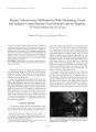
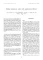
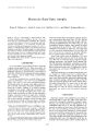
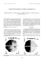
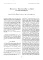
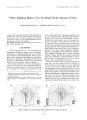
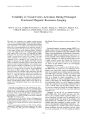
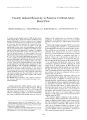
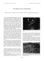
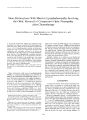
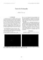
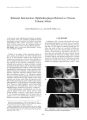
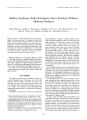
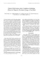
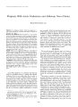
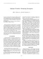
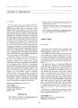
Show Journal of Neum- Opluhulmology 18( 4): 284- 288, 1998. © 1998 Lippincolt Williams & Wilkins, Philadelphia Patient With Kearns- Sayre Syndrome Exhibiting Abnormal Magnetic Resonance Image of the Brain Yuko Kamata, M. D., Yukihiko Mashima, M. D., Masako Yokoyama, M. D., Kortaro Tanaka, M. D., Yu- ichi Goto, M. D., and Yoshihisa Oguchi, M. D. A 33- year- old Japanese man had Kearns- Sayre syndrome ( KSS), which consists of the triad of external ophthalmoplegia, heart block, and " salt- and- pepper" retinopathy. The other systemic manifestations included sensorineural hearing loss, slight generalized muscle weakness, cerebellar ataxia, and elevated levels of cerebrospinal fluid protein. He exhibited a heteroplas-mic mitochondrial DNA deletion of approximately 9 kb between the cytochrome c oxidase subunit 1 and cytochrome b genes. In the authors' experience, this deletion is one of the longest to be observed in such patients. His fundi were characterized bilaterally by white flecks in the inner layers of retina at the midperiphery. Visual evoked potentials showed delayed latency in the PI00 component. The tibial somatosensory evoked potential revealed a marked prolongation of interpeak latency between the N20 and P40 components. Brain magnetic resonance images revealed high- intensity foci in several regions on T2- weighted images. Electrophysiological and magnetic resonance imaging findings suggested an involvement of the white matter of the central nervous system in this patient that was not reflected in the clinical findings. Key words: Brain magnetic resonance imaging- Kearns- Sayre syndrome- Flecked retina- mtDNA deletion- Somatosensory evoked potential- Visual evoked potential. Kearns- Sayre syndrome ( KSS) is a progressive, raul-tisystemic disorder that is characterized by the triad of progressive external ophthalmoplegia, atypical pigmentary degeneration of the retina, and cardiac conduction defects ( 1- 3). More recently, patients with KSS have been reported to show such other clinical manifestations as mental retardation, cerebellar dysfunction, sensorineural hearing loss, concentration of protein higher than 100 mg/ dl in cerebrospinal fluid ( CSF), various endocrine system dysfunctions, and elevated serum or CSF levels of lactate and pyruvate ( 3- 5). This syndrome is a spo- Manuscript received November 17, 1996; accepted June 12, 1998. From the Department of Ophthalmology ( Y. K., Y. M., Y. O.), Department of Neurology ( M. Y., K. T.), Keio University School of Medicine, and Department of Ultrastructural Research, National Institute of Neuroscience, National Center of Neurology and Psychiatry ( Y. G.), Tokyo, Japan. Address correspondence and reprint requests to Yukihiko Mashima, IVf D., Department of Ophthalmology, Keio University School of Medicine, 35 Shinanomachi, Shinjuku- ku, Tokyo 160, Japan. radic condition, with onset before 15 to 20 years of age. Because mitochondrial abnormalities such as ragged red fibers ( 6) and deficiencies in respiratory chain enzymes have been detected in examination of muscle biopsy specimens, it is considered to be a mitochondrial disease ( 7,8). Patients with KSS typically show large- scale het-eroplasmic deletions of mitochondrial DNA ( mtDNA) ( 4,9,10). We report the clinical features of a Japanese man with KSS who exhibited a deletion of 8957 base pairs ( bp) spanning between the cytochrome c oxidase subunit 1 ( CO!) and cytochrome b ( Cyt b) genes, and sparing two replication origins. The size of the deletion was the largest found in 65 Japanese patients with chronic progressive external ophthalmoplegia ( CPEO) or KSS ( 4 and Goto et al., 1995, unpublished data). CASE REPORT After a traffic accident, a 33- year- old Japanese man was transferred to the Keio University Hospital on August 12, 1993. Routine radiographs of the chest and skull revealed no abnormalities, but an electrocardiogram showed complete right bundle branch block and left anterior hemiblock. The patient also evidenced external ophthalmoplegia, ptosis, and hearing loss, all of which were unrelated to the accident. His medical history showed development of bilateral blepharoptosis, abnormal eye movements, and hearing loss at age 10. An operation to correct bilateral ptosis had been performed when he was 11. There was no family history of metabolic, neurologic, or neuromuscular disorders. This unusual patient was therefore admitted to our hospital for detailed evaluation. Ophthalmologic Findings Best- corrected visual acuity was 20/ 25 in the right eye and 20/ 60 in the left eye, and refraction was + 0.5 - 2.0 x 180 in the right eye and + 1.0 - 7.0 x 180 in the left eye; visual acuity was 20/ 40 with correction by contact lens in the left eye. The pupils reacted promptly to light. Bilateral blepharoptosis and incomplete external ophthalmoplegia were present. Intraocular pressure was 12 mm Hg in each eye. The anterior segment and optic 284 KEARNS- SAYRE SYNDROME AND ABNORMAL MR1 285 FIG. 1. A: Photograph of fundus in the right eye shows salt- and-pepper retinopathy with glistening whitish deposits in the superficial and deep intraretinal layers of midperipheral retina with a circinate pattern. B: Fluorescein angiogram in the right eye shows a salt- and- pepper pattern of irregular atrophy of the retinal pigment epithelium with diffuse hyperfluorescence and hypofluo-rescence. media were normal in each eye, but the presence of marked corneal astigmatism ( approximately 7 D) caused anisometropia amblyopia in the left eye. Examination by specular microscopy showed enlarged endothelial cells with polymegethism and pleomorphism in both eyes. Ophthalmoscopic examination revealed bilateral co-nuslike atrophy around the optic discs and atypical pigmentary retinopathy consisting of mottled pigment epithelium, so- called salt- and- pepper retinopathy ( l l ) ( Fig. lA). Optic discs were normal in color. There was no bone spicule pigmentation, as is typically seen in retinitis pigmentosa. Tiny glistening deposits were observed in the superficial and deep intraretinal layers of the midperipheral retina, mainly in the temporal retina, with a circinate pattern present in both eyes. A fluorescein angiogram showed a hypofluorescent ring around the optic disc and a salt- and- pepper pattern of irregular atrophy of the retinal pigment epithelium with diffuse hyperfluorescence and hypofluorescence ( Fig. IB). The white flecks corresponded to mottled hyperfluorescence without dye leakage or nonfluorescence. A 20- J single- flash elec-troretinogram ( ERG) performed after 30 minutes of dark adaptation showed a slight reduction in b- wave amplitude, with attenuated oscillatory potentials. The amplitude of a 30- Hz flicker ERG was slightly reduced in both eyes ( Fig. 2). Visual evoked potentials ( VEPs) to pattern and flash stimulation showed delayed latency ( Fig. 3). Neurologic Evaluation Physical examination showed that the patient was 160 cm tall and weighed 50 kg. He exhibited neurogenic hearing loss, slight generalized muscle weakness, and cerebellar ataxia. There was no evidence of mental retardation or of abnormal reflexes, and muscle tone was normal. Results of laboratory studies showed a serum creatine kinase level of 70 lU/ l ( normal, 67- 210 1U/ 1); aldolase, 3 IU/ 1 ( normal, 2- 5 IU/ 1); lactate, 9.2 mg/ dl ( normal, 3.3- 14.3 mg/ dl); and pyruvate, 0.85 mg/ dl ( normal, 0.3- 0.94 mg/ dl). An echocardiogram showed no abnormalities. An electroencephalogram showed slowing of background activity without paroxysmal discharge. An audiogram revealed bilateral sensorineural hearing loss. The results of needle electromyographic and peripheral nerve conduction studies were within normal limits. Short somatosensory evoked potentials ( SSEP) after stimulation of the tibial nerve showed normal latency of the N20 component but delayed latency of the P40 component ( Fig. 4). Interpeak latency between the P40 and N20 components was markedly prolonged, 31.0 msec in stimulation of the the right limb and 31.4 msec in the left limb ( normal, 12- 22 msec in 30s). A lumbar puncture yielded xanthochromic CSF that showed concentrations of 132 mg/ dl protein ( normal, 15- 45 mg/ dl), 11 mg/ dl lactate, 0.65 mg/ dl pyruvate, and 14.3 mg/ dl Ig ( immunoglobulin) G ( normal, < 5 mg/ dl). A computed tomographic scan of the brain showed focal low- density lesions in the white matter and slight atrophy. A T2- weighted magnetic resonance image ( MRI) of the brain showed symmetrical high- intensity lesions in the white matter, globus pallidus, internal capsule, midbrain, and left cerebellar hemisphere ( Fig. 5). on 49! 8ms 41'. 4ms FIG. 2. A 20- J single- flash electroretinogram ( A) shows a slight reduction in b- wave amplitude. Oscillatory potentials ( B) are slightly attenuated. The amplitude of a 30- Hz flicker electroretinogram ( C) is slightly reduced. ./ Neuw- Ophllmlmol, Vol. IX No. 4, IWX 2H6 Y. KAM ATA ET AL. There was no gadolinium enhancement in these lesions. These lesions were consistently observed in the brain MR1 during a 3- year period of follow- up. Muscle Pathology and mtDNA Analysis A biopsy was performed in the right brachial biceps muscle, and cryostat sections were stained with hematoxylin and eosin, Gomori's trichrome stain, and a set of oxidative enzymes, including succinate dehydrogenase and cytochrome c oxidase. With Gomori's trichrome staining, approximately 1% of fibers showed an intense red band in the perimysium that was consistent with ragged red fibers. Cytochrome c oxidase activity was deficient in the ragged red fibers and the normal-appearing fibers on Gomori's trichrome stain, indicating a focal deficiency of cytochrome c oxidase. MtDNA was extracted from the muscle specimen ( 4), and the deletion junction was localized, as previously described ( 12). Sequencing analysis revealed 8- bp direct repeats of TACTTCTC located in the boundaries of the deletion between the CO I and Cyt b genes ( Fig. 6A). The deletion segment spanned 8,957 bp from nucleotide position 6,484 to 15,440. Evidence of heteroplasmy was found, and the mutant constituted of 25% of total mtDNA ( Fig. 6B). DISCUSSION MtDNA rearrangements are known to cause three phe-notypes of mitochondrial disease: diabetes and deafness, ocular myopathy, and Pearson's syndrome ( 13- 15). Patients with diabetes and deafness often show maternal inheritance of the disorder. However, patients with ocular myopathy or Pearson's syndrome are generally isolated cases that result from new mutations. Among the A R L Lneck size •<•:...•, 150 B R L I 37-, t; FIG. 3. Visual evoked potential to pattern ( A) and flash ( B) stimulation reveals a delay in latency time of P100. The range of normal latency time ( mean ± 2 SD) in 30s is shown. FIG. 4. Tibial short somatosensory evoked potentials in left limb stimulation reveals a normal latency of the N20 component, but delayed latency of the P40 component. Interpeak latency between P40 and N20 is markedly prolonged. mtDNA rearrangement syndromes, CPEO is a milder form, whereas KSS is a severe form that involves multisystem failure. Our patient had a heteroplasmic mitochondrial DNA deletion of approximately 9 kb between the COl and Cyt b genes. There is reportedly no association between the size, site, or populations of deleted mtDNA and respiratory chain enzyme activities in muscles from patients with CPEO or KSS ( 4). In addition to the clinical triad of KSS, our patient exhibited salt- and- pepper retinopathy with white flecks and lesions in the white matter of the brain that were confirmed by a marked delay in SSEPs and VEPs, and high- intensity areas observed on a T2- weighted brain MRI. A flecked retina is a rare manifestation of KSS. White spots or flecks in the retina have been described in only a few patients with CPEO or KSS ( 16- 18). Retinal flecks may occur in association with systemic diseases such as primary hyperoxaluria ( 19), cystinosis ( 20), Sjogren- Larsson syndrome ( 21) and ring 17 chromosome ( 22). Of 61 patients with mitochondrial myopathy, 22 showed pigmentary retinopathy ( 23). Most of the patients had a salt- and- pepper type of retina, with none showing a flecked retina. Fluorescein angiograms and ERGs in our patient revealed a more marked degeneration of the retinal pigment epithelium than of the outer retina. Oscillatory potentials, which are generated more proximally than the a- and b- waves, probably by bipolar cells ( 24), also showed a deterioration. Electroretino-gram findings resembled those in the early stage of diabetic retinopathy ( 25), suggesting the presence of hypoxia of the inner retina. MtDNA defects may involve mainly the neuronal retinal cells in the inner retina and retinal pigment epithelium. However, the association between the white flecks in the retina and the mtDNA deletion in this patient is unclear. Abnormal VEPs and SSEPs have been reported in patients with mitochondrial myopathy, CPEO, or KSS ( 26- 30). Simultaneous recordings of the pattern ERG and the pattern VEPs in patients with mitochondrial myopathy have suggested that central nervous system involvement is predominantly axonal ( 29). Our patient with KSS showed delayed latency in both the flash and ./ Natro- Ophlhainml, Vol. 18, No. 4. 1998 KEARNS- SAYRE SYNDROME AND ABNORMAL MRI 287 FIG. 5. Brain T2- weighted magnetic resonance images. A: symmetrical high- intensity lesions in the white matter ( black arrow); B: symmetrical high- intensity lesions in the internal capsule ( white arrow); C: symmetrical high intensity lesions in the midbrain ( blackarrow); D: high- intensity lesion in the left cerebellar hemisphere ( black arrow). pattern VEPs, which may suggest the presence of lesions of the visual pathway. The patient also showed prolonged N20 to P40 interpeak times in a tibial SSEP. Previous studies of prolonged N20 to P40 conduction indicate that central somatosensory pathways are involved between the lumbar posterior column and the cerebral cortex ( 30). The finding of a normal N20 conduction in the present case indicates that the peripheral somatosensory nerves are not involved. Magnetic resonance imaging may not have been feasible in most of the other studies in patients with KSS, because they may have had metallic pacemakers. Some reports of KSS patients have described areas of high intensity in the white matter of the cerebrum, cerebellum, and brainstem on T2- weighted sequences ( 31- 34). These lesions are consistent with the clinical findings of respiratory arrest, ataxia, and pendular nystagmus associated with KSS ( 14). However, it is not known why the patients with large mtDNA deletions would show lesions of the white matter on MRI. The MRI of the brain in the J Nniro- Ophlhulnwl, Vol. IK No. 4. I99H 288 Y. KAMATA ET AL. FIG. 6. A: Schematic representation of the deleted region of the patient's mitochondrial DNA ( mtDNA). Intact region is shown as a solid area. Deleted fragment is shown as a dotted area. B: Southern blot analysis of mtDNA. Lane 1, noncut mtDNA. Lane 2, PvuU digestion of mtDNA showed a normal 16.6- kb band and a deleted 7.6- kb band. Recognition site of PvuU exists in the non-deleted lesion. Deleted mtDNA constituted 25 percent of total mtDNA. Lane 3, SnaB\ digestion of mtDNA showed a normal 16.6- kb band. Recognition site of SnaB\ exists in the deleted lesion. * Circular deleted mtDNA. present case showed symmetrical, delineated, high-intensity foci in the white matter which were consistently observed on T2- weighted images during a 3- year period of observation. Examination of the CSF did not reveal any possible causes such as demyelination or inflammation. Abnormal metabolism in the deleted mitochondria may be involved in the formation of lesions of the white matter. Findings of MRI, VEP, and SSEP in our patient suggested an involvement of the central nervous system, despite the absence of relevant clinical signs. REFERENCES 1. Kearns TP, Sayre CP. Retinitis pigmentosa, external ophthalmoplegia and complete heart block. Arch Ophthalmol 1958; 60: 280- 9. 2. Kearns TP. External ophthalmoplegia, pigmentary degeneration of the retina, and cardiomyopathy: a newly recognized syndrome. Trans Am Ophthalmol Soc 1965; 63: 559- 625. 3. Bcrcnberg RA, Pellock JM, DiMauro S, ct al. Lumping or splitting? " Ophthalmoplcgia- plus" or Kearns- Sayre syndrome? Ann Neurol 1977; 1: 37- 54. 4. Goto Y, Koga Y, Horai S, Nonaka I. Chronic progressive external ophthalmoplegia: a correlative study of mitochondrial DNA deletions and their phenotypic expression in muscle biopsies. J Neurol Sci 1990; 100: 63- 9. 5. Horwitz SJ, Roessmann U. Kearns- Sayre syndrome with hypoparathyroidism. Ann Neurol 1978; 3: 513- 8. 6. Olson W, Engel WK, Walsh GO, Einaugler R. Oculocraniosomatic neuromuscular disease with " ragged- red" fibers: histochemical and ultrastructural changes in limb muscles of a group of patients with idiopathic progressive external ophthalmoplegia. Arch Neurol 1972; 26: 193- 211. 7. Morgan- Hughes JA, Darveniza P, Landon DN, et al. A mitochondrial myopathy with a deficiency of respiratory chain NADH- CoQ reductase activity. ,/ Neurol Sci 1979; 43: 27- 46. 8. DiMauro S, Bonilla E, Zeviani M, et al. Mitochondrial myopathics. Ann Neurol 1985; 17: 521- 38. 9. Zeviani M, Monies CT, DiMauro S, ct al. Deletions of mitochondrial DNA in Kearns- Sayre syndrome. Neurology I988; 38: 1339- 46. 10. Monies CT, DiMauro S, Zeviani M, ct al. Mitochondrial DNA deletions in progressive external ophthalmoplegia and Kearns- Sayre syndrome. N Engl J Med 1989; 320: 1293- 9. 11. Eagle RC, Hedges TR, Yanoff M. The atypical pigmentary retinopathy of Kearns- Sayre syndrome. A light and electron microscopic study. Ophthalmology 1982; 89: 1433- 40. 12. Goto Y, Nishino I, Horai S, Nonaka I. Deletion of DNA fragments encompassing the deletion junction of mitochondrial genome. Bio-chem Biophys Res Commun 1996; 222: 215- 9. 13. Wallace DC. Mitochondrial DNA mutations in diseases of energy metabolism. .1 Bioenerg Biomembr 1994; 26: 241- 50. 14. Johns DR. Mitochondrial DNA and disease. N Engl .1 Med 1995; 333: 638- 44. 15. Rotig A, Cormier V, Blanche S, et al. Pearson's marrow- pancreas syndrome. A multisystem mitochondrial disorder in infancy. J Clin Invest 1990; 86: 1601- 8. 16. Koerner F, Schlote W. Chronic progressive external ophthalmoplegia. Association with retinal pigmentary changes and evidence in favor of ocular myopathy. Arch Ophthalmol 1972; 88: 155- 66. 17. Bastiaensen LAK. Pigment changes of the retina in chronic progressive external ophthalmoplegia ( CPEO). Acta Ophthalmol 1978;( Suppl) 138: 5- 36. 18. Tsuyama Y, Adachi- Usami E, Takeda N. A case of Kearns- Shy syndrome with later onset. Ophthalmologica 1993; 206: 149- 51. 19. Small KW, Letson R, Scheinman J. Ocular findings in primary hyperoxaluria. Arch Ophthalmol 1990; 108: 89- 93. 20. Kaiser- Kupfer MI, Caruso RC, Minkler DS, Gahl WA. Long- term ocular manifestations in nephropathic cystinosis. Arch Ophthalmol 1986; 104: 706- 11. 21. Gilbert WR, Smith JL, Nyhan WL. The Sjogren- Larsson syndrome. Arch Ophthalmol 1969; 80: 308- 16. 22. Gass JDM, Taney BS. Flecked retina associated with cafe au lait spots, microcephaly, epilepsy, short stature, and ring 17 chromosome. Arch Ophthalmol 1994;! 12: 738- 9. 23. Mullie MA, Harding AE, Petty RKH, Ikeda H, Morgan- Hughes JA, Sanders MD. The retinal manifestations of mitochondrial myopathy. A study of 22 cases. Arch Ophthalmol 1985; 103: 1825- 30. 24. Heynen H, Wachtmcistcr L, van Norren D. Origin of the oscillatory potentials in the primate retina. Vision Res 1985; 25: 1365- 73. 25. Bresnick GH. Electroretinography and color vision in diabetes mellitus. In: Crcpaldi G, Cunha- Vas JG, Fedele D, et al, eds. Microvascular and neurological complications of diabetes. Pa-dova: Livina Press, 1987: 17- 33. 26. Berdjis H, Heider W, Demisch K. ERG and VECP in chronic progressive external ophthalmoplegia ( CPEO). Doc Ophthalmol 1985; 60: 427- 34. 27. Ogasawara S, Nishikawa Y, Yorifuji S, et al. Treatment of Kearns- Sayre syndrome with coenzyme QH). Neurology 1986; 36: 45- 53. 28. Vcrsino M, Piccolo G, Callieco R, et al. Multimodal evoked potentials in progressive external ophthalmoplegia with mitochondrial myopathy. Acta Neurol Scand 1991; 84: 107- 10. 29. Sartucci F, Rossi B, Tognori G, Siciliano G, Guerrini V, Murri L. Evoked potentials in the evaluation of patients with mitochondrial myopathy. Eur Neurol 1993; 33: 428- 35. 30. Schubert M, Zierz S, Dengler R. Central and peripheral nervous system conduction in mitochondrial myopathy with chronic progressive external ophthalmoplegia. Electroencephalogr Clin Neu-rophysiot 1994; 90: 304- 12. 31. Elsas T, Rinck PA, Isaksen C, Nilscn G, Schjctnc OB. Cerebral nuclear magnetic resonance ( MRI) in Kearns syndrome. Acta Ophthalmol 1988; 66: 469- 73. 32. Dcmange P, Gia HP, Kalifa G, Sellier N. MR of Kearns- Sayre syndrome. AJNR Am J Neuroradiol 1989; 10( Suppl): S91. 33. Nakagawa E, Hirano S, Yamauchi H, Goto Y, Nonaka I, Takashima S. Progressive brainstem and white matter lesions in Kcarns- Sayrc syndrome: a case report. Brain Dev 1994; 16: 416- 8. 34. Crisi G, Ferrari G, Merelli E, Cocconcelli P. MRI in a case of Kearns- Sayre syndrome confirmed by molecular analysis. Neuroradiology 1994; 36: 37- 8. ./ Neiim- Oplulmhiwl, Vol. IS, No. 4. 199S |