| OCR Text |
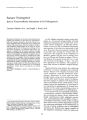
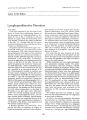
Show Journal of Clinical Neuro-ophthalmology 8(3): 195-201, 1988. © 1988 Raven Press, Ltd., New York Acute Posterior Multifocal Placoid Pigment Epitheliopathy Associated With Cerebral Vasculitis Joel M. Weinstein, M.D., George H. Bresnick, M.D., Carolyn L. Bell, M.D., Robert A. Roschrnann, M.D., Benjamin R. Brooks, M.D., and Charles M. Strother, M.D. Acute multifocal posterior placoid pigment epitheliopathy (APMPPE) is an unusual self-limited retinal disorder that has been associated with various systemic complications. To our knowledge, three prior cases associated with cerebral vasculitis have been described. This article describes a patient with APMPPE and angiographically documented cerebral vasculitis who was notable because of (a) the presence of two different cerebral ischemic events, occurring 1 month apart, and (b) the long latency (3 months) between the onset of ocular symptoms and the second cerebral ischemic event. Recognition of the association between APMPPE and cerebral vasculitis may permit early treatment of eNS involvement and prevention of morbidity. Key Words: Vasculitis-Hemianopia-Pigment epitheliopathy. From the Departments of Ophthalmology (J.M.W., G.H.B.) and Neurology (J.M.W., B.R.B., CM.S.) and Division of Neurosurgery, Department of Surgery (J.M. W.), Division of Rheumatology, Department of Internal Medicine (CL.B., R.A.R.), Department of Radiology (CM.S.), University of Wisconsin School of Medicine, Madison, Wisconsin. Address correspondence and reprint requests to Dr. J. M. Weinstein, Department of Ophthalmology, Clinical Science Center, 600 Highland Avenue, Madison, WI 53792, U.S.A. Presented at the Scientific Meeting of the American Rheumatism Association, June 13, 1987, Washington, DC. U.s.A. 195 Acute posterior multifocal placoid pigment epitheliopathy (APMPPE) was first described by Gass in 1968 (1). Although this disease was initially felt to be a primary disorder of the retinal pigment epithelium, an accumulating body of evidence strongly suggests that the primary lesion is in the small choroidal arterioles and that secondary ischemic changes produce disruption of the pigment epithelium, resulting in typical placoid lesions (2,3). Cerebral vasculitis may accompany APMPPE and has been doumented angiographically in three patients (3-5). CSF pleocytosis has been noted in at least six previous reports (3-8). We report the fourth documented case of CNS vasculitis in association with APMPPE. It is the first case in which two separate ischemic events, separated by a 2-month interval, occurred. Our patient is also unusual in that his second cerebral ischemic event occurred a full 3 months after the onset of his ocular symptoms, suggesting that patients with APMPPE may be at risk for cerebral vasculitis even though their ocular process has begun to heal. CASE REPORT A 23-year-old man was referred for evaluation of a left homonymous hemianopia. He was well until late March 1986 when he experienced fever, headache, cough, and mild myalgias. At that time he noted blurring of central vision in both eyes. He was seen by an ophthalmologist who made the diagnosis of APMPPE on the basis of typical fundus lesions (Figs. 1 and 2) and the characteristic fluorescein angiographic appearance (Fig. 3). No treatment was given. Eight weeks later, the patient's vision had begun to improve, but he noted 196 J. M. WEINSTEIN ET AL. persistent numbness of the right leg, clumsiness of the right hand including difficulty with writing, difficulty hearing in both ears, mild slurring of speech, and impairment of recent memory. He did not seek medical attention for these symptoms. One month later, approximately 12 weeks after the initial onset of visual symptoms, he had a severe headache that lasted all night. When he awoke, he noted left-sided visual loss in both eyes. Past medical history was unremarkable, and a review of systems revealed no other significant complaints. Family history was negative for neurologic, ocular, and rheumatologic disease. On examination, visual acuity was correctable to 20/25 OD and 20/20 OS. Pupillary reactions were normal. There was no afferent pupil defect. Visual field examination on the Goldmann perimeter demonstrated a congruous left homonymous hemianopia (Fig. 4). Ocular motility examination, slit-lamp examination, and intraocular pressure were normal. Fundus examination was consistent with healing APMPPE. The posterior pole demon-strated multiple hypopigmented irregularly shaped flat homogeneous lesions with indistinct borders. Many of the lesions contained pigment at their borders. The patient was admitted to the hospital for further evaluation of his homonymous hemianopia. General physical examination of the heart, lungs, cardiovascular system, abdomen, integument, and musculoskeletal system was within normal limits. Examination of cranial nerves II-XII revealed no further abnormalities. Motor examination revealed normal strength in all extremities but accentuated tone in the right arm and leg. Sensory examination and coordination were normal. Deep tendon reflexes were normal, and plantar reflexes were clearly downgoing. A brief, mental status examination yielded a score of 25 out of a possible 30, but the patient missed all serial sevens and could not perform a three-step command except by continuously repeating the three commands out loud. Reading, writing, and constructional praxis were grossly normal despite the left homonymous hemianopia. Formal neuropsychological FIG. 1. Fundus photograph, right eye. There are multiple, soft, yellow-white opacities (long arrows) at the level of the retinal pigment e.plthelium dlstnbuted around the central fovea throughout the posterior pol S I I . -' . ""'. ~"rl\/ pigmentary changes as a s'ign of.healing (short curved arrow). e. evera eSlons EPITHELIOPATHY AND CEREBRAL VASCULITIS 197 FIG. 2. Fundus photograph, left eye. Multiple yellow, white, and pigmented lesions (arrows) at the level of the retinal pigment epithelium are seen in the papillomacular region and nasal to the disc. testing demonstrated borderline impairment with respect to recent memory function and significant impairment in verbal learning ability. In addition, there were significant problems with attention and concentration despite good effort. Computerized tomography with and without infusion demonstrated two separate lesions. An area of abnormality in the right occipital lobe was consistent with a recent infarction. A second area of abnormality was present in the left basal ganglia. This involved the caudate nucleus and anterior limb of the internal capsule and presumably was the cause of the symptoms of righthanded clumsiness 5 weeks earlier. Magnetic resonance imaging of the occipital lesion was consistent with hemorrhagic infarction (Fig. 5). Cerebral angiography revealed multiple areas of abnormality in the arteries of both cerebral hemispheres (Fig. 6). These were characterized by variation in arterial caliber typical of arteritis. Also present were changes indicating revascularization following arterial obstruction. Cerebrospinal fluid examination demonstrated clear colorless fluid under normal pressure. Protein was 33 mg/dl and glucose was 56 mg/dl. Nineteen white blood cells and eight red blood cells per microliter were present. The white blood cells were 96% lymphocytes. Stains and subsequent cultures of the cerebrospinal fluid were negative for bacteria, fungus, acid-fast bacilli. VORL was negative, and oligoclonal bands were not found. The CSF IgG index was 0.50 (normal, 0.3-0.7). Hematocrit was 47 mlldl and white blood cell count was 19,500/f,d (72% segs, 10% bands, 14% lymphocytes, and 4% monocytes). The following were negative or within normal limits: Wintrobe sedimentation rate, platelet count, serum electrolytes, blood urea nitrogen, serum creatinine and creatinine clearance, prothrombin time, partial thromboplastin time, platelet count, calcium, phosphorus, uric acid, cholesterol, serum albumin and globulin liver enzymes, serum complement (C3 and C4), FTA-ABS, anti-nuclear antibody, antiparietal antibody, anti mitchondrial antibody, anti-smooth-muscle antibody, mononucleosis spot test, and urinalysis. / Clill NeLlrv-0l'htha[IIIVI, Vol. 8. No.3. 1988 198 f. M. WEINSTEIN ET AL. FIG. 3. A: Fluorescein angiogram, right eye. There are multiple hyperfluorescent and hypofluorescent spots scattered throughout the posterior pole (arteriovenous phase). B: Fluorescein angiogram, right eye. Most of the lesions show hyperfluorescence confined to the lesions, indicative of window defects in the retinal pigment epithelium. Some lesions, especially in the papillomacular area, show staining with dye, as does the peripapillary retina, particularly on the nasal side of the disc (late phase). "'( , ! 1";"_-"1 l'O ::::::r_ ,::: :1: , ", ';:: :1: ,: \ ' \ '\ T'i' ........,......,... .,.~,'l:r-t/:. t\,.. "}I~"~··l?~~ .,~.... .~::::=- "-:.::;.~ Q ...... ~ 01 00 I I I I r_ . .. • I • ~ . "~ :f~~_"I'i~ a: ~. . FIG 4. Left homonymous hemianopia demonstrated by Goldmann perimetry. J Clin Neuro-ophthalmol, Vol. 8, No.3, 1988 EPITHELIOPATHY AND CEREBRAL VASCULITIS 199 Serologic studies were performed 5 weeks after the initial onset of neurologic symptoms. Serum antibody titers by complement fixation (LBCFso method) were not significantly elevated for influenza A or B, parainfluenza 1, 2, or 3, adenovirus, respiratory syncytial virus, or cytomegalovirus (complement fixation and latex agglutination methods). Acute and convalescent sera for immunofluorescent antibody showed no evidence of Lyme disease. Cerebrospinal fluid cultures on several human cell lines (human rhabdomyosarcoma, fetal lung, and laryngeal cancer) failed to grow cytomegalovirus, herpes virus, or adenovirus. The patient was given i.v. methylprednisolone, 1 g daily for 3 days and was discharged on prednisone, 80 mg daily. The dose was gradually tapered over the next 4 months. There were no further neurologic sequelae. Ten months after the onset of neurologic symptoms, the patient's visual acuity, hemianopia, and mild memory deficit persisted unchanged. DISCUSSION The acute onset of blurred vision in a young adult accompanied by typical cream-colored, irregularly- shaped placoid lesions in the posterior pole is characteristic of APMPPE (1,6). The lesions become pigmented at an early stage. Within 2-3 weeks after onset, lesions at various stages of development may be seen. The typical fluorescein angiographic findings of discrete areas of early hypofluorescence with later hyperfluorescence further supports the diagnosis (1,6). Ocular manifestations of APMPPE may also include papillitis (2,9-12), iridocyclitis (3,9), peripheral corneal thinning (12), retinal vasculitis (13), and serous retinal detachment (13). CNS manifestations include severe headache (1,4,7-9), multiple episodes of transient cerebral ischemia producing extremity weakness, expressive aphasia and right hemiparesis (3), permanent homonymous hemianopia due to cerebral ischemia (4,5), and cerebrospinal fluid pleocytosis (3-8). The presence of a prodromal respiratory or gastrointestinal illness is also characteristic and was found in 10 of 13 patients with APMPPE examined by Holt et al. (3). Extensive serologic testing for viral and other infectious agents has generally been unsuccessful (3), with the exception of one case associated with adenovirus type 5 infection reported by Azar and associates (14). Histopathologic studies of the retina or other affected tissues in this disease have not, to our knowledge, been performed. Fluores- FIG. 5. Midline sagittal (A) and parasagittal (B) partial saturation magnetic resonance (TR 600, TE 30) images showing evidence of hemorrhagic infarction involving the right occipital lobe. A high signal intensity abnormality is seen along the gyri of the visual cortex. (arrows in A and arrowhead in B). cein angiographic patterns in this disorder have been interpreted by some authors as indicative of choroidal ischemia (2) and by others as representing choroidal/retinal pigment epithelial inflammation (1). The notable features of our patient's illness were (a) the long period (3 months) between onset of ocular symptoms and the second cerebrovascular event; (b) the biphasic nature of the neurologic disease with two discrete cerebrovascular events; and (c) the diffuse vasculitic involvement demon- 1(111I Neuro-ophthalmol. Vol. 8, No.3, 1988 200 J. M. WEINSTEIN ET AL. FIG. 6. A lateral projection of a right internal carotid arteriogram. There are multiple areas of alteration in vascular caliber with associated evidence of vascular recanalization, i.e., neovascularity (arrowheads). strated by angiography and computerized tomography. In prior reports of patients with cerebral vasculitis associated with APMPPE, a single neurologic event occurred between 1 (4) and 8 weeks (5) after the onset of ocular symptoms. Our patient experienced the onset of neurologic symptoms 1 month after blurred vision was noted. He was left with a persistent memory deficit and mild clumsiness of the right hand. A second stroke occurred 2 months later, fully 3 months after the onset of the illness, and resulted in persistent left homonymous hemianopia. This observation and that of Holt et a1. (3), who described a patient with cerebrospinal fluid pleocytosis for at least 2 months after the onset of ocular symptoms, suggest that patients with APMPPE may have active cerebral vasculitis for several months after the onset of ocular symptoms. It might be noted that recurrences of ocular involvement and involvement of the second eye have occasionally been noted after delays of several months or even years (15). Although originally considered to be limited to the eve, systemic involvement in patients with , ,1',:11 mented in recent reports. J Clin Neuro·ophtha'mol. Vul. 8. Nu, 3. 19;;0 In addition to CNS disease, systemic features may include a subclinical nephropathy (10), erythema nodosum (11), or thyroiditis (12). Because there have been no cases of APMPPE from which tissue has been examined pathologically, we are unable to say whether the symptoms and specific abnormalities of nodularity, narrowing of blood vessels, and neovascularity seen angiographically represent a true cerebral vasculitis or some other form of noninflammatory vasculopathy. In patients with CNS involvement due to other vasculitides, e.g., systemic lupus erythematosis, Wegener's granulomatosis, polyarteritis nodosa, and granulomatoid angiitis, CNS dysfunction is usually permanent and in some cases may be progressive, leading to death. Such vasculitides are usually treated with both corticosteroids and cyclophosphamide. In most patients with APMPPE, however, symptoms resolve spontaneously, and aggressive immunosuppressive therapy has not been necessary. The patient described by Smith et a1. (5) experienced meningeal inflammation 1 month after the onset of ocular symptoms. At that time, significant / EPITHELIOPATHY AND CEREBRAL VASCULITIS 201 pleocytosis was present, and the patient was begun on prednisone (60 mg/day orally) with prompt resolution of headache and stiff neck. Three to four weeks later, prednisone was discontinued; within 3 days, the patient experienced a permanent homonymous hemianopia. Steroids were reinstituted once again with prompt resolution of meningeal signs and symptoms suggesting that steroids may suppress disease activity. No further neurological deficit occurred. In addition to the published reports, we are aware of two other cases of APMPPE associated with cerebral vasculitis. Behrens and Miller (personal communication) treated a patient who experienced a transient cerebral ischemic episode 1 month after the onset of her retinal disorder. A 10min episode of left arm paresthesias and periorbital tingling was accompanied by right-sided headache, which had also occurred 2 days previously. There was not prior history of migraine. There was cerebrospinal fluid pleocytosis (10 lymphocytes). Computerized tomography with and without infusion, performed 7 days after the episode, was normal. Sedimentation rate was 3 mmlh. There were no permanent sequelae. C. H. Smith treated a 32-year-old man with APMPPE who developed a right homonymous hemianopia 1 month after the onset of symptoms caused by his retinopathy (Smith, personal communication). Computerized tomography and cerebral angiography confirmed the presence of occipital infarction caused by cerebral vasculitis. The patient experienced partial resolution of his field defect while on prednisone therapy over the next 4 months. Prior reports have not discussed duration of treatment for CNS inflammation in APMPPE. The observations of Smith et al. suggest that steroids are highly effective in suppressing meningeal inflammation due to APMPPE (5). In view of our observations and the review of other reported cases, we suggest that patients with CNS inflammation associated with APMPPE should receive corticosteroid treatment for a minimum of 4 months after the onset of CNS symptoms and that steroids should be tapered with careful monitoring of signs, symptoms, and cerebrospinal fluid parameters. If our patient had received a course of prednisone at the time of his initial neurologic symptoms, the second stroke, which occurred 1 month later, may have been prevented. Acknowledgment: We thank Myles M. Behrens, M.D., for reviewing the manuscript. REFERENCES 1. Gass JDM. Acute posterior multifocal placoid pigment epitheliopathy. Arch Ophtha/mo/ 1968;80:177-85. 2. Deutman AF. Choriocapillaris filling patterns in health and disease. Trans Ophtha/mo/ Soc UK 1980;100:553-8. 3. Holt WS, Regan CDJ. Trempe C. Acute posterior multifocal placoid pigment epitheliopathy. Am 1 Ophtha/mo/ 1976; 81:403-12. 4. Sigelman J. Behrens. M. Hilal S. Acute posterior multifocal placoid pigment epitheliopathy associated with cerebral vasculitis and homonymous hemianopia. Am , Ophthalmo/ 1979;88:919-24. 5. Smith CH, Savino PJ. Beck RW. Schatz NJ. Sergott RC. Acute posterior multifocal placoid pigment epitheliopathy and cerebral vasculitis. Arch Neuro/1983;40:48-50. 6. Ryan SJ. Maumenee AE. Acute posterior multifocal placoid pigment epitheliopathy. Am 1Ophtha/mo/1972;74:1066-74. 7. Bullock JD. Thomas ER, Fletechur RL. Cerebrospinal fluid abnormalities in acute posterior multifocal placoid pigment epitheliopathy. Am 1Ophtha/mo/1977;84:45-9. 8. Fishman GA. Baskin M. Jednick N. Spinal fluid pleocytosis in acute posterior placoid pigment epitheliopathy. AIlIt Ophtha/nlO/ 1977;9:33-6. 9. Savino PJ, Weinberg RJ. Yassin JG. Pilkerton AR. Diverse manifestations of acute posterior multifocal placoid pigment epitheliopathy. Am 1Ophtha/mo/1974;77:659-62. 10. Priluck lA, Robertson DM. Buettner H. Acute posterior multifocal placoid pigment epitheliopathy: urinary findings. Arch Ophtha/mol 1981;99:1560-2. 11. Van Buskirk EB. Lessell S. Friedman E. Pigment epitheliopathy and erythema nodosum. Arch Ophtha/mo/ 1971; 85:369-72. 12. Jacklin HN. Acute posterior multifocal placoid pigment epitheliopathy and thyroiditis. Arch Ophtha/mol 1977; 95:995-7. 13. Bird AC, Hamilton AM. Placoid pigment epitheliopathy presenting with bilateral serous retinal detachments. Br 1 Ophtha/lIllI/ 1972;56:881-6. 14. Azar P. Jr. Gohd RS. Waltman D. Gitter KA. Acute posterior multifocal rlacoid pigment epitheliopathy associated with an adenovirus type 5 infection. Am , Ophtha/mol 1975;80:1003-5. 15. Gass JD. Stereoscopic atlas of maCl//ar diseases. 3rd ed.: St Louis: Mosby Co. 1987:504-10. I Clin Neuro-0l'llthalmo/. Vol. 8. No.3. 1988 [VBlymedisease] |