| OCR Text |
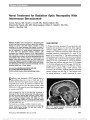
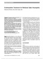
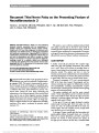
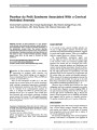
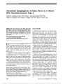
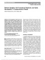
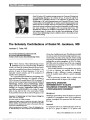
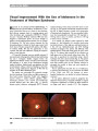
Show Erythropoietin Treatment for Methanol Optic Neuropathy Mohammad Pakravan, MD, Nasrin Sanjari, MD Background: To present the effect of erythropoietin for the treatment of methanol optic neuropathy. Methods: Two patients with methanol optic neuropathy were treated with 10,000 IU of intravenous erythropoietin twice a day for 3 days, 500 mg of methylprednisolone twice a day for 5 days (followed by 2 weeks of oral prednisolone [1 mg/kg per day]), and daily doses of vitamin B12, vitamin B6, and folic acid for 1 month. Results: At presentation, the patients had no perception of light in both eyes, associated with mildly swollen optic discs. Both responded dramatically to the treatment regi-men. In the first patient, visual acuity improved to 20/20 in both eyes within 3 days, whereas in the second patient, visual acuity returned to counting fingers at 6 feet, right eye, and 20/30, left eye, within 3 weeks. Conclusion: Intravenous erythropoietin may be an effective adjuvant when combined with current treatment for patients with methanol optic neuropathy. Journal of Neuro-Ophthalmology 2012;32:325-328 doi: 10.1097/WNO.0b013e318262a7c2 © 2012 by North American Neuro-Ophthalmology Society Methyl alcohol (methanol) is a clear, colorless, and flammable liquid produced by the reaction of hydro-gen with carbon monoxide or carbon dioxide. Methanol is metabolized in the body to formaldehyde by alcohol dehydrogenase, following which formaldehyde is rapidly converted to formic acid, a metabolite that causes the majority of the toxicity associated with methanol (1). Meth-anol is highly toxic for humans if ingested. Ingestion of as little as 10 mL can result in complete and permanent visual loss from bilateral optic neuropathy, and 30 mL can be fatal, although the fatal dose is typically 100-125 mL. Tox-icity usually occurs from accidental ingestion. For example, in some countries, alcoholic beverages are legally prohibited, and homemade alcoholic drinks containing methanol are a source of toxicity (2). A variety of substances are used to treat methanol optic neuropathy. Fomepizole has been found to be safe and effective in the treatment of methanol poisoning, often resulting in both resolution of metabolic acidosis and complete restoration of vision. However, it is expensive and not readily available, particularly in developing countries (3). Ethanol, B-group vitamins, and systemic steroids often are used, but with limited success. In such cases, the final visual acuity is in the range of counting fingers or worse (4). Erythropoietin is a glycoprotein that stimulates red blood cell differentiation by preventing apoptosis of erythroid progenitors in the bone marrow. It has shown that erythropoietin also has neuroprotective and neuroregener-ative properties in the central nervous system (5), and several small case series have documented improvement in vision when used in patients with nonarteritic anterior ischemic optic neuropathy and traumatic optic neuropathy (6,7). We report 2 patients with methanol-induced toxic optic neuropathy who experienced dramatic visual improvement when they were treated with a combination of intravenous erythropoietin, systemic corticosteroids, vitamins, and folic acid. CASE REPORTS Case 1 A 30-year-old man was referred with a chief complaint of bilateral vision loss. He had no history of systemic disease and took no medications. He had drunk about 100 mL of a homemade alcoholic beverage 3 days previously. Sub-sequent analysis of the drink revealed that it contained methanol. We later found that 7 people had consumed the same drink, 3 of whom experienced vision loss, leading to complete blindness in 2, 1 of whom was our patient. A third patient died from severe metabolic acidosis despite Ophthalmic Research Center and Department of Ophthalmology (MP, NS), Labbafinejad Medical Center, Shaheed Beheshti Medical University, Tehran, Iran. Supported by the Ophthalmic Research Center, Shaheed Beheshti Medical University, Tehran, Iran. The authors report no conflicts of interest. Address correspondence to Mohammad Pakravan, MD, Ophthalmic Research Center, Department of Ophthalmology, Labbafinejad Medical Center, Shaheed Beheshti Medical University, Pasdaran Avenue, Boostan 9th Street, Tehran 1666694516, Iran; E-mail: mohpakravan@gmail.com Pakravan and Sanjari: J Neuro-Ophthalmol 2012; 32: 325-328 325 Original Contribution Copyright © North American Neuro-Ophthalmology Society. Unauthorized reproduction of this article is prohibited. treatment with hemodialysis and other supportive measures. The other 3 individuals had no visual or systemic deficits. This patient's visual acuity had begun to decrease within 24 hours after ingestion. At presentation, his vital signs were normal, and he was alert and cooperative. Visual acuity was no light perception (NLP) in both eyes. Pupils were dilated and nonreactive to light. Slit-lamp biomicroscopy revealed no abnormality, and intraocular pressures were normal. Fundus examination revealed mild hyperemic disc swelling bilaterally. Systemic and neurological assessments were unremarkable. Optical coherence tomography (OCT) showed marked thickening of peripapillary retinal nerve fiber layer (RNFL), with average thicknesses of 160 and 171 mm in the right and left eye, respectively (Fig. 1). The patient was admitted to hospital. Complete blood count, erythrocyte sedimentation rate, and C-reactive pro-tein were within normal limits as was a metabolic panel. The patient's blood methanol concentration was 5 mmol/L (normal, 5-15 mmol/L). Magnetic resonance imaging of the brain and orbits was unremarkable. The patient was begun on intravenous methylpredniso-lone (500 mg twice a day) combined with vitamin B12 (100 mg/day), vitamin B6 (100 mg/day), and folic acid (10 mg/day). Because the patient's serum methanol was within the normal range, we chose not to treat him with ethanol. After 2 days, the patient's vision was unchanged. After approval from the ethics committee of the Ophthal-mic Research Center and after obtaining written informed consent, the patient was given infusions of 10,000 IU of intravenous erythropoietin twice a day. After the first 2 infu-sions of erythropoietin, visual acuity improved to 20/200, right eye, and hand movements, left eye. Erythropoietin infusions along with methylprednisolone and vitamins were continued, and 3 days after initiation of erythropoietin, the patient's visual acuity improved to 20/20 in both eyes, pupils were reactive to light, and visual fields were per-formed (Fig. 2). We continued erythropoietin for 3 days, and methylprednisolone for 5 days, followed by oral pred-nisolone (1 mg/kg per day) for 2 weeks. Vitamins were given for 1 month. After 3 weeks, the patient's visual acuity remained 20/20 bilaterally, and the optic discs become mildly pale. OCT showed reduction in the peripapillary RNFL compared with pretreatment values, with average thicknesses of 102 and 114 mm in the right eye and left FIG. 1. Case 1: At presentation, OCT demonstrates thickening of the peripapillary RNFL bilaterally. 326 Pakravan and Sanjari: J Neuro-Ophthalmol 2012; 32: 325-328 Original Contribution Copyright © North American Neuro-Ophthalmology Society. Unauthorized reproduction of this article is prohibited. eye, respectively, and visual fields showed significant improvement. Case 2 A 35-year-old man was referred 1 week after ingestion of a homemade alcoholic beverage containing methanol. He had experienced 24 hours of unconsciousness and severe acid-base imbalance and had been treated with systemic steroids, intravenous ethanol, hemodialysis, and supportive care. Despite improvement in his general condition, he was noted to have no perception of light in either eye. When he arrived at our hospital, his metabolic panel and blood meth-anol level were within normal limits. Visual acuity was NLP in both eyes, with moderately dilated pupils that were non-reactive to light stimulation. Slit-lamp biomicroscopy and intraocular pressures were normal, whereas fundus exami-nation revealed mild swelling of both optic discs (Fig. 3). After obtaining informed consent, the patient was treated with 10,000 IU of intravenous erythropoietin twice a day for 3 days, 500 mg of methylprednisolone twice a day for 5 days (followed by 2 weeks of oral prednisolone [1 mg/kg per day]), vitamin B12 (100 mg/day), vitamin B6 (100 mg/day), and folic acid (10 mg/day) for 1 month. There was no change in the patient's vision over the next 5 days, but 2 weeks later, visual acuity was counting fingers at 6 feet, right eye, and 20/30, left eye. The pupils were sluggishly reactive to light, and the optic discs were pale. DISCUSSION Our 2 patients with methanol optic neuropathy responded dramatically to a combination of intravenous erythropoie-tin, methylprednisolone, vitamins, and folic acid. The effect of methanol poisoning on the optic nerve is complex. The only fundus lesion observed both ophthalmoscopically and angiographically is optic disc edema because of stasis of axoplasmic flow resulting from the inhibition of oxidative metabolism (8). This axoplasmic slowing appears to occur from swelling of the cytoplasm of the astrocytes and oligo-dendroglia in the retrolaminar space as well as from com-pressive obstruction of orthograde axoplasmic flow. The mechanism by which this swelling occurs appears to be a combination of metabolic acidosis and formic acid inhi-bition of cytochrome C oxidase, resulting in histotoxic hyp-oxia (9). In addition, methanol can cause central necrosis of the retrolaminar portion of the optic nerve, and necrosis of the basal ganglia, leading to both blindness and acute encephalopathy (10,11). Necrosis of the optic nerves may, in part, be due to alteration in blood flow (11,12). Although methanol toxicity also leads to retrobulbar demyelination, it is unclear whether this is a primary effect or secondary to axonal damage. Sharpe et al (13) found that optic nerve axons were preserved, yet documented myelin degeneration behind the lamina cribrosa and in cerebral hemispheric white matter. As in our patient, methanol optic neuropathy causes increased thickness of peripapillary RNFL in the acute phase and diffuse thinning chronically (14). Treatment strategies for methanol optic neuropathy are based on detoxification. Fomepizole, an inhibitor of alcohol dehy-drogenase, is very beneficial in treating methanol toxicity, but is not readily available, particularly in developing countries (3). Other treatment protocols, including intra-venous ethanol combined with vitamin B1, B6, and B12, have produced variable results (15). High-dose IV cortico-steroids often are used, as it is believed that this treatment may inhibit demyelination. Abrishami et al (16) adminis-tered high doses of intravenous steroids for 3 days, fol-lowed by oral prednisolone (1 mg/kg) for 11 days in 6 patients with vision ranging from 0.93 to 0.86 logarithm of the minimum angle of resolution (logMAR) (equivalent to 20/150-20/160). Three months after treatment, mean visual acuity ranged from 0.33 to 0.2 logMAR (equivalent to 20/30-20/40). In contrast, Fujihara et al (14) found FIG. 2. Case 1: Automated perimetry performed 3 days after initiation of erythropoietin therapy. FIG. 3. Case 2: One week after ingestion of methanol, both optic discs are swollen. Pakravan and Sanjari: J Neuro-Ophthalmol 2012; 32: 325-328 327 Original Contribution Copyright © North American Neuro-Ophthalmology Society. Unauthorized reproduction of this article is prohibited. that intravenous methylprednisolone given 6 days after ingestion of methanol was not effective in improving vision (14). Group B vitamins are thought to decrease the toxic metabolites of methanol, particularly in the brain (17). In one series of 15 patients with methanol optic neuropathy, the combination of steroids, vitamin B12, and folic acid resulted in visual improvement (18). The ability of erythropoietin to suppress neuronal apoptosis and decrease the inflammatory response has been demonstrated in different models of brain ischemia and inflammation (5). Erythopoietin also has been shown to have beneficial effects on retinal ganglion cells, including reduction in apoptosis and increased survival after experi-mental optic nerve transaction (19), and in the setting of experimental autoimmune encephalomyelitis, chronically elevated intraocular pressure, and diabetic retinopathy (20,21). This has led to reports of systemic and intravitreal injection of erythropoietin to treat patients with nonarteritic anterior ischemic optic neuropathy and traumatic optic neuropathy with beneficial effects (6,7) and without toxicity (6,22). Erythropoietin is readily available and inexpensive, making it a viable treatment option in developing countries. We believe that significant vision improvement of our 2 patients is attributable to the additive effect of erythropoietin when combined standard treatments. However, additional studies are needed to determine whether erythropoietin alone or in combination with other therapeutic agents provide optimal visual recovery in patients with methanol optic neuropathy. ACKNOWLEDGEMENT The authors thank Neil Miller, MD, for his kind guidance and constant supervision and support in completing this study. REFERENCES 1. Barceloux D, Bond GR, Krenzelok EP, Cooper H, Vale JA. Practice guidelines on the treatment of methanol poisoning. J Clin Toxicol. 2002;40:415-446. 2. Prabhakaran V, Ettler H, Mills A. Methanol poisoning: two cases with similar plasma methanol concentrations but different outcomes. Can Med Assoc J. 1993;148:981-984. 3. Brent J, McMartin K, Phillips S, Aaron C, Kulig K. Fomepizole for the treatment of methanol poisoning. N Engl J Med. 2001;344:424-429. 4. Sanaei-zadeh H, Zamani N, Shadnia SH. Outcomes of visual disturbances after methanol poisoning. J Clin Toxicol. 2011;49:102-107. 5. Brines ML, Chezzi P, Keenan S. Erythropoietin crosses the blood-brain barrier to protect against experimental brain injury. Proc Natl Acad Sci. 2000;97:10526-10531. 6. Modarres M, Ghasemi Falavarjani KH, Nazari H, Sanjari MS, Aghamohammadi F, Homaie M, Samiy N. Intravitreal erythropoietin injection for the treatment of non-arteritic anterior ischemic optic neuropathy. Br J Ophthalmol. 2011;95:992-995. 7. Kashkouli MB, Pakdel F, Sanjari MS, Haghighi A, Nojomi M, Homaee MH. Erythropoietin, a novel treatment for traumatic optic neuropathy. Arch Clin Exp Ophthalmol. 2011;249:731-736. 8. Hayreh MS, Hayreh SS, Baumbach G, Cancilla P, Martin- Amat G, Tephly TR, McMartin KE, Makar AB. Methyl alcohol poisoning III. Ocular toxicity. Arch Ophthalmol. 1977;95:1851-1858. 9. Jacobsen D, McMartin KE. Methanol and ethylene glycol poisonings; mechanism of toxicity, clinical course, diagnosis and treatment. Med Toxicol. 1986;1:309-334. 10. Naeser P. Optic nerve involvement in a case of methanol poisoning. Br J Ophthalmol. 1988;72,778-781. 11. Önder F, _Ilker S, Kansu T, Tatar T, Kural G. Acute blindness and putaminal necrosis in methanol intoxication. J Int Ophthalmol. 1999;22:81-84. 12. Sharma M, Volpe NJ, Dreyer EB. Methanol-induced optic nerve cupping. Arch Ophthalmol. 1999;117:286. 13. Sharpe JA, Hostovsky M, Bilbao JM, Rewcastle NB. Methanol optic neuropathy: a histopathological study. Neurology. 1982;32:1093-1100. 14. Fujihara M, Kikuchi M, Kurimot Y. Methanol-induced retinal toxicity patient examined by optical coherence tomography. Jpn J Ophthalmol. 2006;50:239-241. 15. Buzn˘a E, Cernea D. The therapeutic approach in optic neuropathy due to methyl alcohol. Oftalmologia. 1991;35:39-42. 16. Abrishami M, Khalifeh M, Shoayb M. Therapeutic effects of high-dose intravenous prednisolone in methanol-induced toxic optic neuropathy. J Ocul Pharmacol Ther. 2011;3:261-263. 17. Hassanian-Moghadam H, Pajoumand A, Dadgar SM, Shadnia SH. Prognostic factors in methanol poisoning. Hum Exp Toxicol. 2007;26:583-586. 18. Azarmina M, Abrishami M, Ahmadieh H, Soheilian M, Dehghan MH, Mashayekhi A. The results of therapeutic and visual acuity state in patients with optic disc edema due to methanol toxicity. J Ophthalmol Bina. 2000;4:135-139. 19. Weishaupt JH, Rohde GP, Ölking E, Siren AL, Ehrenreich H, Bähr LD. Effect of erythropoietin axotomy-induced apoptosis in rat retinal ganglion cells. Invest Ophthalmol Vis Sci. 2004;45:1514-1522. 20. King CE, Rodger J, Batlet C, Esmaieli T, Dunlop SA, Beazley LD. Is both neuroprotective and neurogenerative following optic nerve transaction? Exp Neurol. 2007;205:48-55. 21. Biotang, Inc. http://www.biotangusa.com/bt/recombinant-humanerythropoietin- alpha-fc-chimera-10ug.html. Accessed February 7, 2012. 22. Lagreze WA, Feltgen N, Bach M, Jehle T. Feasibility of intravitreal erythropoietin injection in humans. Br J Ophthalmol. 2009;93:1667-1671. 328 Pakravan and Sanjari: J Neuro-Ophthalmol 2012; 32: 325-328 Original Contribution Copyright © North American Neuro-Ophthalmology Society. Unauthorized reproduction of this article is prohibited. |