OCR Text |
Show Clinical Neuro-ophthalmic Findings in Familial Dysautonomia Carlos E. Mendoza-Santiesteban, MD, Thomas R. Hedges III, MD, Lucy Norcliffe-Kaufmann, PhD, Floyd Warren, MD, Shantan Reddy, MD, Felicia B. Axelrod, MD, Horacio Kaufmann, MD Background: 
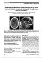
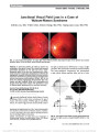
To define the clinical neuro-ophthalmic abnor-malities of patients with familial dysautonomia (FD). Methods: Sixteen patients (32 eyes) with the clinical and molecular diagnoses of FD underwent thorough neuro-ophthalmic clinical evaluation. Results: Visual acuity ranged from 0.05 to 1.0 decimal units and was reduced in 15 of 16 patients. Mild to moderate corneal opacities were found in most patients but were visually significant in only 2 eyes. Red-green color vision was impaired in almost all cases. Depression of the central visual fields was present on automated visual fields in all patients, even in those with normal visual acuity. Temporal optic nerve pallor was present in all cases and was associated with retinal nerve fiber layer loss in the papillomacular region. Various ocular motility abnormalities also were observed. Conclusion: Patients with FD have a specific type of optic neuropathy with predominant loss of papillomacular nerve fibers, a pattern similar to other hereditary optic neuropa-thies caused by mutations either in nuclear or in mitochon-drial DNA, affecting mitochondrial protein function. Defects of eye movements, particularly saccades, also appear to be a feature of patients with FD. Journal of Neuro-Ophthalmology 2012;32:23-26 doi: 10.1097/WNO.0b013e318230feab © 2012 by North American Neuro-Ophthalmology Society Familial dysautonomia (FD), also known as the Riley- Day syndrome or hereditary sensory and autonomic neuropathy type III, is an autosomal recessive disorder that impairs the development of specific sensory and autonomic neurons during embryogenesis (1-5). The most common mutation, located on the long arm of chromosome 9 (9q31) (6,7), results in a splicing abnor-mality and a deficiency of the IkB kinase complex- associated protein (IKAP or Elp1) (8-12). The splicing abnormality is tissue specific: neurons produce mostly mutant IKAP messenger RNA (mRNA) and little protein product, whereas other cells produce both normal and mutant mRNAs in different ratios (13). IKAP is widely expressed throughout the body, with its expression being highest in neural tissue and the retina (14). Patients with FD have a complex neurological pheno-type with decreased pain and temperature perception, impaired sense of taste, abnormal swallowing, gait ataxia, decreased/absent myotatic reflexes, and extremely labile blood pressure. Vision deteriorates with age in patients with FD, and by adulthood, most patients have severe visual loss (15-18). Visual impairment has dramatic con-sequences for these patients as they lack proprioceptive afferents and thus rely heavily on vision for activities of daily living. Dry eye secondary to lacrimal deficiency, myopia, corneal anesthesia, corneal abrasions, and ulcer-ations were believed to be the main cause of visual deterioration (15,17). However, based on clinical obser-vations, the degree of visual impairment cannot be explained by corneal complications alone, and visual loss occurs in patients without severe corneal damage. Early case reports describe exotropia, pupillary dysfunction, and optic nerve atrophy, but it is not known whether these neuro-ophthalmic features occur in all patients (18-24). Our aim was to define the neuro-ophthalmic phenotype of patients with FD. Dysautonomia Center (CEM-S, LN-K, FBA, HK), NYU Langone Medical Center, New York University, New York, New York; New England Eye Center (CEM-S, TRH), Tufts Medical Center, Tufts University, Boston, Massachusetts; and Department of Ophthalmol-ogy (FW, SR), NYU Langone Medical Center, New York University, New York, New York. Supported in part by the Dysautonomia Foundation, Inc, the National Institutes of Health (U54-NS065736-01), the Massachusetts Lions Clubs/Research to Prevent Blindness Challenge Grant, and the Carl Zeiss Meditec. The authors report no conflicts of interest. Address correspondence to Thomas R. Hedges, MD, New England Eye Center, Tufts Medical Center, Tufts University, 800 Washington Street, Box 450, Boston, MA 02111; E-mail: thedges@tuftsmedicalcenter.org Mendoza-Santiesteban et al: J Neuro-Ophthalmol 2012; 32: 23-26 23 Original Contribution Copyright © North American Neuro-Ophthalmology Society. Unauthorized reproduction of this article is prohibited. METHODS Subjects We evaluated 32 eyes in 16 patients with FD, 6 men and 10 women with a mean age of 26.8 years (range, 12- 61 years). All patients had typical clinical features, and the diagnosis was confirmed by genetic testing. All patients signed an informed consent and the NYU Institutional Review Board approved the study. Procedure Best-corrected visual acuity (BCVA) was assessed indepen-dently for each eye using Snellen optotypes with optimum refractive correction, and the results were presented as decimal units. Color vision was estimated using Ishihara plates (Ishihara 16 plates, Tokyo, Japan), and Hardy-Rand-Rittler color vision test (American Optics, Richmond, VA). Pupillary reflexes were assessed using a standard bright light (Halogen illuminator lamp HPX 3.5 V; Welch Allyn, Skaneateles Falls, NY). Ductions, fixation, saccades, pursuit, convergence, vestibulo-ocular reflexes, and optokinetic responses were assessed. Visual fields were obtained using a Humphrey Field Analyzer (HFA 750i; Carl Zeiss, Dublin, CA), with the 30-2 program and SITA Fast strategy using optimal refractive correction. Anterior segments were examined by slit-lamp biomicroscopy. Ocular fundus examination was performed with a binocular indirect ophthalmoscopy (Keeler Vantage, London, United Kingdom) and slit-lamp biomicroscopy (Haag Streit BQ 900; Haag Streit, Koeniz, Switzerland) to evaluate the posterior pole using a noncontact lens of +78 diopters and red-free light with particular attention of the retinal nerve fiber layer. In some patients, mydriatic color and red-free fundus photographs (TRC 50 IX; Topcon, Tokyo, Japan) were acquired. RESULTS The main neuro-ophthalmic findings are summarized in Table 1. Myopic refractive defects were present in 27 of 32 eyes. Most patients had a history of corneal lesions. We classified the corneal opacities as absent or mild (17 eyes), moderate (13 eyes), and severe (2 eyes). In none of the eyes was the opacity severe enough to preclude fundus examina-tion through indirect ophthalmoscopy or non-contact lens biomicroscopy, even in the 2 severely affected eyes. Band keratopathy was present in 1 eye. BCVA ranged from 0.05 to 1.0 decimal units. Most of the patients had acuity less than 0.5 decimal units, which did not appear to be due to corneal opacities or other surface abnormalities except in 2 eyes. A red-green color vision defect was found bilaterally in all patients examined with variable degrees of severity, and in 4 eyes of 2 patients, blue-yellow color deficiency also was detected. Central or cecocentral visual field defects were pre-dominant and found in 26 eyes. Four eyes had generalized, deep field depression, and this was associated with poor visual acuity (Table 1). Two eyes had normal visual fields. Retinal nerve fiber layer loss was detected in all eyes, accompanied by temporal pallor of the optic disc. Twenty-five eyes showed only pallor in the temporal region of the optic nerve together with a wedge-shaped loss of fibers in the papillomacular bundle (Table 1). In 7 eyes, there was also generalized optic nerve pallor, but this was always more evident in the temporal portion of the disc (Fig. 1). Retinal vascular tortuosity was present in 6 eyes. Ocular motility was impaired in various ways (Table 1). While ductions and versions were full in all patients, exo-phoria was seen in almost all cases (30 eyes). This was TABLE 1. Neuro-ophthalmic findings in patients with FD Age, y No. of Patients 0-20 7 20-40 6 40 3 Total 16 Corneal opacities No. of Eyes Absent or mild 17 Moderate 13 Severe 2 Total 32 Visual acuity* 0.6-1.0 2 0.25-0.5 19 0.05-0.2 11 Total 32 Color vision† 15/15-11/15 6 10/15-6/15 14 5/15-1/15 12 Total 32 Visual field loss Mild/nonspecific 2 Generalized depression 4 Central-cecocentral 26 Total 32 Pupillary reflex Normal 4 Hyporeflective 28 Nonreactive 0 Total 32 Optic nerve Normal 0 Temporal pallor 25 Temporal + diffuse pallor 7 Total 32 Ocular motility Exophoria 30 Saccadic dysmetria 18 Saccadic intrusion 24 *Decimal notation †Ishihara color plates (Tokyo, Japan). 24 Mendoza-Santiesteban et al: J Neuro-Ophthalmol 2012; 32: 23-26 Original Contribution Copyright © North American Neuro-Ophthalmology Society. Unauthorized reproduction of this article is prohibited. associated with limited convergence. There were no limita-tions of excursions. Saccades tended to be dysmetric; more often they were hypermetric with corrective refixation sac-cades. In some cases, there was adduction slowing, which was confirmed by testing with the optokinetic drum. Pur-suit movements were interrupted by repeated saccadic intrusions in almost all cases. DISCUSSION The main neuro-ophthalmic finding in patients with FD was a characteristic optic neuropathy. Visual acuity was reduced in most patients and appeared to be worse in the older patients. Corneal opacities were present in many eyes, but, in almost all eyes, the central corneas appeared clear enough by slit-lamp biomicroscopy to allow for good vision. The predominant red-green color deficiency that was almost always detected is consistent with optic neuropathy, although some blue color deficits in some eyes may suggest additional retinal dysfunction. The central and cecocentral visual field defects seen in most patients are very similar to those seen in patients with hereditary, toxic, and nutritional deficiency optic neuropathies. In some cases, the central and cecocentral visual field defects were profound and associated with poor visual acuity and color perception. Also, in almost all cases, there was optic nerve head pallor, which was confined to or most prominent in the temporal portion of the optic nerve head and associated with a wedge-shaped area of retinal nerve fiber loss in the papillomacular bundle. The abnormalities in ocular motility suggest that the disease process in FD can affect ocular motor control mechanisms. The dysmetric saccades, saccadic intrusions, and disrupted pursuit eye movements that we observed might indicate loss of the fine-tuning control of eye movements. Whether these defects are due to abnormalities at the level of the extraocular muscle or due to a supra-nuclear control deficit cannot be determined by our studies. There were few retinal findings in our patients. The macular area was always normal with no pigment epithelial changes. In some patients, the fundi showed myopic changes associated with myopic refractive errors greater than −7.00 diopters. Retinal vascular tortuosity was seen in 8 eyes, as previously reported (16,21). Few authors have described optic neuropathy in FD patients. Rizzo et al (19) studied 3 patients with FD and found optic neuropathy, which they believed was "a rare manifestation of this rare disease." They did suspect that it appeared later in the lives of affected patients (19). Diamond et al (20) described a group of FD patients who had abnormal visual evoked potentials and optic nerve pal-lor. Other case reports describing the optic neuropathy in patients with FD have also been published without addi-tional details (21-24). We detected optic nerve damage in all patients, some as young as 11 years old and all with loss of retinal nerve fibers in the papillomacular bundle. Involvement of central visual field, deficient color perception, and the temporal optic disc pallor that we have observed in patients with FD is strikingly similar to the findings in other hereditary optic neuropathies, such as Leber hereditary optic neuropathy, dominant optic atrophy, and some recessive optic neuropathies. All these optic neuropathies have in common mitochondrial protein dysfunction caused by mutations either in nuclear or mitochondrial DNA (25,26). This suggests that the primary mutation in FD may affect mitochondrial protein synthesis in the nervous system. REFERENCES 1. Norcliffe-Kaufmann L J, Axelrod F, Kaufmann H. Afferent baroreflex failure in familial dysautonomia. Neurology. 2010;23:1904-1911. 2. Smith AA, Dancis J. Physiologic studies in familial dysautonomia. J Pediatr. 1963;63:838-840. 3. Smith AA, Dancis J. Peripheral sensory deficits in familial dysautonomia. J Pediatr. 1964;65:1035-1036. 4. Gold-von Simon G, Axelrod FB. Familia dysautonomia: update and recent advances. Curr Probl Pediatr Adolesc Health Care. 2006;36:218-237. 5. Axelrod FB. Hereditary sensory and autonomic neuropathies: familial dysautonomia and other HSANs. Clin Auton Res. 2002;12(suppl 1):2-14. FIG. 1. Fundus appearance of a patient with FD demonstrating bilateral temporal optic disc pallor and loss of the retinal nerve fiber layer in the papillomacular bundle. Mendoza-Santiesteban et al: J Neuro-Ophthalmol 2012; 32: 23-26 25 Original Contribution Copyright © North American Neuro-Ophthalmology Society. Unauthorized reproduction of this article is prohibited. 6. Blumenfeld A, Slaugenhaupt SA, Axelrod FB, Lucente DE, Maayan C, Liebert CB, Ozelius LJ, Trofatter JA, Haines JL, Breakefield XO, Gusella JF. Localization of the gene for familial dysautonomia on chromosome 9 and definition of DNA markers for genetic diagnosis. Nat Genet. 1993;4:160-164. 7. Axelrod FB. Autonomic and sensory disorders. In: Emory AE, Rimoin DL, eds. Principles and Practice of Medical Genetics. 3rd edition. Edinburgh, Scotland: Churchill Livingstone, 1996:397-411. 8. Slaugenhaupt SA, Blumenfeld A, Gill SP, Leyne M, Mull J, Cuajungo MP, Liebert CB, Chadwick B, Idelson M, Reznik L, Robbins C, Makalowska I, Brownstein M, Krappmann D, Scheidereit C, Maayan C, Axelrod FB, Gusella JF. Tissue-specific expression of a splicing mutation in the IKBKAP gene causes familial dysautonomia. Am J Hum Genet. 2001;68:598-605. 9. Anderson SL, Coli R, Daly IW, Kichula EA, Volpi SA, Ekstein J, Rubin BY. Familial dysautonomia is caused by mutations in the IKAP gene. Am J Hum Genet. 2001;68:753-758. 10. Dong J, Edelmann L, Bajwa AM, Kornreich R, Desnick R. Familial dysautonomia: detection of the IKBKAP IVS20+6/C and R696P mutations and frequencies among Ashkenazi Jews. Am J Med Genet. 2002;110:253-257. 11. Slaugenhaupt SA, Mull J, Leyne M, Cuajungco MP, Gill SP, Hims M, Quintero F, Axelrod FB, Gusella JF. Rescue of a human mRNA splicing defect by the plant cytokinin kinetin. Hum Mol Genet. 2004;13:429-436. 12. Sugarman EA, Allitto BA. Familial dysautonomia mutation frequency: clinical testing of greater than 2700 specimens confirms high frequency in Ashkenazi Jews. Am J Hum Genet. 2002;71(suppl):387. 13. Cuajungco MP, Leyne M, Gill SP, Mull J, Lu W, Zagzag D, Axelrod FB, Gusella JF, Maayan CH, Slaugenhaupt SA. Tissue-specific reduction in splicing efficiency of IKBKAP due to the major mutation associated with familial dysautonomia. Am J Hum Genet. 2003;72:749-758. 14. Mezey E, Parmalee A, Szalayova I, Gill SP, Cuajungco MP, Leyne M, Slaugenhaupt SA, Michael J, Brownstein MJ. Of splice and men: what does the distribution of IKAP mRNA in the rat tell us about the pathogenesis of familial dysautonomia? Brain Res. 2003;983:209-214. 15. Riley CM, Day RL, McGreeley D, Langford WS. Central autonomic dysfunction with defective lacrimation. Report of 5 cases. Pediatrics. 1949;3:468-477. 16. Liebman SD. Ocular manifestations of Riley-Day syndrome. Arch Ophthalmol. 1956;56:719-725. 17. Kroop IG. The production of tears in familial dysautonomia: preliminary report. J Pediatr. 1956;58:328-329. 18. Smith AA, Dancis J, Breinin G. Ocular responses to autonomic drugs in familial dysautonomia. Invest Ophthalmol. 1965;4:358-361. 19. Rizzo JF, Lessell S, Liebman SD. Optic atrophy in familial dysautonomia. Am J Ophthalmol. 1986;102:463-467. 20. Diamond GA, D'Amico RA, Axelrod FB. Optic nerve dysfunction in familial dysautonomia. Am J Ophthalmol. 1987;104:645-648. 21. Axelrod FB, D'Amico R. Familial dysautonomia. In: Fraunfelder FT, Roy FH, eds. Current Ocular Therapy. 4th edition. Philadelphia, PA: WB Saunders, 1995:413-415. 22. Groom M, Kay MD, Corrent GF. Optic neuropathy in familial dysautonomia. J Neuro-Ophthalmol. 1997;17:101-102. 23. Newman NJ, Biousse V. Hereditary optic neuropathies. Eye (Lond). 2004;18:1144-1160. 24. Axelrod FB, Solomon J, D'Amico R. Familial dysautonomia. In: Fraunfelder FT, Hampton Roy F, eds. Current Ocular Therapy, 5th edition. Philadelphia, PA: WB Saunders, 2000: 285-288. 25. Carelli V, Ross-Cisneros FN, Sadun AA. Mitochondrial dysfunction as a cause of optic neuropathies. Prog Retin Eye Res. 2004;1:53-89. 26. Carelli V, Ross-Cisneros FN, Sadun AA. Optic nerve degeneration and mitochondrial dysfunction: genetic and acquired optic neuropathies. Neurochem Int. 2002;6:573-584. 26 Mendoza-Santiesteban et al: J Neuro-Ophthalmol 2012; 32: 23-26 Original Contribution Copyright © North American Neuro-Ophthalmology Society. Unauthorized reproduction of this article is prohibited. |