| Title |
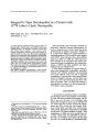
Stargardts Type Maculopathy in a Patient with 11778 Lebers Optic Neuropathy |
| Creator |
Yen, May-Yung; Wei, Yau-Huei; Liu, Jorn-Han |
| Affiliation |
Department of Ophthalmology, Taipei Veterans General Hospital, Taiwan, ROC. |
| Abstract |
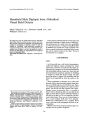
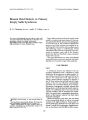
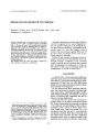
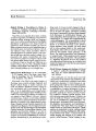
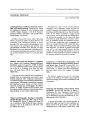
A 13-year-old boy presented with a 2-month history of blurred vision. Visual acuity was 20/200 in both eyes. Ophthalmoscopic examination revealed normal discs and "beaten bronze atrophy" in the maculae. Subsequently, progressive vision loss with optic atrophy occurred over the next few years. Fluorescein angiographic findings were compatible with Stargardt's maculopathy. Because his cousin developed sequential vision loss diagnosed as Leber's hereditary optic neuropathy, molecular genetic analysis was performed on blood mitochondrial DNA (mtDNA) from our patient, his cousin with vision loss, and another three asymptomatic cousins. The results showed that they all harbored homoplasmic G to A point mutations at nucleotide position 11778 of the ND4 gene in mtDNA. |
| Subject |
Adolescent; DNA Mutational Analysis; DNA, Mitochondrial/genetics; Fluorescein Angiography; Fundus Oculi; Humans; Macular Degeneration/diagnosis; Macular Degeneration/genetics; Macular Degeneration/physiopathology; Male; Optic Atrophies, Hereditary/diagnosis; Optic Atrophies, Hereditary/genetics; Optic Atrophies, Hereditary/physiopathology; Pedigree; Point Mutation; Visual Acuity |
| Format |
application/pdf |
| Publication Type |
Journal Article |
| Collection |
Neuro-Ophthalmology Virtual Education Library: Journal of Neuro-Ophthalmology Archives: https://novel.utah.edu/jno/ |
| Publisher |
Lippincott, Williams & Wilkins |
| Holding Institution |
Spencer S. Eccles Health Sciences Library, University of Utah |
| Rights Management |
© North American Neuro-Ophthalmology Society |
| Setname |
ehsl_novel_jno |
| ID |
224717 |
| Reference URL |
https://collections.lib.utah.edu/ark:/87278/s62n87cj/224717 |