John A. Moran Eye Center Neuro-Ophthalmology Collection: A variety of lectures, videos and images relating to topics in Neuro-Ophthalmology created by faculty at the Moran Eye Center, University of Utah, in Salt Lake City.
NOVEL: https://novel.utah.edu/
TO
Filters: Collection: "ehsl_novel_jmec"
| Identifier | Title | Description | Subject | ||
|---|---|---|---|---|---|
| 1 |
 |
2-7 | Abducting (Dissociated) Nystagmus | Example of a patient with abducting (dissociated) nystagmus. Patient has a subtle internuclear ophthalmoplegia. Right eye has right-beating jerk nystagmus, with smaller oscillations in the left eye. Disease/Diagnosis: Abducting Nystagmus | Abducting Nystagmus; Dissociated Nystagmus |
| 2 |
 |
2-10 | Binocular Pendular Nystagmus | Example of a patient with binocular pendular nystagmus. Patient has somewhat dissociated nystagmus, with nystagmus seen more prominently in the left eye. Patient shows an occasional jerk nystagmus to the right in the right eye. Left eye oscillations are mostly pendular. | Binocular Pendular Nystagmus; Pendular Nystagmus; Acquired Pendular Nystagmus |
| 3 |
 |
2-11 | Brun's Nystagmus | Observation of patient with Brun's Nystagmus. Shows patient gazing to the right and the nystagmus beating in the direction of the gaze. | Brun's Nystagmus; Cyclical |
| 4 |
 |
2-1 | Congenital Nystagmus | Example of patients with congenital nystagmus. First patient's nystagmus are mostly jerk and not pendular. Second patient's nystagmus are mostly pendular. Both patients show a uniform horizontal oscillation. Second patient also shows differences in frequency of oscillations depending on gaze, includ... | Congenital Nystagmus |
| 5 |
 |
NOVEL_Moran_3a-30 | Congenital Nystagmus | Patient with congenital nystagmus (no audio) | Congenital Nystagmus |
| 6 |
 |
2-21 | Convergence Retraction Nystagmus (Parinaud's Syndrome) | Examples of patients with convergence retraction nystagmus. Shows saccadic oscillations in patients looking upwards and following downwards moving targets. Also shows a side-view of the retracting movements of the globes. | Convergence Retraction Nystagmus; Parinaud's Syndrome; Dorsal Midbrain Syndrome; Lid Retraction |
| 7 |
 |
2-14 | Dissociated Nystagmus | Example of a patient with dissociated nystagmus. Demonstrates difference in movements between each eye. | Dissociated Nystagmus |
| 8 |
 |
2-6 | Downbeat Nystagmus | Example of patients with downbeating jerk nystagmus. Demonstrates how oscillations grow more prominent when the patient gazes down or laterally. Discusses some causes, including Arnold-Chiari malformation, infarction, and demyelination. | Downbeat Nystagmus |
| 9 |
 |
NOVEL_Moran_3a-1 | Downbeat Nystagmus | Example of patient with downbeat nystagmus. Patient is led through instructions of where to gaze. (no audio) | Downbeat Nystagmus |
| 10 |
 |
NOVEL_Moran_3a-9 | Downbeat Nystagmus | Example of patient with downbeat nystagmus. Patient is led through instructions of where to gaze. | Downbeat Nystagmus |
| 11 |
 |
1-8 | Internuclear Ophthalmoplegia (2 Examples) | Two examples of patients with internuclear ophthalmoplegia. First patient has a right internuclear ophthalmoplegia. Patient had subacute bacterial endocarditis with a bacterial abscess in the brain stem. Ductions and gaze to the right look good, but when gazing to the left, the right eye does not ad... | Internuclear Ophthalmoplegia; Abducting Nystagmus |
| 12 |
 |
2-2 | Latent Nystagmus | Example of a patient with latent nystagmus. Demonstrates a lack of oscillations in forward gaze, followed by the occlusion of each eye, showing how this generates a jerking oscillation in the non-occluded eye away from the occluded eye. | Latent Nystagmus; Fusional Maldevelopment Nystagmus Syndrome |
| 13 |
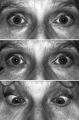 |
Figure-13 | Light-near Dissociation | Light-near dissociation in a 51-year-old woman with multiple sclerosis who experienced double vision for 1 week. Her pupils are 5 mm in diameter in room light (top), react poorly in response to direct light reaction (middle), but constrict promptly in response to near stimulation (bottom). She also ... | Nystagmus, Etiology, Pathologic; Nystagmus, Physiopathology, Pathologic; Reflex, Pupillary |
| 14 |
 |
2-9 | Monocular Pendular Nystagmus | Example of a patient with monocular pendular nystagmus, with discussion of situations in which this condition is seen: acquired disorder of the visual-sensory pathway, and acquired disorder of the brain stem (e.g. multiple sclerosis). | Monocular Pendular Nystagmus; Sensory Nystagmus; Pendular Nystagmus; Acquired Pendular Nystagmus |
| 15 |
 |
2-12 | Periodic Alternating Nystagmus | Example of a patient with periodic alternating nystagmus, showing an alternation between left-beats and right-beats as the patient maintains forward gaze. Nystagmus maintain horizontal direction regardless of position of gaze. | Periodic Alternating Nystagmus |
| 16 |
 |
2-16 | Physiologic (End-Gaze) Nystagmus | Demonstration of physiological nystagmus, where oscillations do not represent pathology, but occur when the patient's gaze is drawn too far laterally. | End-Gaze Nystagmus; Physiologic Nystagmus |
| 17 |
 |
2-8 | Rebound Nystagmus | Example of a patient with rebound nystagmus, where the oscillations alternate direction as the patient shifts gaze in different directions. Discussion of relationship to disease and disorders of the cerebellum, including degenerations of the cerebellum, infarction, and demyelination. | Rebound Nystagmus; Gaze-Evoked Nystagmus |
| 18 |
 |
NOVEL_Moran_3a-17 | Retraction Nystagmus | Patient with retraction nystagmus (no audio) | Retraction Nystagmus |
| 19 |
 |
NOVEL_Moran_3a-36 | Rotary Downbeat | Patient with rotary downbeat nystagmus (no audio) | Rotary Downbeat Nystagmus |
| 20 |
|
2-13 | Rotary Nystagmus | Example of a patient with rotary nystagmus, showing occasional counterclockwise rotary movements of both eyes. Seen more in intrinsic disorders of the brainstem. | Rotary (Torsional) Nystagmus |
| 21 |
 |
2-4 | See-saw Nystagmus | Example of a patient with see-saw nystagmus, showing how one eye elevates as the other depresses, with the elevating eye intorting as the depressing eye extorts. Shows vertical oscillations with pendular waveforms. Suggests a large structural lesion in the pericellar region (associated with bi-tempo... | See-saw Nystagmus |
| 22 |
 |
Katz_seesaw | See-saw Nystagmus | 7-year-old female whose mother noticed her eyes "bouncing" for 2 months. Visual acuity 20/70 OD and 20/40 OS, reduced color vision OU, and no afferent pupillary defect. See-saw nystagmus documented with videography. Manual perimetry revealed a complete right homonymous hemianopia. MRI revealed a lar... | See-saw Nystagmus |
| 23 |
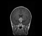 |
CorT1_with_11.jpg | See-saw Nystagmus MRI 1 | MRI; See-saw Nystagmus | See-saw Nystagmus |
| 24 |
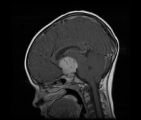 |
SagT1_with_11.jpg | See-saw Nystagmus MRI 2 | MRI; See-saw Nystagmus | See-saw Nystagmus |
| 25 |
 |
1-13 | Spasm of the Near Reflex | Example of patient with spasm of the near reflex and voluntary nystagmus. Discussion of similar-looking conditions (e.g. six nerve palsy, limitation of abduction, lateral rectus muscle problems) and how to tell them apart from spasm of the near reflex by observing the myosis evoked by the near respo... | Spasm of the Near Reflex; Spasm of the Near Triad; Voluntary Nystagmus |