The Emory Eye Center Neuro-Ophthalmology Collection contains a variety of lectures, videos and images relating to the discipline of neuro-ophthalmology created by faculty at Emory University in Atlanta, GA.
NOVEL: https://novel.utah.edu/
TO
Filters: Collection: ehsl_novel_eec
1 - 25 of 5
| Title | Creator | Subject | Description | ||
|---|---|---|---|---|---|
| 1 |
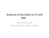 |
Anatomy of the Orbits on CT and MRI | Valérie Biousse, MD | Anatomy of the Orbit; Anatomy of the Optic Nerve | MRI and CT imaging of the orbit. |
| 2 |
 |
Pulsatile Proptosis Profile | Valérie Biousse, MD | Sphenoid Bone; Hypoplasia; Neurofibromatosis; Pulsatile Proptosis | See the PowerPoint description for Pulsatile Proptosis From Sphenoid Wing Hypoplasia in Neurofibromatosis Type 1 at: http://content.lib.utah.edu/cdm/ref/collection/ehsl-eec/id/64 See also Pulsatile Proptosis Full Face video: http://content.lib.utah.edu/cdm/ref/collection/ehsl-eec/id/14 |
| 3 |
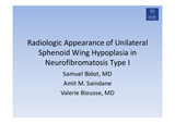 |
Radiologic Appearance of Unilateral Sphenoid Wing Hypoplasia in Neurofibromatosis Type I | Samuel Bidot, MD; Amit M. Saindane, MD; Valérie Biousse, MD | Sphenoid Bone; Hypoplasia; Neurofibromatosis; Pulsatile Proptosis | MRI features of greater wing sphenoid hypoplasia in the setting of neurofibromatosis type 1. - Figure 1 : Orbital MRI with contrast showing right greater sphenoid wing hypoplasia. The lack of bone tissue leads to herniation of the right temporal lobe into the orbit, pushing forward the orbital conte... |
| 4 |
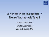 |
Pulsatile Proptosis from Sphenoid Wing Hypoplasia in Neurofibromatosis Type 1 | Samuel Bidot, MD; Amit M. Saindane, MD; Valérie Biousse, MD | Sphenoid Bone; Hypoplasia; Neurofibromatosis; Pulsatile Proptosis | Clinical and radiologic features of greater wing sphenoid hypoplasia in the setting of neurofibromatosis type 1. Figure 1 : slit lamp examination showing Lisch nodules; Figure 2 : orbit CT scan (1); Figure 3 : orbit CT scan (2) with annotations. For visual examples of this disorder, please see the... |
| 5 |
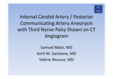 |
Internal Carotid Artery / Posterior Communicating Artery Aneurysm with Third Nerve Palsy Shown on CT Angiogram | Samuel Bidot, MD; Amit M. Saindane, MD; Valérie Biousse, MD | Optic Canal; Optic Nerve; Sphenoid Bone; Chiasmatic Groove; Orbit | Internal Carotid Artery / Posterior Communicating Artery Aneurysm with Third Nerve Palsy Shown on CT Angiogram ; anatomic description of vascular and bony findings on the CTA. - Figure 1 : 51 year-old man complaining of painful binocular diplopia. Orange arrows indicate the direction of gaze. In p... |
1 - 25 of 5