The Emory Eye Center Neuro-Ophthalmology Collection contains a variety of lectures, videos and images relating to the discipline of neuro-ophthalmology created by faculty at Emory University in Atlanta, GA.
NOVEL: https://novel.utah.edu/
TO
Filters: Collection: ehsl_novel_eec
1 - 25 of 5
| Title | Creator | Subject | Description | ||
|---|---|---|---|---|---|
| 1 |
 |
Cotton Wool Spots in Giant Cell Arteritis | Rahul A. Sharma, MD, MPH; Valérie Biousse, MD | Hypertension; Disc Edema; Occipital Lobe Hemorrhage | This is a case of cotton wool spots in a patient with temporal artery-biopsy proven temporal arteritis.; ; A 66-year-old woman presents with isolated painless vision loss related to a left optic neuropathy in her left eye. She denies systemic symptoms to suggest giant cell arteritis.; Her examinatio... |
| 2 |
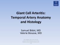 |
Giant Cell Arteritis: Temporal Artery Anatomy and Histology | Samuel Bidot, MD; Valérie Biousse, MD | Giant Cell Arteritis; Superficial Temporal Artery | Gross anatomy and histology of the normal superficial temporal artery.; Histopathology of the superficial temporal artery involved by active and healed GCA; Summary of the main histopathologic findings in GCA |
| 3 |
 |
Classic Pathology Findings in Giant Cell Arteritis | Andre Aung, MD; Corrina Azarcon, MD; Wael A. Alsakran, MD; Valérie Biousse, MD | Giant Cell Arteritis; Temporal Artery Biopsy; Pathology | An 80-year-old Caucasian woman presented with a 10 day history of headaches, intermittent binocular diplopia, and jaw pain. Temporal artery biopsy confirmed a diagnosis of giant cell arteritis. Pathology findings were classic for giant cell arteritis with numerous inflammatory cells in the tunica me... |
| 4 |
 |
Choroidal Infarction in Giant Cell Arteritis | Wael A. Alsakran, MD; Andre Aung, MD; Valérie Biousse, MD | Giant Cell Arteritis; Ancillary Testing | An 80-year-old Caucasian woman presented with a 10-day history of headaches, intermittent binocular diplopia, and jaw pain. Temporal artery biopsy confirmed a diagnosis of giant cell arteritis. Examination showed characteristic large area of choroidal ischemia that is well-known to be associated wit... |
| 5 |
 |
MRI Findings in Giant Cell Arteritis | Wael A. Alsakran, MD; Andre Aung, MD; Valérie Biousse, MD | Giant Cell Arteritis; Imaging | Case 1. An 80-year-old Caucasian woman presented with a 10-day history of headaches, intermittent binocular diplopia, and jaw pain. Temporal artery biopsy confirmed a diagnosis of giant cell arteritis. MRI with contrast showed enhancement of bilateral optic nerve sheaths in addition to enhancement o... |
1 - 25 of 5