This collection consists of short (2-5 minute) introductory videos on neuro-ophthalmological condition pathophysiology, diagnosis and treatment modalities by Dr. Andrew Lee.
Andrew G. Lee, MD, Professor of Ophthalmology, Weill Cornell Medicine; Chairman, Department of Ophthalmology, The Methodist Hospital, Houston, TX; Adjunct Professor of Ophthalmology, Baylor College of Medicine; Adjunct Professor of Ophthalmology, University of Iowa Hospitals and Clinics and the University of Buffalo; Clinical Professor of Ophthalmology, Department of Ophthalmology and Visual Sciences, University of Texas Medical Branch at Galveston; UT MD Anderson Cancer Center; and Texas A and M College of Medicine.
NOVEL: https://novel.utah.edu/
TO
Filters: Collection: ehsl_novel_lee
1 - 25 of 13
| Title | Description | Subject | ||
|---|---|---|---|---|
| 1 |
 |
DWI - Diffusion Weighted Imaging | Dr. Lee lectures medical students on diffusion weighted imaging. | Imaging; MRI; DWI; Edema |
| 2 |
 |
Electroretinogram (ERG) | Dr. Lee lectures medical students on electroretinogram (ERG). | Electroretinogram (ERG); Retinopathy; Measurement; Diagnosis |
| 3 |
 |
Fat Suppression on T1 | 1. MRI a. T1 i. Fat Suppression 1. Fat suppression is crucial to properly visualize pathology | MRI; T1; Fat; Suppression |
| 4 |
 |
Frequency Doubling Technology Perimetry | Summary: Frequency Doubling Technology (FDT): an easily portable, fast, and inexpensive instrument used to detect visual field loss • similar to Humphrey visual field, but uses frequency doubling to create an illusion • not used in neuro-ophthalmology, but reasonable as an automated perimetry sc... | FDT; Visual Field Loss; Automated Perimetry Screen |
| 5 |
 |
Hydroxychloroquine Toxicity and Screening | Dr. Lee lectures medical students on hydroxychloroquine toxicity. | Hydroxychloroquine Toxicity; Bulls-eye Maculopathy; Plaquenil |
| 6 |
 |
MRI in Neuro-ophthalmology | Dr. Lee lectures medical students on the use of MRI imaging in neuro-ophthalmology. | MRI; Imaging |
| 7 |
 |
Macular Ganglion Cell Layer | Dr. Lee lectures medical students on | Ganglion Cell; Optical Coherence Tomography (OCT); Neuro-Ophthalmology |
| 8 |
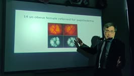 |
Magnetic Resonance Imaging (MRI) Neuroop Pearls | Dr. Lee lectures medical students on neuroop pearls. | MRI; Papilledema; Optic Neuritis |
| 9 |
 |
Optical Coherence Tomography (OCT) in Neuro-Ophthalmology | Summary: • Optical Coherence Tomography (OCT) in Neuro-ophthalmology o Measures peripapillary retinal nerve fiber layer thickness o Scale is color coded and the black line represents the patient's peripapillary retinal nerve fiber layer thickness -Green zone is good • 5th to 95th percentile by a... | Optical Coherence Tomography (OCT); Papilledema; Fiber; Thickening |
| 10 |
 |
Positron Emission Tomography (PET) Scan | Dr. Lee lectures medical students on PET scans. | Neuroimaging |
| 11 |
 |
Spasms: When to Image Them | Summary: • Benign essential blepharospasm o Bilateral, simultaneous spasm of the eyelids, looks like excessive blinking o Idiopathic, benign, does not require imaging • Meige Syndrome o Spasm of both the eyelids and the face, can descend to the lower face • Hemifacial spasm o Spasm of one enti... | Blepharospasm; MEIGE; Myokymia |
| 12 |
 |
Ultrasound in Neuro-Ophthalmology | Dr. Lee lectures medical students on ultrasound in neuro-ophthalmology. | Ultrasound; Neuro-Ophthalmology; Imaging |
| 13 |
 |
Venous Sinus Imaging | Dr. Lee lectures medical students on the imaging of venous sinus disease. | Imaging; Thrombosis; Stenosis; Sinus |
1 - 25 of 13