The Health Education Assets Library (HEAL) is a collection of over 22,000 freely available digital materials for health sciences education. The collection is now housed at the University of Utah J. Willard Marriott Digital Library.
TO
| Title | Description | Subject | Collection | ||
|---|---|---|---|---|---|
| 201 |
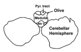 |
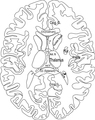
MRI Atlas: Brain (Axial) - Scan 1 - Labeled Outline | Anatomical structures of the brain are identified in this labeled outline from the UCLA Interactive Neurosciences MRI Atlas. | UCLA Interactive Neuroscience | |
| 202 |
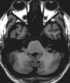 |
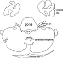
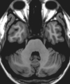
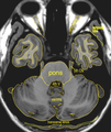
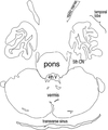
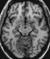
MRI Atlas: Brain (Axial) - Scan 2 | Anatomical structures of the brain are depicted in this scan from the UCLA Interactive Neurosciences MRI Atlas. | Flocculus; CN8; Vermis; Cerebellar Hemisphere; Transverse Sinus | UCLA Interactive Neuroscience |
| 203 |
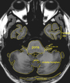 |
MRI Atlas: Brain (Axial) - Scan 2 - Labeled | Anatomical structures of the brain are identified in this scan with labeled outline from the UCLA Interactive Neurosciences MRI Atlas. | UCLA Interactive Neuroscience | |
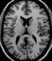
| 204 |
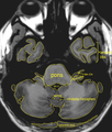 |
MRI Atlas: Brain (Axial) - Scan 2 - Labeled (Enlarged) | Anatomical structures of the brain are identified in this scan with labeled outline from the UCLA Interactive Neurosciences MRI Atlas. | UCLA Interactive Neuroscience | |
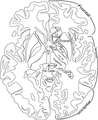
| 205 |
 |
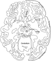
MRI Atlas: Brain (Axial) - Scan 2 - Labeled Outline | Anatomical structures of the brain are identified in this labeled outline from the UCLA Interactive Neurosciences MRI Atlas. | UCLA Interactive Neuroscience | |
| 206 |
 |
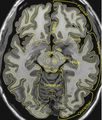
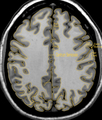
MRI Atlas: Brain (Axial) - Scan 3 | Anatomical structures of the brain are depicted in this scan from the UCLA Interactive Neurosciences MRI Atlas. | CN 5; Vermis; Transverse Sinus | UCLA Interactive Neuroscience |
| 207 |
 |
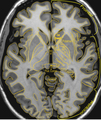
MRI Atlas: Brain (Axial) - Scan 3 - Labeled | Anatomical structures of the brain are identified in this scan with labeled outline from the UCLA Interactive Neurosciences MRI Atlas. | UCLA Interactive Neuroscience | |
| 208 |
 |
MRI Atlas: Brain (Axial) - Scan 3 - Labeled (Enlarged) | Anatomical structures of the brain are identified in this scan with labeled outline from the UCLA Interactive Neurosciences MRI Atlas. | UCLA Interactive Neuroscience | |
| 209 |
 |
MRI Atlas: Brain (Axial) - Scan 3 - Labeled Outline | Anatomical structures of the brain are identified in this labeled outline from the UCLA Interactive Neurosciences MRI Atlas. | UCLA Interactive Neuroscience | |
| 210 |
 |
MRI Atlas: Brain (Axial) - Scan 4 | Anatomical structures of the brain are depicted in this scan from the UCLA Interactive Neurosciences MRI Atlas. | Gyrus Rectus; Insula of Reil; Interpeduncular Fossa; Optic Tract; Cerebral Peduncle; Red Nucleus; PAG | UCLA Interactive Neuroscience |
| 211 |
 |
MRI Atlas: Brain (Axial) - Scan 4 - Labeled | Anatomical structures of the brain are identified in this scan with labeled outline from the UCLA Interactive Neurosciences MRI Atlas. | UCLA Interactive Neuroscience | |
| 212 |
 |
MRI Atlas: Brain (Axial) - Scan 4 - Labeled Outline | Anatomical structures of the brain are identified in this labeled outline from the UCLA Interactive Neurosciences MRI Atlas. | UCLA Interactive Neuroscience | |
| 213 |
 |
MRI Atlas: Brain (Axial) - Scan 4 - Labeled Outline | Anatomical structures of the brain are identified in this scan with labeled outline from the UCLA Interactive Neurosciences MRI Atlas. | UCLA Interactive Neuroscience | |
| 214 |
 |
MRI Atlas: Brain (Axial) - Scan 5 | Anatomical structures of the brain are depicted in this scan from the UCLA Interactive Neurosciences MRI Atlas. | Insula of Reil; Claustrum; Head of Caudate; Internal Capsule (Anterior Limb); Internal Capsule (Posterior Limb); Globus Pallidus Externus; Globus Pallidus Internus; Posterior Commissure | UCLA Interactive Neuroscience |
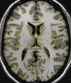
| 215 |
 |
MRI Atlas: Brain (Axial) - Scan 5 - Labeled (Enlarged) | Anatomical structures of the brain are identified in this scan with labeled outline from the UCLA Interactive Neurosciences MRI Atlas. | UCLA Interactive Neuroscience | |
| 216 |
 |
MRI Atlas: Brain (Axial) - Scan 5 - Labeled Outline | Anatomical structures of the brain are identified in this scan with labeled outline from the UCLA Interactive Neurosciences MRI Atlas. | UCLA Interactive Neuroscience | |
| 217 |
 |
MRI Atlas: Brain (Axial) - Scan 5 - Labeled Outline | Anatomical structures of the brain are identified in this labeled outline from the UCLA Interactive Neurosciences MRI Atlas. | UCLA Interactive Neuroscience | |
| 218 |
 |
MRI Atlas: Brain (Axial) - Scan 6 | Anatomical structures of the brain are depicted in this scan from the UCLA Interactive Neurosciences MRI Atlas. | Cingulate Gyrus (Rostrum); Head of Caudate; Anterior Nucleus of Thalamus (Splenium); Insula of Reil | UCLA Interactive Neuroscience |
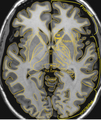
| 219 |
 |
MRI Atlas: Brain (Axial) - Scan 6 - Labeled | Anatomical structures of the brain are identified in this scan with labeled outline from the UCLA Interactive Neurosciences MRI Atlas. | UCLA Interactive Neuroscience | |
| 220 |
 |
MRI Atlas: Brain (Axial) - Scan 6 - Labeled (Enlarged) | Anatomical structures of the brain are identified in this scan with labeled outline from the UCLA Interactive Neurosciences MRI Atlas. | UCLA Interactive Neuroscience | |
| 221 |
 |
MRI Atlas: Brain (Axial) - Scan 6 - Labeled Outline | Anatomical structures of the brain are identified in this labeled outline from the UCLA Interactive Neurosciences MRI Atlas. | UCLA Interactive Neuroscience | |
| 222 |
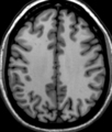 |
MRI Atlas: Brain (Axial) - Scan 7 | Anatomical structures of the brain are depicted in this scan from the UCLA Interactive Neurosciences MRI Atlas. | Superior Frontal Gyrus; Cingulate Gyrus; Centrum Semiovale; Pre-central Gyrus; Central Sulcus; Post-central Gyrus | UCLA Interactive Neuroscience |
| 223 |
 |
MRI Atlas: Brain (Axial) - Scan 7 - Labeled | Anatomical structures of the brain are identified in this scan with labeled outline from the UCLA Interactive Neurosciences MRI Atlas. | UCLA Interactive Neuroscience | |
| 224 |
 |
MRI Atlas: Brain (Axial) - Scan 7 - Labeled Outline | Anatomical structures of the brain are identified in this labeled outline from the UCLA Interactive Neurosciences MRI Atlas. | UCLA Interactive Neuroscience | |
| 225 |
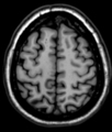 |
MRI Atlas: Brain (Axial) - Scan 8 | Anatomical structures of the brain are depicted in this scan from the UCLA Interactive Neurosciences MRI Atlas. | Superior Frontal Gyrus; Pre-central Gyrus; Central Sulcus; Post-central Gyrus; Superior Frontal Sulcus; Mid-frontal Gyrus | UCLA Interactive Neuroscience |