The Health Education Assets Library (HEAL) is a collection of over 22,000 freely available digital materials for health sciences education. The collection is now housed at the University of Utah J. Willard Marriott Digital Library.
TO
| Title | Description | Subject | Collection | ||
|---|---|---|---|---|---|
| 201 |
 |
Hassall's corpuscle in thymus (mouse) | Electron microscopy. The centre of a small Hassall's corpuscle consists of darker-stained cells (1) which are keratinizing and represent degenerating cytoplasmic remnants (2). (3) is an infiltrating monocyte and (*) indicate the presence of free keratohyalin granules from disintegrated epithelial ce... | thymic corpuscle (Hassalls); lymphoid tissue ; epithelioreticular cell (ERC) | Poja Histology Collection - Lymphatic Tissues and Organs Subset |
| 202 |
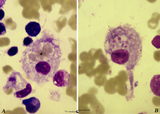 |
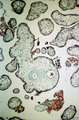
Hemophagocytosis by reticular cells in bone marrow smear (human) | Stain: May-Grnwald-Giemsa (MGG). (A) shows a reticular cell that has phagocytosed several erythrocytes (*). The cell is surrounded by darkly stained expulsed nuclei. In (B) a similar cell has phagocyized blood platelets (arrows). | Poja Histology Collection - Blood & Bone Marrow Subset | |
| 203 |
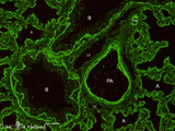 |
Heparan sulfates in the terminal sac period of the lung (mouse, fetus) | Stain: fluorescence microscopy with anti-heparan sulphate antibody (a phage-display antibody, HS4E4). Heparan sulfates are linear polysaccharides that belong to the group of the glycosaminoglycans (GAGs). These GAGs are found associated with basement membranes as shown here. The sulphated saccharide... | Bronchioli; Air spaces; Terminal sac period | Poja Histology Collection - Respiratory System Subset |
| 204 |
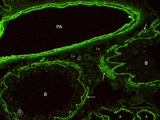 |
Heparan sulfates in the terminal sac-period of the lung (mouse, fetus) | Stain: fluorescence microscopy with anti-heparan sulphate antibody (a phage-display antibody, LKiv69). Heparan sulfates are linear polysaccharides that belongs to the group of the glycosaminoglycans (GAGs). The sulphated saccharide domains provide numerous docking sites for protein ligands. These GA... | Bronchioli; Air spaces; Terminal sac period | Poja Histology Collection - Respiratory System Subset |
| 205 |
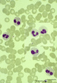 |
Heterozygotic Pelger-Hut's nuclear anomaly of neutrophilic granulocytes in peripheral blood smear (human) | Stain: May-Grnwald-Giemsa (MGG). The majority of neutrophils has bilobed nuclei, the lobes being rounder than normal and the chromatin is more condensed. About 30% of the cells appear as band forms (imitating shift to the left). A characteristic 'pince-nez' shape is common. Note the fine dust-like g... | Poja Histology Collection - Blood & Bone Marrow Subset | |
| 206 |
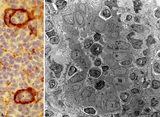 |
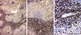
High endothelial venules (HEV) (rat lymph node, human palatine tonsil) | Stain: (A) Anti-laminin-antibody and immunoperoxidase staining using diaminobenzidin (DAB)/ hematoxylin counterstained on frozen section of lymph node; (B) electron microscopy (palatine tonsil). (A): Using an antibody against laminin brown-stained basement membranes are outlined demonstrating postc... | palatine tonsil; high endothelial venule (HEV); laminin; immunohistochemistry | Poja Histology Collection - Lymphatic Tissues and Organs Subset |
| 207 |
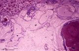 |
Hilum side of lymph node (human) | Stain: Azan. Site of the hilum of a lymph node includes part of the medulla and medullary cords (1) and medullar sinuses (2). In the hilum (H) dilated efferent lymphatic vessels (3) and draining veins (4) and entering arteries (5) are present. (6) represents adipose tissue embedding the efferent ves... | hilum; lymphatic vessels; medullary cords; paracortex | Poja Histology Collection - Lymphatic Tissues and Organs Subset |
| 208 |
 |
Homozygotic Pelger-Huet's nuclear anomaly of granulocytes in peripheral blood smear (human) | Stain: May-Grnwald-Giemsa (MGG). In this case of homozygotic Pelger-Hut anomaly the neutrophil (1) as well as the eosinophil (2) have non-lobed nuclei with condensed chromatin. Characteristic fine dust-like granules are present in the neutrophil, while the eosinophil has separate recognizable coarse... | Poja Histology Collection - Blood & Bone Marrow Subset | |
| 209 |
 |
Homozygotic Pelger-Hut's nuclear anomaly of neutrophilic granulocytes in peripheral blood smear (human) | Stain: May-Grnwald-Giemsa (MGG). The neutrophils in this patient with homozygotic Pelger-Hut's anomaly have non-lobed nuclei. The chromatin is condensed and fine dust-like granules are present. | Poja Histology Collection - Blood & Bone Marrow Subset | |
| 210 |
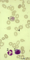 |
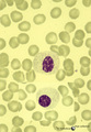
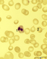
Howell-Jolly bodies in reticulocytes in peripheral blood smear (human) | Stain: May-Grnwald-Giemsa (MGG). The Howell-Jolly-bodies (1) in the reticulocyte are remnants of the nucleus (nuclear chromatin granules) of an erythroblast during anomalous development due to vitamin B12 deficiency, folic acid deficiency or in megaloblastic and hemolytic anemias. (2) Neutrophil wit... | Poja Histology Collection - Blood & Bone Marrow Subset | |
| 211 |
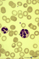 |
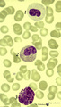
Hypersegmented neutrophil in peripheral blood smear of a hyperthermic patient (human) | Stain: May-Grnwald-Giemsa (MGG). In hyperthermic patients the neutrophils (1) show hypersegmentation of the nuclear lobe (>5) and a pyknotic appearance. The nucleus appears like a bunch of grapes. (2) immature band neutrophil (juvenile unsegmented neutrophil). Hypersegmentation may also occur in meg... | Poja Histology Collection - Blood & Bone Marrow Subset | |
| 212 |
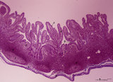 |
Ileum with Peyer's patches ('gut-associated lymphatic tissue' or GALT) (human) | Stain: Azan. Survey ileum (see also Digestive System: Ileum) A large amount of non-encapsulated diffuse lymphatic tissue or mucosa-associated lymphatic tissue (MALT) is located in the subepithelial lamina propria/submucosa of the ileum and called 'gut-associated lymphatic tissue' (GALT). These so-c... | follicle; lymph node; germinal center | Poja Histology Collection - Lymphatic Tissues and Organs Subset |
| 213 |
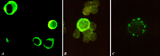 |
Immunohistochemical identification of splenic B cells (rat) | Stain: Immunofluorescence of FITC-labeled anti-Mark-1 antibody against rat B cells, carried out on spleen cells after fixation (A, B) or non-fixated spleen cells (C). (A) shows (young) plasma cells producing antibodies. (B) a green-stained B cell. The surrounding negative cells are likely T cells.... | immunofluorescence; B lymphocyte; capping | Poja Histology Collection - Lymphatic Tissues and Organs Subset |
| 214 |
 |
Immunohistochemistry of B cells in splenic white pulp (rat) | Stain: Immunohistochemistry of Vector red for Mark-1-antibody-stained B cells. (1) indicates the PALS area populated with unstained T cells located around the central artery of the white pulp. (2) indicates the positively red stained B cells in the germinal centre of the follicle. (3) the B-cells i... | marginal zone; immunohistochemistry; follicle | Poja Histology Collection - Lymphatic Tissues and Organs Subset |
| 215 |
 |
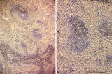
Immunohistochemistry of ED1-positive subset of macrophages in spleen (rat) | Stain: Immunoperoxidase staining using diaminobenzidin (DAB)/ hematoxylin counterstained on frozen section with the anti-macrophage antibody ED1. Most of the labeled (brown) macrophages are found in the red pulp (2) up to the marginal zone border (B). The PALS area (1) contains sparsely spread ED1 ... | ED1 ; macrophages; immunohistochemistry; marginal zone | Poja Histology Collection - Lymphatic Tissues and Organs Subset |
| 216 |
 |
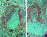
Immunohistochemistry of laminin in cortex lymph node (rat) | Anti-laminin-antibody and immunoperoxidase staining with diaminobenzidin (DAB) and hematoxylin counterstained on frozen section. Using an antibody against laminin brown-stained basement membranes (BM) are outlined demonstrating postcapillary venules (1) in the paracortical areas, as well as larger b... | paracortex; high endothelial venule (HEV); laminin; immunohistochemistry | Poja Histology Collection - Lymphatic Tissues and Organs Subset |
| 217 |
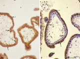 |
Immunohistochemistry of tertiary villi (human placenta, midpregnancy ) | Stain: Immunoperoxidase staining with diaminobenzidin (DAB) and hematoxylin counterstaining for keratin 7 (OVTL 12-30 antibody) at the left (A) and for human anti-chorionic gonadotrophin (hCG-DAKO 231antibody) at the right (B). Tertiary villi show a keratin-positive reaction (brown-stained) in all ... | placenta; chorionic villi; HCG; syncytiotrophoblast; keratin 7; immunohistochemistry | Poja Histology Collection - Placenta |
| 218 |
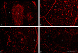 |
Immunohistochemistry of thymus (rat) | Stain: Alexa-594 red immunofluorescence. (1) Medulla. (2) Cortex. (A): Staining with a monoclonal antibody against vimentin illustrates that vimentin is localised in the stromal cells of the connective tissue of the capsule, of the septa or trabeculae that invade the cortex (2) and overwhelmingly ... | vimentin; laminin; fibronectin; ED2 monoclonal antibody | Poja Histology Collection - Lymphatic Tissues and Organs Subset |
| 219 |
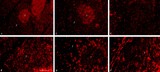 |
Immunohistochemistry with cellular markers in thymus (rat) | Stain: Alexa-594 red immunofluorescence. (1) medulla. (2) cortex. (A-survey, B-cortex): ER13 antibody stains for MHC-class II antigens on reticular cell types in medulla and cortex. (C): ED1 monoclonal antibody stains a single chain glycoprotein of 110 kDa on the lysosomal membrane of myeloid cel... | ER13 antibody; ED1 monoclonal antibody; ED2 monoclonal antibody; ER2 monoclonal antibody | Poja Histology Collection - Lymphatic Tissues and Organs Subset |
| 220 |
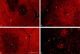 |
Immunohistochemistry with cellular markers in thymus (rat) | Stain: Alexa 594 red immunofluorescence. (1) medulla; (2) cortex; (3) septa. (A): Strong CD8 staining of the thymic cortical lymphocytes (single CD8+ and double positive CD4+CD8+ thymocytes). (B): OX19 staining for almost all T cells, equivalent to T1 human marker and Ly-1 mouse marker. Note that... | cyclophosphamide; CD8 monoclonal antibody; OX19 monoclonal antibody; ER1 monoclonal antibody | Poja Histology Collection - Lymphatic Tissues and Organs Subset |
| 221 |
 |
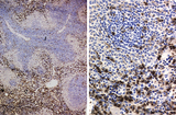
Immunoperoxidase stained CD3 positive T cells in spleen (rat) | Stain: Immunoperoxidase staining using diaminobenzidin (DAB)/ hematoxylin counterstained on frozen section with anti CD3 antibodies. Survey (A) and detail (B) show that CD3 marker for mature T cells stains positively (brown) in the PALS area (1) around the central arteries, while the adjacent follic... | CD3 lymphocytes; immunohistochemistry; PALS | Poja Histology Collection - Lymphatic Tissues and Organs Subset |
| 222 |
 |
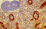
Immunoperoxidase stained CD8 positive T cells in spleen (rat) | Stain: Immunoperoxidase staining using diaminobenzidin (DAB)/ hematoxylin counterstained on frozen section with anti-CD8 antibodies. A, B and C show brown-stained CD8-positive T cells or Tc cells at different enlargements. The CD8 Tc/s cells are localised predominantly in the PALS area (1), while th... | CD8 lymphocytes; immunohistochemistry; PALS | Poja Histology Collection - Lymphatic Tissues and Organs Subset |
| 223 |
 |
Intervillous space with villi (human placenta, midpregnancy, cross-section) | Stain: Trichrome (Goldner). In the middle a thick stem villus (cross-sections of left artery (1) and right vein (2) of the umbilical circulation in the thick embryonic connective tissue) with ramifications into terminal villi (tertiary). Most tertiary villi of varying diameters contain capillaries a... | placenta; chorionic villi; fibrinoid; cytotrophoblast; syncytiotrophoblast | Poja Histology Collection - Placenta |
| 224 |
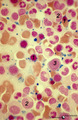 |
Iron (Perls) staining of sideroblasts in bone marrow smear (human) | Stain: Perl's stain for iron (Prussian blue). The intense blue stained hemosiderin in the erythroblasts (1) is visible as siderotic granules (pathologic siderosomes). These pathologic siderosomes are sometimes distributed in a circle around the nucleus (ringed sideroblasts). The myeloid cell types (... | Poja Histology Collection - Blood & Bone Marrow Subset | |
| 225 |
 |
Karyorrhexis of polychromatic erythroblast in bone marrow smear (human) | Stain: May-Grnwald-Giemsa (MGG). The nucleus of the polychromatic erythroblast (1) shows abnormal karyorrhexis due to vitamin B12 deficiency or folic acid deficiency. Normochromic normocyte (2, normal erythrocyte), poikilocytosis and oval macrocytes are seen. | Poja Histology Collection - Blood & Bone Marrow Subset |