Home
Browse
Ask Us
Chat
Harmful Language Statement
Log in
Advanced Search
Year
1944
1945
1946
1947
1948
1949
1950
1951
1952
1953
1954
1955
1956
1957
1958
1959
1960
1961
1962
1963
1964
1965
1966
1967
1968
1969
1970
1971
1972
1973
1974
1975
1976
1977
1978
1979
1980
1981
1982
1983
1984
1985
1986
1987
1988
1989
1990
1991
1992
1993
1994
1995
1996
1997
1998
1999
2000
2001
2002
2003
2004
2005
2006
2007
2008
2009
2010
2011
2012
2013
2014
2015
2016
2017
2018
2019
2020
2021
2022
2023
TO
1944
1945
1946
1947
1948
1949
1950
1951
1952
1953
1954
1955
1956
1957
1958
1959
1960
1961
1962
1963
1964
1965
1966
1967
1968
1969
1970
1971
1972
1973
1974
1975
1976
1977
1978
1979
1980
1981
1982
1983
1984
1985
1986
1987
1988
1989
1990
1991
1992
1993
1994
1995
1996
1997
1998
1999
2000
2001
2002
2003
2004
2005
2006
2007
2008
2009
2010
2011
2012
2013
2014
2015
2016
2017
2018
2019
2020
2021
2022
2023
Type
Image
281
Image/StillImage
22
Image/MovingImage
3
Format
image/jpeg
685
Collection
College of Nursing
2
Discovery and Innovation at Universit...
1
Health Education Assets Library (HEAL)
379
History of the Health Sciences
172
NOVEL
3
NOVEL - Daniel Gold Collection
59
NOVEL - Emory Eye Center
2
NOVEL - Frank B. Walsh Collection
5
NOVEL - J. Lawton Smith Lecture Colle...
21
NOVEL - Moran Eye Center
37
NOVEL - William F. Hoyt Collection
2
Photo Archives
1
Utah COVID-19
1
More
Filters:
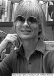
Format:
"image/jpeg"
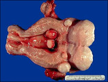
201
-
225
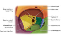
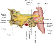
of
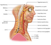
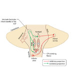
685
<
4
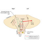
5
6
7
8
9
10
11
12
13
>
Gallery view
Number of results to display per page
10
25
50
100
200
Sort by Relevance
Sort by Title A-Z
Sort by Title Z-A
Sort by Date Ascending
Sort by Date Descending
Sort by Last Modified Ascending
Sort by Last Modified Descending
Title
Date
Type
Setname
201
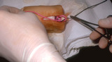
Excision: suturing
1997-01-01
ehsl_heal
202
Excision: suturing
1997-01-01
ehsl_heal
203
Excision: suturing
1997-01-01
ehsl_heal
204
Excision: suturing
1997-01-01
ehsl_heal
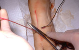
205
Excision: suturing vertical mattress
1997-01-01
ehsl_heal
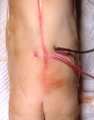
206
Excision: vertical mattress suture
1997-01-01
ehsl_heal
207
Eyelid Anatomy
2017
Image
ehsl_novel_gold
208
Ezell, Annette S., M.S.
Image
ehsl_con
209
Ezell, Annette S., M.S.
Image
ehsl_con
210
Fibroid uterus
1998-01-01
ehsl_heal
211
Fibroma
1998-01-01
ehsl_heal
212
Fibrous septae
1997-01-01
ehsl_heal
213
Figure 17: Bony Structures Relevant to the Orbit
2022
Image
ehsl_novel_gold
214
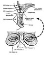
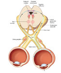
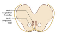
Figure 1: Oculosympathetic Pathway for Pupillary Dilation
2022
Image
ehsl_novel_gold
215
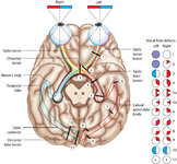
Figure 24: Typical Visual Field Defects Associated with Discrete Lesions Along the Visual Pathways
2022
Image
ehsl_novel_gold
216
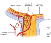
Figure 27: Vascular Supply of the Optic Nerve Head, Choroid and Retina
2022
Image
ehsl_novel_gold
217
Figure 2: Parasympathetic Pathway for Pupillary Constriction
2022
Image
ehsl_novel_gold
218
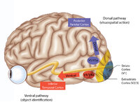
Figure 43: How the Brain Makes Sense of What It Sees - The Dorsal and Ventral Visual Pathways, and a 3 Tiered Approach to Vision
2022
Image
ehsl_novel_gold
219
Figure 46: The Course of the 6th (VI) Nerve
2022
Image
ehsl_novel_gold
220
Figure 50: Anatomy and Physiology of the Saccadic Pathways
2022
Image
ehsl_novel_gold
221
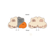
Figure 51: Lateral Medullary Lesion Causing Saccadic Dysmetria
2022
Image
ehsl_novel_gold
222
Figure 51: Lateral Medullary Lesion Causing Saccadic Dysmetria (Supplement)
Image
ehsl_novel_gold
223
Figure 51: Lateral Medullary Lesion Causing Saccadic Dysmetria (Supplement)
Image
ehsl_novel_gold
224
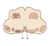
Figure 53: Vascular Distribution and Anatomy Relevant to the Lateral Medullary (Wallenberg) Syndrome
2022
Image
ehsl_novel_gold
225
Figure 53: Vascular Distribution and Anatomy Relevant to the Lateral Medullary (Wallenberg) Syndrome (Supplement)
Image
ehsl_novel_gold
201
-
225
of
685
<
4
5
6
7
8
9
10
11
12
13
>