John A. Moran Eye Center Neuro-Ophthalmology Collection: A variety of lectures, videos and images relating to topics in Neuro-Ophthalmology created by faculty at the Moran Eye Center, University of Utah, in Salt Lake City.
NOVEL: https://novel.utah.edu/
TO
Filters: Collection: "ehsl_novel_jmec"
| Title | Description | Type | ||
|---|---|---|---|---|
| 176 |
 |
Color Vision Testing | Demonstration of color vision examination. | Text |
| 177 |
 |
Stereoacuity Testing | Demonstration of examination for stereoacuity. | Text |
| 178 |
 |
Amsler Grid Testing | Demonstration of Amsler Grid examination. | Text |
| 179 |
 |
Near Reflex and Accomodation | Description of testing the near reflex and accomodation. | |
| 180 |
 |
Dilation Lag | Two examples of dilation lag (Horner's syndrome). In the first example, the right pupil dilates much faster than the left pupil when the light is turned out. In the second example, the left pupil dilates much faster than the right pupil when the light is turned out. Discussion of methods of document... | Image/MovingImage |
| 181 |
 |
Parinaud's Syndrome | Two examples of patients with Parinaud's syndrome, a dorsal midbrain syndrome. Discussion of hallmarks of this syndrome, including convergence retraction nystagmus, vertical gaze palsies, light-near dissociation, and Collier's Sign. Discussion of age-dependent disorders associated with this syndrome... | Image/MovingImage |
| 182 |
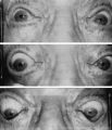 |
Argyll Robertson Pupils | Argyll Robertson pupils in an elderly man treated for tabes dorsalis in 1952. His pupils are small and slightly irregular, constrict poorly in response to light stimulation (top), dilate poorly in darkness (middle), but constrict promptly in response to near stimulation (bottom). | Image |
| 183 |
 |
Sector Palsies and Light-Near Dissociation | Example of patient with bilateral Adie's pupils. Exam is performed with a slit-lamp. Shows iris stroma and focal segments of iris sphincter that retain their contractilty. Suggests post-ganglionic parasympathetic denervation. | Image/MovingImage |
| 184 |
 |
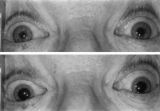
Left-sided Dilation Lag in a Man with Horner's Syndrome | Left-sided dilation lag in a 29-year-old man with Horner's syndrome caused by a posterior mediastinal ganglioneuroma. Note that the degree of anisocoria is greater after 5 seconds in darkness (top) compared with findings after 15 seconds in darkness (bottom). | Image |
| 185 |
 |
RAPD Present | This clip demonstrates the technique used to determine that Relative Afferent Pupillary Defect (RAPD) is present in a patient. | Image/MovingImage |
| 186 |
 |
Normal Light Reflex without RAPD | This clip demonstrates the examination of the Relative Afferent Pupillary Defect (RAPD.) Demonstration of gauging the size of the pupil in light, testing light reflexes, swinging flashlight test for optic nerve abnormality. | Image/MovingImage |
| 187 |
 |
Pupil Exam | Demonstration of pupil examination. | Text |
| 188 |
 |
Enhanced Mydriasis in Response to Hydroxyamphetamine | Enhanced mydriasis in response to hydroxyamphetamine in a 77-year-old woman with a long-standing, preganglionic, right-sided Horner's syndrome that occurred following cervical neck dissection for thoracic outlet syndrome 30 years earlier. Miosis of the right pupil is apparent in room light (top). Th... | Image |
| 189 |
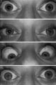 |
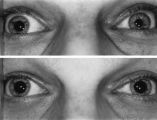
Pupil Signs in a 32-year-old Woman with Right-sided Adie's Pupil | Pupil signs in a 32-year-old woman with right-sided Adie's pupil. The right pupil is larger than the left pupil (top), reacts poorly to direct light stimulation (second panel), and better in response to near stimulation (third panel). The right pupil also shows a supersensitive response 30 minutes a... | Image |
| 190 |
 |
Transillumination - Lisch Nodules | Demonstration of transillumination of the Lisch nodules on a patient with neurofibromatosis. Shows how Lisch nodules that were not very visible in slit-lamp examination are better seen with transillumination, which may therefore be useful in detecting Lisch nodules earlier in children where they are... | Image/MovingImage |
| 191 |
 |
Transillumination - Ciliary Body Neurofibromas | Example of transillumination on a patient with neurofibromatosis, but without Lisch nodules. Shows suspected neurofibromas in the ciliary body. | Image/MovingImage |
| 192 |
 |
Optic Disc: Anatomy, Variants, Unusual discs | Discussion of viewing the optic disc. Includes development of direct ophthalmoscope. Covers normal optic disc and nerve fiber; nerve fiber loss and defects; cilioretinal arteries; venous anomolies; papilledema; pseudopapilledema; myopic disc; hyperoptic disc; little red discs; megallopapilla; myelin... | Text |
| 193 |
 |
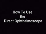
How to Use the Direct Ophthalmoscope in an Exam | Demonstration of using the direct ophthalmoscope to examine the optic disc. Covers hand placement , which eye to use, and distance from patient. | Image/MovingImage |
| 194 |
 |
Tour of the Fundus | This clip demonstrates the funduscopic examination technique. | Image/MovingImage |
| 195 |
 |
Normal Optic Disc | Overview of the structure and function of the normal optic disc. | Text |
| 196 |
 |
Tour of the Direct Ophthalmoscope | This clip describes the parts and operation of the ophthalmoscope as an ocular examination tool. Includes adjustment of aperture size and adjustment of lenses. | Image/MovingImage |
| 197 |
 |
Exophthalmometry | Demonstration of exophthalmometry examination. | Text |
| 198 |
 |
The Orbital Exam | Comprehensive demonstration of the entire orbital examination. | |
| 199 |
|
Progressive Supranuclear Palsy | Example of patient with progressive supranuclear palsy. Discussion of difference between saccadic movement in supranuclear palsy and nystagmus. Shows saccadic intrusions in forward gaze, pursuit, saccades, and doll's head maneuver. | Image/MovingImage |
| 200 |
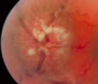 |
3-35a - Papilledema Stages | Grading Papilledema: Stage 4 Stage 4 = Complete obliteration of the cup and complete obscuration of at least some vessels on the surface of the disc. There may be small dilated capillaries on the disc that resemble telangiectasia. It is not the NFL infarcts or hemorrhages but the obscuration of the ... | Image |