John A. Moran Eye Center Neuro-Ophthalmology Collection: A variety of lectures, videos and images relating to topics in Neuro-Ophthalmology created by faculty at the Moran Eye Center, University of Utah, in Salt Lake City.
NOVEL: https://novel.utah.edu/
TO
Filters: Collection: "ehsl_novel_jmec"
| Title | Description | Type | ||
|---|---|---|---|---|
| 176 |
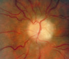 |
3-66d - Shunt Vessels (Post-papilledema) | The retino-choroidal collaterals seen with chronic papilledema begin with a "Hairnet" of telangiectasias that gradually winnow down to one or more large collateral tortuous draining channel. The presence of these vessels is evidence of long standing disc swelling. When the CSF pressure is lowered, t... | Image |
| 177 |
 |
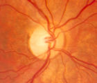
4-35 - Cupped Optic Nerve | Atrophic Glaucoma Atrophic glaucomatous discs show thinning of the neuro-retinal rim, "saucerization" (which is shallow cupping), evidence of peripapillary atrophy, and pallor of the very narrow neuroretinal rim. Notice that there is severe atrophy of the nerve fiber layer. | Image |
| 178 |
 |
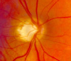
4-52b - Dominant Optic Neuropathy | A son presented with bilateral optic atrophy of unknown etiology after he failed a school visual exam. When looking for dominant optic atrophy, look at the parents. Mother was examined to find similar kind of atrophy. 4-52a mother, 4-52b son. | Image |
| 179 |
 |
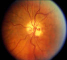
4-60a - Dominant Optic Neuropathy | A son presented with bilateral optic atrophy of unknown etiology after he failed a school visual exam. When looking for dominant optic atrophy, look at the parents. Mother was examined to find similar kind of atrophy. 4-60a mother, 4-60b son. | Image |
| 180 |
 |
Basal Encephaloceles | Text | |
| 181 |
 |
Dissection of the Carotid Artery | ||
| 182 |
 |
Mimics of Atrophy | Text | |
| 183 |
 |
Shunt Vessel Meningioma | RETINO-CHOROIDAL (OPTO-CILIARY) COLLATERAL VESSELS: (also known as Retinal-choroidal venous collaterals, opticociliary veins or ciliary shunt vessels) Retino-choroidal collaterals are potential telangiectatic connections between the retina and choroidal circulation. Although sometimes called "shunts... | Image |
| 184 |
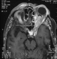 |
Shunt Vessel Meningioma - MRI | Meningiomas block venous egress and open potential venous channels known as retinochoroidal (optociliary) collateral vein. This meningioma extends from the back of the globe through the optic canal. | Image |
| 185 |
 |
Herpes Zoster Ophthalmicus with Third Nerve Palsy | Images showing presentation of Herpes Zoster (Zoster Ophthalmicus). | Text |
| 186 |
 |
Papilledema 2013 | Discussion of papilledema, the swelling due to increased pressure. | Text |
| 187 |
 |
Normal Eye Movements | This is an examination of a person with normal eye movements. Notice the patient has normal excursions. He has normal pursuit and saccades (horizontally and vertically). | Text |
| 188 |
 |
Nutritional Amblyopia | Example of patient with amblyopia with nutritional causes. | Text |
| 189 |
 |
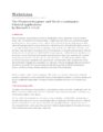
Retino-choroidal Vessels or Optociliary Veins or Ciliary Shunt | Overview of retino-choroidal collaterals, which are potential telangiectatic connections between the retina and choroidal circulation. Although sometimes called "shunts", these collaterals are between the retinal venous circulation and the choroidal venous circulation. | Text |
| 190 |
 |
Optic Disc Pallor Pseudo and Real | Discussion of the causes of optic disc pallor. | Text |
| 191 |
 |
Retinitis Pigmentosa Disease of Rods | Discussion of retinitis pigmentosa which is a retinal/choroidal degeneration caused by various genetic defects. | Text |
| 192 |
 |
Optic Disc: Anatomy, Variants, Unusual discs | Discussion of viewing the optic disc. Includes development of direct ophthalmoscope. Covers normal optic disc and nerve fiber; nerve fiber loss and defects; cilioretinal arteries; venous anomolies; papilledema; pseudopapilledema; myopic disc; hyperoptic disc; little red discs; megallopapilla; myelin... | Text |
| 193 |
 |
Normal Optic Disc | Overview of the structure and function of the normal optic disc. | Text |
| 194 |
 |
Test Duane | ||
| 195 |
 |
Optic Nerve Tumors Benign and Malignant | Discussion of optic nerve tumors including meningioma and glioma. | Text |
| 196 |
 |
Stargardt's Disease | Discussion of Stargardt's disease, an inherited maculopathy which frequently presents with a loss of central vision. | Text |
| 197 |
 |
The Electroretinogram and Electro-oculogram: Clinical Applications | The global or full-field electroretinogram (ERG) is a mass electrical response of the retina to photic stimulation. The ERG is a test used worldwide to assess the status of the retina in eye diseases in human patients and in laboratory animals used as models of retinal disease. | Text |
| 198 |
 |
Visually Evoked Potentials | Detailed explanation of visually evoked potentials. The terms visually evoked potential (VEP), visually evoked response (VER) and visually evoked cortical potential (VECP) are equivalent. They refer to electrical potentials, initiated by brief visual stimuli, which are recorded from the scalp overl... | Text |
| 199 |
 |
The Electro-oculogram: Clinical Applications | The electrooculogram measures the potential that exists between the cornea and Bruch's membrane at the back of the eye. The potential produces a dipole field with the cornea approximately 5 millivolts positive compared to the back of the eye, in a normally illuminated room. Although the origin of th... | Text |
| 200 |
 |
Basic Headache | Presentation covering an overview of headache and migraine. | Text |