The Health Education Assets Library (HEAL) is a collection of over 22,000 freely available digital materials for health sciences education. The collection is now housed at the University of Utah J. Willard Marriott Digital Library.
TO
Filters: Collection: "ehsl_heal" Format: image
| Title | Description | Subject | Collection | ||
|---|---|---|---|---|---|
| 176 |
 |
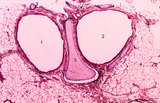
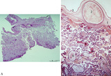
Transitional zone in the middle part of nasal vestibulum (comparable with red zone or vermilion border of lip, human) | Stain: Azan. At the right slightly cornified squamous epithelium (1) with small dermal papillae (2) and numerous blood vessels (3). To the left the transition to pseudostratified epithelium (4). In the submucosa branching seromucous nasal glands with draining ducts (5). | Stratified epithelium; Pseudostratified epithelium; Nasal glands; Seromucous glands | Poja Histology Collection - Respiratory System Subset |
| 177 |
 |
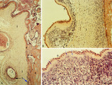
Survey of bronchus and lung parenchym (human, adult) | Stain: Azan. Lumina of bifurcated bronchi (1, 2). Thin plates of hyaline cartilage (3) and connective tissue surround the bronchi. Arrows (→) indicate lung alveoli with black-stained patches of carbon deposits. (4) Lung parenchyma with alveoli. | Lung parenchym | Poja Histology Collection - Respiratory System Subset |
| 178 |
 |
Chorioamnionitis (human) | Histologically chorioamnionitis describes the progression of the inflammatory process. Bacteria firstly colonized the chorioamniotic surface. In first two days polymorphonuclear granulocytes (PMN) migrate to the chorion (chorionitis) marginate and adhere to the bottom of the chorionic plate (stage 1... | chorioamnionitis; placenta; trophoblast | Poja Histology Collection - Placenta |
| 179 |
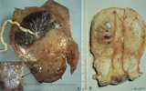 |
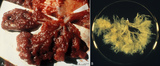
Partial hydatidiform mole and invasive mole (human) | (A) Macroscopy: The partial mole (1) occupies a large part of the placenta and is distinct from the normal chorionic plate where the umbilical cord (2) inserts eccentrically with branches of the umbilical vessels. The inset shows a circumscript area with swollen transparent grape-like vesicles (chor... | partial hydatidiform mole ; chorioadenoma | Poja Histology Collection - Placenta |
| 180 |
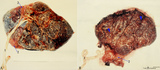 |
Normal term placenta (human) | (A) Left (fetal surface): 30-90 cm long umbilical cord (1), remnant of ruptured transparent amnion (2→) , near the eccentric attachment of the umbilical cord to the chorion plate ramification of the umbilical vessels (3) (arteries are smaller than the veins). (B) Right (maternal surface). The su... | placenta; amnion; chorion plate; cotyledon | Poja Histology Collection - Placenta |
| 181 |
 |
Survey and detail of the chorionic plate and intervillous space (human placenta, full-term) | Stain: (A) Perjodic acid-Schiff reaction (PAS); (B) Hematoxylin-azophloxine. (A) At the top the chorionic plate (1) with cross-sections of umbilical vessels (2). At (3) the folded amnion covering the chorionic plate. Ramifications of thicker stem villi (6) demonstrate free-floating terminal villi ... | placenta; chorionic plate; amnion; terminal villi | Poja Histology Collection - Placenta |
| 182 |
 |
Fetal cotyledons (normal term human placenta) | After dissection of one placental lobe several cotyledons (2) are visible in (A). Each fetal cotyledon (2) consists of a main stem villus (1) and all its branches. (B) After trypsinization of a cotyledon the tree of arborisation of the stem villus and its branches becomes visible. (By courtesy... | placenta; cotyledon; villus; trypsinization | Poja Histology Collection - Placenta |
| 183 |
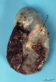 |
Macroscopy of fetus (human) | The fetus (1)is completely wrapped in a shiny transparent amnion (2) and closely associated with the brown-coloured placenta (3). (By courtesy of the Museum of Anatomy and Pathology, University Medical Center, St. Radboud University, Nijmegen, The Netherlands) | fetus; placenta; amnion | Poja Histology Collection - Placenta |
| 184 |
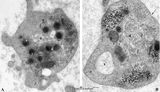 |
Platelets (peripheral blood, human) | Electron microscopy. These anucleate cells (2-4 μm) are derived from cytoplasmic fragments of a megakaryocyte and when free floating in the peripheral blood they develop thin extensions. These platelets contain among others few mitochondria (1), microtubules (2), glycogen (3), small vacuoles (4, op... | Poja Histology Collection - Blood & Bone Marrow Subset | |
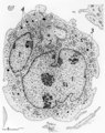
| 185 |
 |
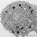
Megakaryocyte (bone marrow, rabbit) | Electron microscopy. A survey of a megakaryocyte demonstrates very well the differences in diameter between this giant polyploid cell (1) and the other young white blood cells (2-5). In the intermediate zone the (dark) granules (of the future platelets) are close associated with the electron-light i... | Poja Histology Collection - Blood & Bone Marrow Subset | |
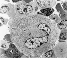
| 186 |
 |
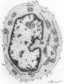
Megakaryocyte (peripheral blood, human) | Electron microscopy. A small part of a megakaryocyte (see also inset) shows two nuclear segments (1) and two areas of cytoplasm. Seen at this magnification and close to the perinuclear area, the granular population can be divided into homogeneous electron-dense (2) and electron-grey (3) granules. Th... | Poja Histology Collection - Blood & Bone Marrow Subset | |
| 187 |
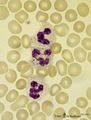 |
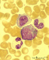
Neutrophilic granulocytes in peripheral blood smear (human) | Stain: May-Grnwald-Giemsa (MGG). The three neutrophilic granulocytes display segmented and lobulated nuclei. The lobes are connected with thin chromatin strands (→). The cytoplasm is ample filled with fine, dust-like granules and the majority are specific granules filled with enzymes such as lysoz... | Poja Histology Collection - Blood & Bone Marrow Subset | |
| 188 |
 |
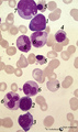
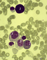
Multiple myeloma, plasma cell leukemia in bone marrow smear (human) | Stain: May-Grnwald-Giemsa (MGG). Mutiple myeloma (Kahler's disease) is characterized by proliferation of abnormal plasma cells (1, myeloma cells) in the bone marrow. In the great majority of patients secretion of a single homogenous immunoglobulin product (monoclonal component) occurs. Note the ecce... | Poja Histology Collection - Blood & Bone Marrow Subset | |
| 189 |
 |
Pleiomorph normal bone marrow smear (human) | Stain: May-Grnwald-Giemsa (MGG). The normal bone marrow with pleiomorphism, i.e. contains all stages of the erythroblastic differentiation series (1-5) as well as the stages of the myeloblastic series (6). (1) Basophilic erythroblast. (2),(3),(4) Polychromatic erythroblasts. (5) Orthochromatic er... | Poja Histology Collection - Blood & Bone Marrow Subset | |
| 190 |
 |
Lymphocyte | Scheme electron microscopy. A lymphocyte with an indented nucleus and (1) nucleolus shows a juxta-nuclear Golgi area (2) and centriole (3), several mitochondria (4) and sparsely profiles of rough endoplasmic reticulum (5) in the cytoplasm. Few dense-stained granules (6) are present. The granules con... | Poja Histology Collection - Blood & Bone Marrow Subset | |
| 191 |
 |
Small, medium and large lymphocytes in blood smear (human) | Stain: May-Grnwald-Giemsa (MGG). The picture shows three activation stages of lymphocytes as (1) small, (2) medium and (3) large lymphocytes. In that range the nucleus becomes less condensed and more transparent, while the amount of cytoplasm increases. The large lymphocyte also contains some granul... | Poja Histology Collection - Blood & Bone Marrow Subset | |
| 192 |
 |
Myeloid cells in bone marrow smear (human) | Stain: May-Grnwald-Giemsa (MGG). (1) promyelocyte is the largest cell in the myeloid series. It has a transparent nucleus with nucleoli and ample cytoplasm with many azurophilic granules. (2) myelocyte. (3) metamyelocyte with an indented nucleus. (4) beginning of nucleus segmentation in the band neu... | Poja Histology Collection - Blood & Bone Marrow Subset | |
| 193 |
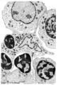 |
Erythroblastic island in bone marrow | Scheme electron microscopy. Different stages of maturing erythroblasts (2) are exposed in this scheme. The centrally localized phagocytic reticular cell (1) has many long cytoplasmic extensions that form a network with similar cells within the bone marrow. Its nucleus is irregular. The cytoplasm has... | Poja Histology Collection - Blood & Bone Marrow Subset | |
| 194 |
 |
Peroxidase activity in neutrophilic granulocyte (peripheral blood, guinea pig) | Electron microscopy (peroxidase reaction with diaminobenzidin staining). The elongated nucleus (3) of this strongly ameboid phagocytic cell has several projections. These nuclear extensions are interconnected by thin heterochromatin strands. The cytoplasm exhibits moderate amounts of organelles, gly... | Poja Histology Collection - Blood & Bone Marrow Subset | |
| 195 |
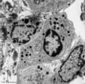 |
Plasma cells with Russell bodies (nose septum, rat) | Electron microscopy. Accumulation of few plasma cells. These mature plasma cells exhibit in their cytoplasm's dilated rough endoplasmic reticula (1) filled with electron-grey material (antibodies) as well as electron-dense granules (3) of varying sizes confirming their secretory activities. (2) indi... | Poja Histology Collection - Blood & Bone Marrow Subset | |
| 196 |
 |
Pro-megakaryocyte | Scheme electron microscopy. The pro-megakaryocyte (20-80 m) develops from a basophilic megakaryoblast (up to 50 m) that originates from a committed stem cell. The large pro-megakaryocyte starts with the early synthesis of the characteristic granules of platelets. Apart from a bilobed nucleus (1) and... | Blood; Bone Marrow; Electron microscopy; Pro-megakaryocyte; Megakaryocyte | Poja Histology Collection - Blood & Bone Marrow Subset |
| 197 |
 |
A bare megakaryocyte nucleus and myelocytes in bone marrow smear (human) | Stain: May-Grnwald-Giemsa (MGG). (1) Indicates a bare large-sized polyploid nucleus of a megakaryocyte which has totally shed the cytoplasm. These cells are often seen in normal marrow and are ultimately removed by macrophages. There is no cytoplasm left, nor platelets are left around the nucleus. T... | Poja Histology Collection - Blood & Bone Marrow Subset | |
| 198 |
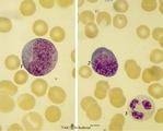 |
Promyelocyte and basophilic erythroblast in bone marrow smear (human) | Stain: May-Grnwald-Giemsa (MGG). The composition shows a (pro)myelocyte (1) along a basophilic erythroblast (2). The (pro)myelocyte nucleus is less condensed, contains nucleoli and the light basophilic cytoplasm is filled with azurophilic granules. The cytoplasm of the erythroblast is strong basophi... | Poja Histology Collection - Blood & Bone Marrow Subset | |
| 199 |
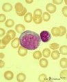 |
Myeloblast in bone marrow smear (human) | Stain: May-Grnwald-Giemsa (MGG). The myeloblast (1) shows a very transparent nucleus and several distinct nucleoli. The slightly basophilic cytoplasm is limited to a small rim in contrast to a promyelocyte. No granules are yet visible. (2) small lymphocyte. | Blood; Bone Marrow; Myeloblast; Lymphocyte | Poja Histology Collection - Blood & Bone Marrow Subset |
| 200 |
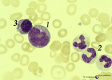 |
Promonocyte in bone marrow smear (human) | Stain: May-Grnwald-Giemsa (MGG). The promonocyte (1) has an irregular nucleus and shows the first step towards indentation (arrow). The cell has a characteristic greyish-blue cytoplasm. (2) Represents three young neutrophils. (3) A plasmacytoid lymphocyte. | Poja Histology Collection - Blood & Bone Marrow Subset |