The Health Education Assets Library (HEAL) is a collection of over 22,000 freely available digital materials for health sciences education. The collection is now housed at the University of Utah J. Willard Marriott Digital Library.
TO
Filters: Collection: ehsl_heal
| Title | Description | Subject | Collection | ||
|---|---|---|---|---|---|
| 151 |
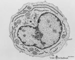 |
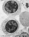
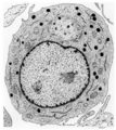
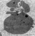
Plasmablast | Scheme electron microscopy. This plasmablast (up to 20 μm) shows an indented large nucleus (1) and a distinct nucleolus (2). In addition to free ribosomes long profiles of rough endoplasmic reticulum (RER) (3) are being developed concomitant with Golgi packages (4). The cytoplasm is scanty with few... | Poja Histology Collection - Blood & Bone Marrow Subset | |
| 152 |
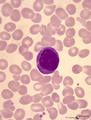 |
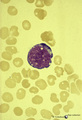
Plasmablast in peripheral blood smear (human) | Stain: May-Grnwald-Giemsa (MGG). The oval shaped cell with a slightly excentric nucleus and coarsely clumped chromatin has a basophilic blue cytoplasm in which the Golgi area usually remains unstained (white area). | Poja Histology Collection - Blood & Bone Marrow Subset | |
| 153 |
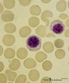 |
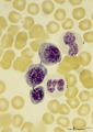
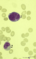
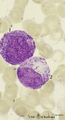
Plasmacytoid lymphocytes (activated B cells) in peripheral blood smear (human) | Stain: May-Grnwald-Giemsa (MGG). Two different plasmacytoid lymphocytes or activated young B cells (up to 15 μm) contain a dark-stained nucleus and a slightly basophilic cytoplasm with a kind of a 'nuclear hof' indicating the Golgi area. These cells will develop into plasma cells. | Poja Histology Collection - Blood & Bone Marrow Subset | |
| 154 |
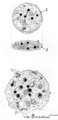 |
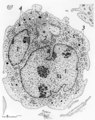
Platelets | Scheme electron microscopy. These disc-shaped anucleate cells (2-4 m) are derived from cytoplasmic fragments of the megakaryocyte. They contain few mitochondria, a canalicular system, a smooth tubular system, marginal localized microtubules, glycogen and different types of granula. A circumferential... | Poja Histology Collection - Blood & Bone Marrow Subset | |
| 155 |
 |
Platelets (peripheral blood, human) | Electron microscopy. These anucleate cells (2-4 μm) are derived from cytoplasmic fragments of a megakaryocyte and when free floating in the peripheral blood they develop thin extensions. These platelets contain among others few mitochondria (1), microtubules (2), glycogen (3), small vacuoles (4, op... | Poja Histology Collection - Blood & Bone Marrow Subset | |
| 156 |
 |
Platelets (peripheral blood, human) | Electron microscopy. These disc-shaped cells (2-4 m) without nucleus are derived from cytoplasmic fragments of the megakaryocyte. Free floating in the peripheral blood they develop thin extensions. They contain among others few mitochondria, smooth tubular systems, marginal localized microtubules, g... | Poja Histology Collection - Blood & Bone Marrow Subset | |
| 157 |
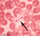 |
Platelets in peripheral blood smear (human) | Stain: May-Grnwald-Giemsa (MGG). The platelets or thrombocytes are small cell fragments released from megakaryocytes. The platelets (→) measure 1-3 μm in diameter. They contain fine azurophilic granules which may be dispersed throughout the cytoplasm or concentrated in the centre; in the latter c... | Poja Histology Collection - Blood & Bone Marrow Subset | |
| 158 |
 |
Pleiomorph bone marrow smear (human) | Stain: May-Grnwald-Giemsa (MGG). Low power view of a normal bone marrow smear. The bone marrow is pleiomorph and contains all cell types such as a large reticulum cells (1) with many granules or lysosomes (phagocytosis); myeloid cells (2); and erythroblasts (3). | Poja Histology Collection - Blood & Bone Marrow Subset | |
| 159 |
 |
Pleiomorph normal bone marrow smear (human) | Stain: May-Grnwald-Giemsa (MGG). The normal bone marrow with pleiomorphism, i.e. contains all stages of the erythroblastic differentiation series (1-5) as well as the stages of the myeloblastic series (6). (1) Basophilic erythroblast. (2),(3),(4) Polychromatic erythroblasts. (5) Orthochromatic er... | Poja Histology Collection - Blood & Bone Marrow Subset | |
| 160 |
 |
Polychromatic erythroblasts (bone marrow, rabbit) | Electron microscopy. Shown are two polychromatic erythroblasts or intermediate normoblasts between other erythroblasts and reticulocytes (2). Few mitochondria and polysomes are present but there is a decrease in amount of all organelles and nuclear clumping starts to take place. During this maturing... | Poja Histology Collection - Blood & Bone Marrow Subset | |
| 161 |
 |
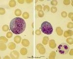
Polychromatic erythroblasts and neutrophilic granulocytes in bone marrow smear (human) | Stain: May-Grnwald-Giemsa (MGG). Three polychromatic erythroblasts or normoblasts (1) and two neutrophils (2). The erythroblasts show light basophilic cytoplasm and well condensed nuclear chromatin. The lower neutrophil (more mature) has a more segmented nucleus than the upper one. | Poja Histology Collection - Blood & Bone Marrow Subset | |
| 162 |
 |
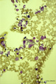
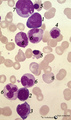
Polycythemia in bone marrow smear (human) | Stain: Hematoxylin and eosin. Low power view. Polycythemic phase. Hypercellular marrow as a result of excessive proliferation of erythroid cells (numerous dark nuclei) and of the megakaryocytic lineage (tendency to cluster). (1) fat cells in bone marrow. (2) megakaryocytes. (3) erythropoietic cell t... | Poja Histology Collection - Blood & Bone Marrow Subset | |
| 163 |
 |
Pro-megakaryocyte | Scheme electron microscopy. The pro-megakaryocyte (20-80 m) develops from a basophilic megakaryoblast (up to 50 m) that originates from a committed stem cell. The large pro-megakaryocyte starts with the early synthesis of the characteristic granules of platelets. Apart from a bilobed nucleus (1) and... | Blood; Bone Marrow; Electron microscopy; Pro-megakaryocyte; Megakaryocyte | Poja Histology Collection - Blood & Bone Marrow Subset |
| 164 |
 |
Proerythroblast and eosinophilic metamyelocyte in bone marrow smear (human) | Stain: May-Grnwald Giemsa (MGG). The late proerythroblast (1) has a deep blue-stained basophilic cytoplasm and so called ears (→, slight bulging) of sidewards accumulated ribosomes (in electron microscopy) in the cytoplasm. The eosinophilic metamyelocyte (2) has characteristic large, solitary brow... | Poja Histology Collection - Blood & Bone Marrow Subset | |
| 165 |
 |
Proerythroblasts in bone marrow smear (human) | Stain: May-Grnwald-Giemsa (MGG). The two proerythroblasts (1) are large cells with condensed nuclear chromatin and a deep blue-stained basophilic cytoplasm with so-called ears (→, bulging) where ribosomes (in electron microscopy) are aggregated or shifted towards the periphery of the cell. (2) smu... | Poja Histology Collection - Blood & Bone Marrow Subset | |
| 166 |
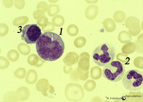 |
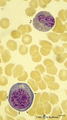
Promonocyte in bone marrow smear (human) | Stain: May-Grnwald-Giemsa (MGG). The promonocyte (1) has an irregular nucleus and shows the first step towards indentation (arrow). The cell has a characteristic greyish-blue cytoplasm. (2) Represents three young neutrophils. (3) A plasmacytoid lymphocyte. | Poja Histology Collection - Blood & Bone Marrow Subset | |
| 167 |
 |
Promonocyte in bone marrow smear (human) | Stain: May-Grnwald-Giemsa (MGG). The young promonocyte (1) shows clear nucleoli and a distinct indentation of the nucleus (arrow). (2) Neutrophilic myelocyte. | Poja Histology Collection - Blood & Bone Marrow Subset | |
| 168 |
 |
Promyelocyte | Scheme electron microscopy. The large promyelocyte (15-25 μm) contains a nucleus (1) with one or two nucleoli, the rough endoplasmic reticulum (4) is present and show many expanded profiles. Distinct also are numerous ribosomes and a well-developed Golgi area (2) where 'non-specific', primary or az... | Poja Histology Collection - Blood & Bone Marrow Subset | |
| 169 |
 |
Promyelocyte and basophilic erythroblast in bone marrow smear (human) | Stain: May-Grnwald-Giemsa (MGG). The composition shows a (pro)myelocyte (1) along a basophilic erythroblast (2). The (pro)myelocyte nucleus is less condensed, contains nucleoli and the light basophilic cytoplasm is filled with azurophilic granules. The cytoplasm of the erythroblast is strong basophi... | Poja Histology Collection - Blood & Bone Marrow Subset | |
| 170 |
 |
Promyelocyte and myelocyte in bone marrow smear (human) | Stain: May-Grnwald-Giemsa (MGG). The large promyelocyte (1) has a large nucleus with fine disperse chromatine and visible nucleoli. The cell contains primary, azurophilic granules. In the myelocyte (2) the nucleus is located eccentrically at one side of the cell, and the amount of cytoplasm is relat... | Poja Histology Collection - Blood & Bone Marrow Subset | |
| 171 |
 |
Promyelocyte in bone marrow smear (human) | Stain: May-Grnwald-Giemsa (MGG). The promyelocyte is the largest cell in the myeloid series in bone marrow. The nucleus is still transparent with fine chromatin and nucleoli are well visible. The cytoplasm contains ample azurophilic, primary granules. | Poja Histology Collection - Blood & Bone Marrow Subset | |
| 172 |
 |
Promyelocyte in bone marrow smear (human) | Stain: May-Grnwald-Giemsa (MGG). The large promyelocyte contains nucleoli, coarse chromatin pattern, and abundant azurophilic granules in a basophilic cytoplasm. (2) A neutrophilic band form leukocyte with folded nucleus. | Poja Histology Collection - Blood & Bone Marrow Subset | |
| 173 |
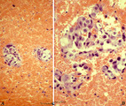 |
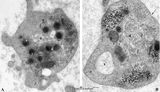
Prostatic carcinoma metastasis in bone marrow of sternum (human) | Stain: Hematoxylin and eosin. Survey (A) and detail (B) of a prostate carcinoma metastasized to the bone marrow, (sternum aspiration). Clusters of tumour cells; clearly foreign to the marrow. Common primary tumours that metastasize to the bone marrow include breast, prostate, lung, thyroid, kidney, ... | Poja Histology Collection - Blood & Bone Marrow Subset | |
| 174 |
 |
Reactive promyelocyte during sepsis or chemotherapy in peripheral bood smear (human) | Stain: May-Grnwald-Giemsa (MGG). Neutrophilic promyelocyte with toxic granulation and vacuolar degeneration. Note that nucleoli are still visible. | Poja Histology Collection - Blood & Bone Marrow Subset | |
| 175 |
 |
Reticulocyte (peripheral blood, human) | Electron microscopy. An anucleate reticulocyte with fimbriated processes (asymmetrical folding of the cell membrane) after nuclear extrusion. The cytoplasm contains a heavy amount of hemoglobin, electron-dense lysosomes, vesicular remnants as well as scattered clumped polysomes. | Poja Histology Collection - Blood & Bone Marrow Subset |