The Health Education Assets Library (HEAL) is a collection of over 22,000 freely available digital materials for health sciences education. The collection is now housed at the University of Utah J. Willard Marriott Digital Library.
TO
Filters: Collection: ehsl_heal
| Title | Description | Subject | Collection | ||
|---|---|---|---|---|---|
| 151 |
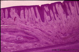 |
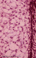
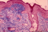
Gingiva - 'attached' gingiva of decalcified alveolar bone, human | Stain: Hematoxylin and eosin. Stratified squamous epithelium with parakeratosis (reddish) and deep papillae of the lamina propria. At the bottom, lamellar bone tissue (alveolar bone) with thickened periosteum (part of the periodontal ligament). At the bottom dentin. | oral cavity | Poja Histology Collection - Oral Cavity Subset |
| 152 |
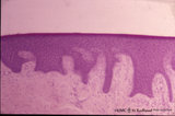 |
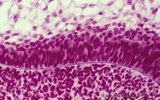
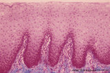
Gingiva - 'free' gingiva of decalcified alveolar bone, human | Stain: Hematoxylin and eosin. Thick layer of stratified squamous epithelium with parakeratosis (reddish) and broad papillae of the lamina propria. At the left, dense collagen tissue (projections of the the periodontal ligament). | oral cavity | Poja Histology Collection - Oral Cavity Subset |
| 153 |
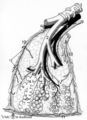 |
Gross scheme of a lung lobule and its vascularization (human) | Scheme of branching of intrapulmonary bronchus. A small intrapulmonal bronchus with cartilage rings (1) divides into bronchioli (2), at (3) the terminal bronchioli that continues into respiratory bronchioli (4) followed by alveolar ducts (5) and at (6) the alveoli. A respiratory bronchiolus with alv... | Intrapulmonary bronchus; Bronchioli; Alveolar ducts; Lymphatic plexus; Visceral pleura | Poja Histology Collection - Respiratory System Subset |
| 154 |
 |
Heparan sulfates in the terminal sac period of the lung (mouse, fetus) | Stain: fluorescence microscopy with anti-heparan sulphate antibody (a phage-display antibody, HS4E4). Heparan sulfates are linear polysaccharides that belong to the group of the glycosaminoglycans (GAGs). These GAGs are found associated with basement membranes as shown here. The sulphated saccharide... | Bronchioli; Air spaces; Terminal sac period | Poja Histology Collection - Respiratory System Subset |
| 155 |
 |
Heparan sulfates in the terminal sac-period of the lung (mouse, fetus) | Stain: fluorescence microscopy with anti-heparan sulphate antibody (a phage-display antibody, LKiv69). Heparan sulfates are linear polysaccharides that belongs to the group of the glycosaminoglycans (GAGs). The sulphated saccharide domains provide numerous docking sites for protein ligands. These GA... | Bronchioli; Air spaces; Terminal sac period | Poja Histology Collection - Respiratory System Subset |
| 156 |
 |
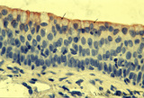
Keratin 7 in the bronchial epithelium of the lung (human, adult) | Stain: anti-keratin 7 antibody (Pan-Ck 7) immunoperoxidase staining (with aminoethylcarbazole (AEC) substrate). The epithelium of the bronchus (1) stains dark brown-red after the reaction with AEC indicating a positive reaction for cytokeratin 7. This antibody can be used in the study of development... | Bronchiolus; Immunoperoxidase; Immuno-reaction | Poja Histology Collection - Respiratory System Subset |
| 157 |
 |
Keratin 7 in the lining alveolar epithelium of the lung (human, adult) | Stain: anti-keratin 7 antibody (Pan-Ck 7) immunoperoxidase staining (with aminoethylcarbazole (AEC) substrate). Both alveolar epithelial cell types I (1) and II (2) stain dark brown-red after the reaction with AEC indicating a positive reaction for cytokeratin 7. Note the white (non-stained) capilla... | Pneumocyte I; Pneumocyte II; Imunoperoxidase; Immuno-reaction | Poja Histology Collection - Respiratory System Subset |
| 158 |
 |
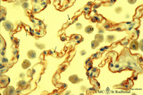
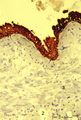
Keratin 7 staining in dysplastic epithelium of a bronchus (human, adult) | Stain: anti-keratin 7 antibody (Pan-Ck 7) immunoperoxidase staining (with aminoethylcarbazole (AEC) substrate). A red-brown staining with AEC indicates a positive reaction for cytokeratin 7. Disturbances in the growth and maturing might result in dysplasia of the epithelium with a changed reaction p... | Immunoperoxidase; Immuno-reaction | Poja Histology Collection - Respiratory System Subset |
| 159 |
 |
Keratin 7 staining in squamous metaplasia and dysplasia of the epithelium of a bronchus (human, adult) | Stain: anti-keratin 7 antibody (Pan-Ck 7) immunoperoxidase staining (with aminoethylcarbazole (AEC) substrate). A red-brown staining with AEC indicates a positive reaction for cytokeratin 7. Note that the top layer only reacts weakly positive for keratin 7 (↓). Upon squamous metaplasia the bronch... | Squamous metaplasia; Immunoperoxidase; Immuno-reaction | Poja Histology Collection - Respiratory System Subset |
| 160 |
 |
Keratin 7 staining in squamous metaplasia of the epithelium of a bronchiolus (human, adult) | Stain: anti-keratin 7 antibody (Pan-Ck 7) immunoperoxidase staining (with aminoethylcarbazole (AEC) substrate). A red-brown staining with AEC indicates a positive reaction for cytokeratin 7. Note that the top layer only is stained positively for keratin 7 (↓). Upon squamous metaplasia the bronchio... | Bronchiolus; Squamous metaplasia; Immunoperoxidase; Immuno-reaction | Poja Histology Collection - Respiratory System Subset |
| 161 |
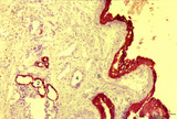 |
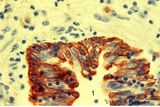
Keratin staining of bronchus epithelium (human, adult) | Stain: Anti-keratin-5-7-14-19 (OVTL 12/5 monoclonal antibody with immunoperoxidase staining on frozen sections). The pseudostratified epithelium (1) is stained keratin-positively (brown product) as well as the epithelial derivates such as the bronchial glands (2) and their excretory ducts (3). Other... | Bronchial epithelium; Keratin antibodies; Tumor diagnosis | Poja Histology Collection - Respiratory System Subset |
| 162 |
 |
Keratin staining of bronchus epithelium (human, adult) | Stain: Anti-keratin-5-7-14-19 (OVTL 12/5 monoclonal antibody with immunoperoxidase staining on frozen sedctions). The pseudostratified epithelium (1) reacts positively (brown-red product), the nuclei of the basal cells are well visible (arrows). Other structures react negatively (blue stain) such as... | Tumor diagnosis; Bronchial epithelium; Lung parenchym; Keratin antibodies | Poja Histology Collection - Respiratory System Subset |
| 163 |
 |
Lamellar body of a type II alveolar cell in the lung (dog) | Electron microscopy. Extrusion of surfactant material out of a type II alveolar cell (pneumocyte II, great alveolar cell). After fusion of a multilamellar body (1) (cytosome) with the apical cell membrane (note microvilli 2) the electron-dense lamellar content or surfactant (consisting of phosphatid... | Type II alveolar cell; Pneumocyte II; Multilamellar bodies; Cytosomes; Tubular myelin | Poja Histology Collection - Respiratory System Subset |
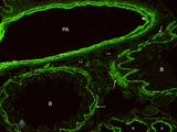
| 164 |
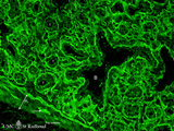 |
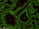
Laminin in the canalicular period of the lung (mouse, embryo) | Stain: Fluorescence microscopy with anti-laminin antibody. Laminin is a component of the basement membrane. Strong fluorescence visualizes the course of all basement membranes, especially around the bronchioles (B) and their branches in the developing lung tissue. In between the distal air spaces (t... | Bronchioli; Canalicular period | Poja Histology Collection - Respiratory System Subset |
| 165 |
 |
Late cap stage in tooth development - human, embryo | Stain: Azan. From top to bottom: Top side stellate reticulum (enamel pulp) consisting of a network of ectoderm-derived cells; Right side outer dental epithelium with part of the fibrous tooth follicle. This epithelium will further develop downwards as the outer layer of the Hertwig's epithelial root... | oral cavity | Poja Histology Collection - Oral Cavity Subset |
| 166 |
 |
Late cap stage in tooth development - human, embryo | Stain: Azan. The stellate reticulum (enamel pulp) consists of a network of ectoderm-derived branched cells and fluid-filled spaces (a.o. proteoglycans). It is a specialized avascular layer as a support and protection for the inner dental epithelial cells. At the right side one layer of cuboidal oute... | oral cavity; tooth development; outer dental epithelium; stellate reticulum | Poja Histology Collection - Oral Cavity Subset |
| 167 |
 |
Late cap stage in tooth development - human, embryo | Stain: Azan. From top to bottom: At the top stellate reticulum (enamel pulp) consisting of a loose network of ectoderm-derived cells; Darker stained cell layers of the stratum intermedium; Columnar inner dental epithelium (presecreetory ameloblasts) at the distal side (secretion area) oriented towar... | oral cavity | Poja Histology Collection - Oral Cavity Subset |
| 168 |
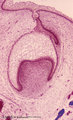 |
Late cap stage of tooth development - human, embryo; low magnification | Stain: Azan. From top to bottom: Stratified ectoderm with a distinct basal layer (red line) of cuboid cells; Dental lamina giving rise to the cap stage (center) and to the primordium of permanent tooth (right); Odontogenic organ or enamel organ (future deciduous tooth surrounded by fibrous tooth fol... | oral cavity; dental lamina | Poja Histology Collection - Oral Cavity Subset |
| 169 |
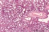 |
Late terminal sac period of developing lung (human, fetus, low magnification) | Stain: Hematoxylin and eosin. Cross-sections of a bronchus (1) with cartilage rings (2) closely associated with pulmonary arteries (3). The alveolar appearance is evident, and the open spaces represent future alveolar duct systems as well as alveolar sacs. The cellular septa are still thick due to n... | Lung development; Terminal sac period | Poja Histology Collection - Respiratory System Subset |
| 170 |
 |
Lip (human), external skin surface | Stain: Azan. Keratinized squamous epithelium with one hair follicle associated with sebaceous gland. Richly vascularized connective tissue. Note the thin red cornified layer on the surface. | oral cavity | Poja Histology Collection - Oral Cavity Subset |
| 171 |
 |
Lip (human), mucoserous labial glands in mucous inner surface | Stain: Azan. Mixed labial glands with serous demilunes (von Ebner-Giannuzzi), a few myoepithelial cells, and a striated duct (right upper corner). | oral cavity; lining mucosa; labial glands | Poja Histology Collection - Oral Cavity Subset |
| 172 |
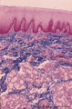 |
Lip (human), mucous inner surface | Stain: Azan. Non-keratinized epithelium with high, narrow dermal papillae and many capillaries. Mixed labial glands (seromucous) and few skeletal muscle fibers (orbicularis oris) in the submucosa. | oral cavity; lining mucosa; labial glands | Poja Histology Collection - Oral Cavity Subset |
| 173 |
 |
Lip (human), mucous inner surface | Stain: Azan. Non-keratinized epithelium with high, narrow dermal papillae and many capillaries. Note lymphocytic infiltrates and the capillaries in the dermal papillae. | oral cavity; lining mucosa | Poja Histology Collection - Oral Cavity Subset |
| 174 |
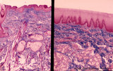 |
Lip (human), outer (left) and inner side (right) | Stain: Azan. Left image: keratinized squamous epithelium with thin red cornified layer, hair follicle, sebaceous glands and skeletal muscle fibers (orbicularis oris). Right image: non-keratinized epithelium with high, narrow dermal papillae and more capillaries. Mixed labial glands (seromucous), f... | oral cavity; lining mucosa | Poja Histology Collection - Oral Cavity Subset |
| 175 |
 |
Lip (human), region between red zone (vermilion border) and mucosa inner surface | Stain: Azan. Bundles of skeletal muscle fibers (musculus orbicularis oris), highly vascularized lamina propria. Note the narrow dermal papillae on the left side (inner lip) and the broad irregular papillae on the right side (red zone of the lip). | oral cavity; lining mucosa; red zone; vermilion border | Poja Histology Collection - Oral Cavity Subset |