The Health Education Assets Library (HEAL) is a collection of over 22,000 freely available digital materials for health sciences education. The collection is now housed at the University of Utah J. Willard Marriott Digital Library.
TO
| Title | Description | Subject | Collection | ||
|---|---|---|---|---|---|
| 151 |
 |
PAC's with and without aberrant conduction - marquette | PAC's with and without aberrant conduction - marquette | Knowledge Weavers ECG | |
| 152 |
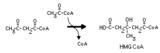 |
Hydroxymethylglutaryl CoA synthase reaction | This irreversible reaction occurs in the mitochondria, where it is the first step in ketone body synthesis. It also occurs in the cytoplasm, where it leads to isoprenoid and steroid synthesis. | Biosynthesis | Knowledge Weavers Fatty Acids |
| 153 |
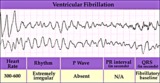 |
Ventricular fibrillation - marquette | Ventricular fibrillation - marquette | Knowledge Weavers ECG | |
| 154 |
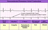 |
Nonconducted PAC - marquette | Nonconducted PAC - marquette | Knowledge Weavers ECG | |
| 155 |
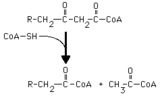 |
Thiolase reaction | Thiolase (3-ketoacyl CoA thiolase) cleaves a long chain fatty acyl CoA, forming acetyl CoA and a long chain fatty acyl CoA that is two carbons shorter. | Knowledge Weavers Fatty Acids | |
| 156 |
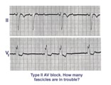 |
Trifascicular block: RBBB, LAFB, and mobitz II 2nd degree AV block | A nice example of trifascicular block: Lead V1 shows RBBB; Lead II is mostly negative with an rS morphology suggesting left anterior fascicular block. Since Mobitz II 2nd degree AV block is more often located in the bundle branch system, the only location left for this block is the left posterior ... | Knowledge Weavers ECG | |
| 157 |
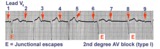 |
2nd degree AV block, type I, with junctional escapes | Junctional escapes are passive, protective events whenever the heart rate slows below that of the escape mechanism. In this example of 2nd degree AV block, type I, the escapes occur following the non-conducted P waves. Arrows indicate the position of the P waves. Note that the escape beats have a... | Knowledge Weavers ECG | |
| 158 |
 |
Atrial flutter with 2:1 AV conduction | Atrial flutter with 2:1 AV block is one of the most frequently missed ECG rhythm diagnoses because the flutter waves are often hard to find. In this example two flutter waves for each QRS are best seen in lead III and V1. The ventricular rate at 150 bpm should always prompt us to consider atrial fl... | Knowledge Weavers ECG | |
| 159 |
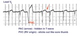 |
Identification of PVC's and PAC's | PVC's usually stick out like sore thumbs; PAC's are often difficult to see because they are hidden in the preceding ST-T wave. The PVC in this example is mostly negative in lead V1 suggesting RV origin; i.e., most of activation is moving in posterior direction towards the left ventricle. | Knowledge Weavers ECG | |
| 160 |
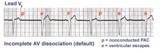 |
AV dissociation by default | The nonconducted PAC's set up a long pause which is terminated by ventricular escapes; note the wider QRS morphology of the escape beats indicating their ventricular origin. Incomplete AV dissociation occurs during the escape beats, since the atria are still under the control of the sinus node. | Knowledge Weavers ECG | |
| 161 |
 |
Lead error: V1 and V3 are transposed! | In the precordial leads the V1 and V3 chest electrodes are interchanged. Experienced ECG interpreters should be able to spot this lead placement error. | Knowledge Weavers ECG | |
| 162 |
 |
Infero-posterior MI & RBBB: Frontal Plane Leads + V1 | Infero-posterior MI & RBBB: Frontal Plane Leads + V1 | Knowledge Weavers ECG | |
| 163 |
 |
WPW Type Pre-excitation: Precordial Leads | WPW Type Pre-excitation: Precordial Leads | Knowledge Weavers ECG | |
| 164 |
 |
Acute anterior MI | Acute anterior MI | Knowledge Weavers ECG | |
| 165 |
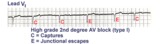 |
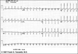
2nd degree AV block, type I with escapes and captures | Often in the setting of 2nd degree AV block the pauses caused by nonconducted P waves are long enough to enable escape pacemakers from the junction or ventricles to take over. This example illustrates junctional escapes, labled E and captures, labled C. Note that the PR intervals for the captures ... | Knowledge Weavers ECG | |
| 166 |
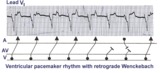 |
Ventricular paced rhythm with retrograde wenckebach | Retrograde atrial captures from a ventricular paced rhythm are occurring with increasing R-P intervals; i.e., retrograde Wenckebach. The ladder diagram indicates that after the blocked retrograde event, a single sinus P wave is seen dissociated from the ventricular rhythm. | Wenckebach AV Block | Knowledge Weavers ECG |
| 167 |
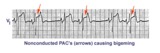 |
Nonconducted PAC's: an unusual bigeminy | Occasionally nonconducted PAC's can create interesting rhythms. In this example every other sinus beat is followed by an early, nonconducted PAC. The resulting pause sets up a bigeminal rhythm. Note the distortion of the T waves caused by the nonconducted PAC's. | Knowledge Weavers ECG | |
| 168 |
 |
Atrial flutter with 2:1 AV conduction: leads II, III, V1 | In leads II and III, the one of the flutter waves occurs at the end of the QRS complex and might be mistaken for part of the QRS itself; i.e., the S wave. In lead V1, the two flutter waves for every QRS are more easily identified. | Knowledge Weavers ECG | |
| 169 |
 |
Accelerated IVR with AV dissociation - marquette | Accelerated IVR with AV dissociation - marquette | Knowledge Weavers ECG | |
| 170 |
 |
Old inferior MI | Old inferior MI | Knowledge Weavers ECG | |
| 171 |
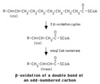 |
Beta-oxidation of a delta-9 fatty acyl CoA | Enoyl CoA isomerase is required to move the double bond in a Delta-9 fatty acyl CoA to a position where it can continue in beta-oxidation. | Knowledge Weavers Fatty Acids | |
| 172 |
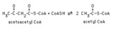 |
Thiolase reaction with acetoacetyl CoA | Thiolase (3-ketoacyl CoA thiolase) cleaves acetoacetyl CoA, forming two molecules of acetyl CoA. | Knowledge Weavers Fatty Acids | |
| 173 |
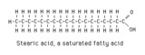 |
Stearic acid structure | Stearic acid is a typical long chain saturated fatty acid. | Knowledge Weavers Fatty Acids | |
| 174 |
 |
Diagram: type I vs. type II 2nd degree AV block | In type I 2nd degree AV block the PR progressively lengthens until a nonconducted P wave occurs. The PR gets longer by smaller and smaller increments; this results in gradual shortening of the RR intervals. The RR interval of the pause is usually less than the two preceding RR intervals. The RR i... | Knowledge Weavers ECG | |
| 175 |
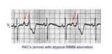 |
PAC's with RBBB aberration | These PAC's, indicated by arrows, enter the ventricles and find the right bundle refractory. They therefore conduct with RBBB aberrancy. In most normal hearts the right bundle recovery time is longer than the left bundle's; most aberrancy, therefore, has aRBBB morphology. In some diseased hearts t... | Knowledge Weavers ECG |