The Health Education Assets Library (HEAL) is a collection of over 22,000 freely available digital materials for health sciences education. The collection is now housed at the University of Utah J. Willard Marriott Digital Library.
TO
Filters: Collection: "ehsl_heal"
| Title | Description | Subject | Collection | ||
|---|---|---|---|---|---|
| 151 |
 |
Adrenal cortical hyperplasia | Patient with ectopic ACTH production and diffuse adrenal cortical hyperplasia. | ACTH | HEAL Reviewed Collection |
| 152 |
 |
Adrenal gland neoplasm | Adrenal gland neoplasm | Knowledge Weavers Pathology | |
| 153 |
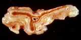 |
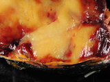
Adrenal gland unfixed normal | Adrenal gland unfixed normal. Cut surface. Photograph. Multimedia. | Adrenal Glands; Endocrinology; Anatomy | Slice of Life |
| 154 |
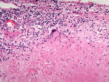 |
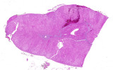
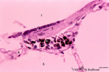
Adrenal gland with granulomatous reaction (gumma) to syphilis | This high power image (20X) shows a granuloma with a giant cell in the adrenal gland. The granuloma is a gumma formed during tertiary syphilis | gumma | HEAL Reviewed Collection |
| 155 |
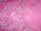 |
Adrenal gland with granulomatous reaction (gumma) to syphilis | This medium power image shows a granuloma in the adrenal gland. The granuloma is a gumma formed during tertiary syphilis. | Gumma | HEAL Reviewed Collection |
| 156 |
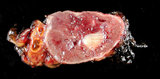 |
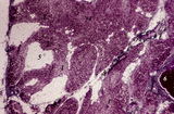
Adrenal gland with pheochromocytoma | Patient with clinical symptoms of hypertension. Image shows adjacent normal adrenal gland with paraganglioma (pheochromocytoma) involving adrenal medulla. | Adrenalin; Noradrenalin | HEAL Reviewed Collection |
| 157 |
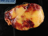 |
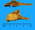
Adrenal myelolipoma | Gross photograph with ruler showing residual normal adrenal and mixture of yellow adipose tissue and red myeloid areas of myelolipoma. | benign | HEAL Reviewed Collection |
| 158 |
 |
Adrenal myelolipoma | Gross photograph showing residual normal adrenal and mixture of yellow adipose tissue and red myeloid areas of myelolipoma. | benign | HEAL Reviewed Collection |
| 159 |
 |
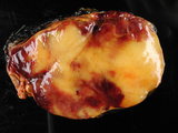
Adrenal myelolipoma | Close up showing residual normal adrenal and mixture of yellow adipose tissue and red myeloid areas of myelolipoma. | Benign | HEAL Reviewed Collection |
| 160 |
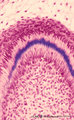 |
Advanced bell stage with ameloblasts and odontoblasts in tooth development - human embryo | Stain: Azan. From top to bottom: Stellate reticulum consisting of a non-vascularized network of ectoderm-derived cells; Cell layers of the stratum intermedium; Columnar (presecretory) ameloblasts with their upper side (nuclear area) in close contact with the stratum intermedium, and at the distal si... | oral cavity; predentin | Poja Histology Collection - Oral Cavity Subset |
| 161 |
 |
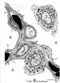
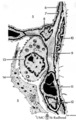
Afferent lymph vessel in lymph node (human) | Stain: Azan. Left (A) and right (B): part of the cortex of a lymph node with the capsule (1) and subcapsular (or marginal) sinus (2) filled with lymphocytes. Left (A): (3) show perpendicularly localized reticular cells and fibres (blue) in the sinus. (4) indicate afferent lymph vessel with valves ... | lymph vessel; subcapsular sinus; follicle; cortex | Poja Histology Collection - Lymphatic Tissues and Organs Subset |
| 162 |
 |
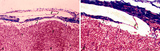
Age involution of thymus (human) | Stain: Azan. A: Although the adipose tissue in the thymus of a patient of 65 years is predominant it still contains areas of remnants (arrows 1+2) of cortical (2) and medullary portions (1) of the thymus. B+C: Degradation of thymic tissues is less progressed in adults and shows less replacement of... | thymus age; thymus involution; adipose cells; lymphoid tissue | Poja Histology Collection - Lymphatic Tissues and Organs Subset |
| 163 |
 |
Age involution of thymus (human, postpuberal) | Stain: Azan. The size of the thymus is age-dependent, and undergoes a continuous process of involution, starting at postpuberal age. Due to depletion and reduced production of cortical thymocytes, as well as a gradual atrophy of the epithelial cells, the clear distinction between medulla (1) and cor... | thymus age; adipose cells; thymus involution; lymphoid tissue | Poja Histology Collection - Lymphatic Tissues and Organs Subset |
| 164 |
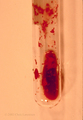 |
Agglutination of Red Blood Cells | agglutination of RBCs at room temperature in CBC tube | Ec0400; Cold Agglutinin Disease; CAD | Albert Einstein College of Medicine Gallery of Hematology Images |
| 165 |
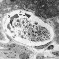 |
Aggregation of platelets (liver, rat) | Electron microscopy. Within a liver sinusoid (S) close to a small bile ductile (B) in the periportal area of the liver some erythocytes (E) and an aggregation of platelets (3) are present. (2) represents the space of Disse to which the liver parenchyma cells border (1). These disc-shaped anucleate p... | Poja Histology Collection - Blood & Bone Marrow Subset | |
| 166 |
 |
Air-blood barrier in the lung (mammals) | Scheme electron microscopy. (1, ↓) Represents type I pneumocytes lining alveolar spaces (A). Cell (2) represents a free alveolar macrophage. The type II pneumocyte (3) is adherent to type I pneumocyte extensions (note junctional connection), and contains multilamellar bodies (surfactant). A myofib... | Pneumocyte type I ; Pneumocyte type II | Poja Histology Collection - Respiratory System Subset |
| 167 |
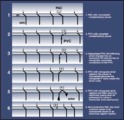 |
All about premature beats | All about premature beats | Knowledge Weavers ECG | |
| 168 |
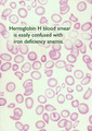 |
Alpha Thalassemia (3 alpha gene deletion) | peripheral smear of Hb H (three alpha gene deletion) | Hypochromic; Microcytic Anemia; Fb010T | Albert Einstein College of Medicine Gallery of Hematology Images |
| 169 |
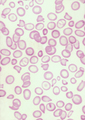 |
Alpha Thalassemia (3 alpha gene deletion) | peripheral smear of Hb H (three alpha gene deletion) | Microcytic Anemia; Fb0100 | Albert Einstein College of Medicine Gallery of Hematology Images |
| 170 |
 |
Alveolar cell type II (pneumocyte II) in an alveolus (dog) | Electron microscopy. At (X) the alveolar space, the bulging alveolar cell type II shows the characteristic multilamellar bodies (*, cytosomes) that contain precusor material of surfactant. The lamellar bodies are responsible for the vacuolated appearance of these cells, and they give rise to surfact... | Pneumocyte II; Alveolar cell type II | Poja Histology Collection - Respiratory System Subset |
| 171 |
 |
Alveolar cells in the lung (mammals) | Scheme electron microscopy. (5) alveolar space; (6) type I Pneumocyte; (7) basal lamina; (8) myofibroblast; (9) collagen and elastin fibers; (10) mesothelial cell of the visceral pleura; (11) capillary with erythrocyte; (12) endothelial cell lining the capillary; (13) type II pneum... | Pneumocyte type I ; Pneumocyte type I I | Poja Histology Collection - Respiratory System Subset |
| 172 |
 |
Alveolar duct in the lung (mouse) | Stain: PAS and hematoxylin. Part of an alveolar duct lumen (1) that shows bronchiolar characteristics such as cuboidal epithelium (2) covering a bundle of smooth muscle (3) and connective tissue containing macrophages (4) with black pigment deposits. Note PAS-positivity of these cuboidal cells (Clar... | Alveolar ducts; Clara cells | Poja Histology Collection - Respiratory System Subset |
| 173 |
 |
Alveolar macrophage in lung (rat) | Electron microscopy. A wandering alveolar macrophage (1) migrates through an alveolar pore from an alveolar space (A1) into another (A2). Note the dark lysosomal structures and abundant organelles in its cytoplasm. The thin lining type I alveolar cell is hardly discernable (thin arrows →). Interst... | Pneumocyte I; Alveolar macrophages | Poja Histology Collection - Respiratory System Subset |
| 174 |
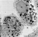 |
Alveolar macrophages in lung (human, adult) | Electron microscopy. Two macrophages in the alveolar space (N = nucleus). Note the heterogeneity of many lysosomal structures containing among others carbon particles and the abundancy of organelles. | Alveolar macrophages | Poja Histology Collection - Respiratory System Subset |
| 175 |
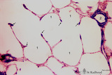 |
Alveolar sac in the lung (human) | Stain: Azan. Characteristic alveolar tips (arrows) of neighbouring thin-walled alveoli (1). The tips contain elastin masked by collagen (blue-stained). At (2) small pulmonary arteries. | Alveolar tips | Poja Histology Collection - Respiratory System Subset |