The Health Education Assets Library (HEAL) is a collection of over 22,000 freely available digital materials for health sciences education. The collection is now housed at the University of Utah J. Willard Marriott Digital Library.
| Title | Description | Subject | Collection | ||
|---|---|---|---|---|---|
| 126 |
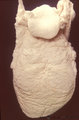 |
Tongue (macroscopy, dorsal side, human) | At the bottom (apex) of the picture the dorsal side is covered with numerous closed packed, small, conical filiform papillae. Interspersed among them are the larger fungiform papillae. Posteriorily the circumvallatae papillae are arranged in a V-shape row. Behind the V-row the area is covered with l... | oral cavity; papillae | Poja Histology Collection - Oral Cavity Subset |
| 127 |
 |
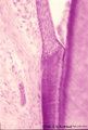
Tongue (ventral part, human) | Stain: Hematoxylin and eosin. Non-keratinized squamous epithelium, followed by a small lamina propria and the intrinsic striated lingual muscles. | oral cavity; lingual muscles | Poja Histology Collection - Oral Cavity Subset |
| 128 |
 |
Tongue Papillae Scheme - tongue, human, adult | A. Scheme survey dorsal surface of tongue (human, adult). Magnification 35x. B. Scheme of filiform and fungiform papillae (tongue, human, adult). Magnification 35x. 1. foliate papillae (rudimentary in human); 2. circumvallate papillae; 3. foramen cecum; 4. palatine tonsils; 5. filiform ... | oral cavity; papillae | Poja Histology Collection - Oral Cavity Subset |
| 129 |
 |
Tooth ('free' gingiva of decalcified tooth, human, adult) | Stain: Hematoxylin and eosin. Oral side (left) with stratified squamous epithelium, at the right side part of a tooth (dentin), at the bottom side part of the alveolar bone. The epithelium shows slight parakeratosis with many narrow deep papillae of the lamina propria (oral side). On the tooth-relat... | oral cavity; junctional epithelium; alveolar bone; gingival sulcus | Poja Histology Collection - Oral Cavity Subset |
| 130 |
 |
Tooth ('free' gingiva of decalcified tooth, human, adult) | Stain: Hematoxylin and eosin. On top part of the gingival sulcus, at the left stratified junctional epithelium normally attached to the surface of the enamel (dissolved due to decalcification). Top left shows foci of chronic inflammatory cell infiltration, left side faint stained bundle of fibers (p... | oral cavity; acellular cementum; junctional epithelium | Poja Histology Collection - Oral Cavity Subset |
| 131 |
 |
Tooth ('free' gingiva of decalcified tooth, human, adult) | Stain: Hematoxylin and eosin. On top the gingival sulcus; at the left stratified junctional epithelium normally attached to the surface of the enamel (dissolved due to decalcification). Below the epithelium a focus of chronic inflammatory cell infiltration, followed by a bundle of fibers of the per... | oral cavity; acellular cementum; junctional epithelium | Poja Histology Collection - Oral Cavity Subset |
| 132 |
 |
Uvula (soft palate, human; low magnification) | Stain: Azan. At the right (oral side, non-keratinized stratified squamous epithelium); at the left (nasal side, same epithelium). In the submucosa mainly mucous uvular glands are localized. At the bottom, striated muscle of the uvular muscle. The uvular tip is richly vascularized. | oral cavity | Poja Histology Collection - Oral Cavity Subset |
| 133 |
 |
Uvula (soft palate, human) | Stain: Azan. Detail of the uvula at the nasal side shows the border between the non-keratinized stratified squamous epithelium (right, oral side) and pseudostratified columnar epithelium (left, nasal side). The lamina propria is composed of dense connective tissue. At the bottom part of a blood-fil... | oral cavity; lining mucosa | Poja Histology Collection - Oral Cavity Subset |
| 134 |
 |
Uvula (soft palate, human) | Stain: Azan. Detail of the uvula at the oral side; non-keratinized stratified squamous epithelium. Note richly vascularized lamina propria. In the submucosa mainly mucous uvular glands with draining ducts. | oral cavity; lining mucosa; mucous glands | Poja Histology Collection - Oral Cavity Subset |