The Health Education Assets Library (HEAL) is a collection of over 22,000 freely available digital materials for health sciences education. The collection is now housed at the University of Utah J. Willard Marriott Digital Library.
TO
Filters: Collection: "ehsl_heal"
| Title | Description | Subject | Collection | ||
|---|---|---|---|---|---|
| 126 |
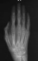 |
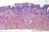
Hand X-ray | This radiograph of a child's hand with advanced polyarticular JRA shows subarticular osteopenia and fusion of the metacarpal bones. | Polyarticular Juvenile Rheumatoid Arthritis | HEAL Reviewed Collection |
| 127 |
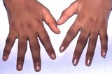 |
Interphalangeal Polyarthritis | In rheumatoid arthritis the proximal interphalangeal joints are more affected than the distal ones. | Polyarticular Juvenile Rheumatoid Arthritis; Interphalangeal Joint | HEAL Reviewed Collection |
| 128 |
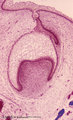 |
Late cap stage of tooth development - human, embryo; low magnification | Stain: Azan. From top to bottom: Stratified ectoderm with a distinct basal layer (red line) of cuboid cells; Dental lamina giving rise to the cap stage (center) and to the primordium of permanent tooth (right); Odontogenic organ or enamel organ (future deciduous tooth surrounded by fibrous tooth fol... | oral cavity; dental lamina | Poja Histology Collection - Oral Cavity Subset |
| 129 |
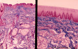 |
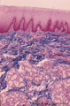
Lip (human), outer (left) and inner side (right) | Stain: Azan. Left image: keratinized squamous epithelium with thin red cornified layer, hair follicle, sebaceous glands and skeletal muscle fibers (orbicularis oris). Right image: non-keratinized epithelium with high, narrow dermal papillae and more capillaries. Mixed labial glands (seromucous), f... | oral cavity; lining mucosa | Poja Histology Collection - Oral Cavity Subset |
| 130 |
 |
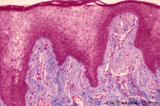
Lip (human), region between red zone (vermilion border) and mucosa inner surface | Stain: Azan. Bundles of skeletal muscle fibers (musculus orbicularis oris), highly vascularized lamina propria. Note the narrow dermal papillae on the left side (inner lip) and the broad irregular papillae on the right side (red zone of the lip). | oral cavity; lining mucosa; red zone; vermilion border | Poja Histology Collection - Oral Cavity Subset |
| 131 |
 |
Lip (human), mucous inner surface | Stain: Azan. Non-keratinized epithelium with high, narrow dermal papillae and many capillaries. Mixed labial glands (seromucous) and few skeletal muscle fibers (orbicularis oris) in the submucosa. | oral cavity; lining mucosa; labial glands | Poja Histology Collection - Oral Cavity Subset |
| 132 |
 |
Lip (human), mucoserous labial glands in mucous inner surface | Stain: Azan. Mixed labial glands with serous demilunes (von Ebner-Giannuzzi), a few myoepithelial cells, and a striated duct (right upper corner). | oral cavity; lining mucosa; labial glands | Poja Histology Collection - Oral Cavity Subset |
| 133 |
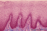 |
Lip (human), mucous inner surface | Stain: Azan. Non-keratinized epithelium with high, narrow dermal papillae and many capillaries. Note lymphocytic infiltrates and the capillaries in the dermal papillae. | oral cavity; lining mucosa | Poja Histology Collection - Oral Cavity Subset |
| 134 |
 |
Lip (human), transitional zone (red zone or vermilion border) | Stain: Azan. Slightly cornified epithelium with high irregular dermal papillae and many capillaries. Note the epithelium is thicker, but less cornified than the epidermis. The red color of the lips is due to the rich vascularity of the lamina propria and the lucidity of the epithelium. | oral cavity; lining mucosa; red zone; vermilion border | Poja Histology Collection - Oral Cavity Subset |
| 135 |
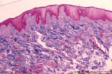 |
Lip (human), transitional zone (red zone or vermilion border) | Stain: Azan. Slightly cornified epithelium with high irregular dermal papillae and many capillaries. Fat cells and skeletal muscle cells are located in the submucosa. | oral cavity; lining mucosa; red zone; vermilion border | Poja Histology Collection - Oral Cavity Subset |
| 136 |
 |
Lymph Nodes of Head and Neck (Labeled) | Lymph nodes. Head. Neck. | Submental; Submandibular; Deep Cervical; Jugulodigastric; Mastoid; Occipital; Parotid | Royal College of Surgeons in Ireland Illustrations |
| 137 |
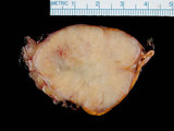 |
Lung carcinoma metastatic to adrenal gland | This is a gross photograph of a non-small cell carcinoma metastatic to the adrenal gland. | HEAL Reviewed Collection | |
| 138 |
 |
Molluscum contagiosum | Photomicrograph showing cytopathic changes of pox virus (Molluscum contagiosum) in HIV patient. | AIDS | HEAL Reviewed Collection |
| 139 |
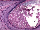 |
Molluscum contagiosum | Photomicrograph showing cytopathic changes of pox virus (Molluscum contagiosum) in HIV patient. | AIDS | HEAL Reviewed Collection |
| 140 |
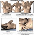 |
Monthly Breast Self-Exam (Labeled) | Monthly breast self-exam. | Cancer Screening | Royal College of Surgeons in Ireland Illustrations |
| 141 |
 |
Odontoblast in tooth development - mammalian embryo | Scheme electronmicroscopy. A columnar and irregular shaped cell (mesenchymal origin) with a taperwise odontoblastic process within the predentin. The bottom side is adjacent to the future pulp cells. The cell body contains many organelles and especially a well-developed Golgi area with prosecretoy g... | oral cavity | Poja Histology Collection - Oral Cavity Subset |
| 142 |
 |
Parotid gland (human) | Stain: Mallory trichrome. Survey: at the left bottom a large interlobular duct (in lumen remnants of secretion products) within a septum of dense connective tissue. At the top thinner (blue) septum, a thick (red-bluish) septum at the left. In the center three (intralobular) striated ducts between th... | oral cavity; serous gland | Poja Histology Collection - Oral Cavity Subset |
| 143 |
 |
Papillae circumvallatae of the tongue (dorsal side, human) | Stain: Azan. Three broad papillae with taste buds facing the grooves in which the serous von Ebner glands drain. Striated skeletal muscles (musculus verticalis linguae and musculus longitudinalis superior) and lightly stained mucous glands (posterior lingual glands). | oral cavity; von Ebner; lingual muscles; lingual glands | Poja Histology Collection - Oral Cavity Subset |
| 144 |
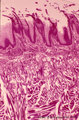 |
Papillae filliformes of the tongue (dorsal side, human) | Stain: Heidenhain light bordeaux. Threadlike keratinized extensions of the stratified epithelium. Primary connective tissue papillae with 2 to 3 secondary papillae. The skeletal muscle fibers are arranged in three directions. | oral cavity; filiform papillae | Poja Histology Collection - Oral Cavity Subset |
| 145 |
 |
Parotid gland (human) | Stain: Azan. The parotid gland: in most species the gland is composed entirely of serous acini. At the right a small (intralobular) striated duct; centrally one large interlobular duct with blood vessels. Scattered a few (white) fat cells. | oral cavity; serous gland | Poja Histology Collection - Oral Cavity Subset |
| 146 |
 |
Papillae circumvallatae of the tongue (dorsal side, human) | Stain: Azan. A broad papilla with taste buds (lightly stained spots) facing the grooves in which the serous glands (von Ebner) drain. | oral cavity; von Ebner; circumvallate papillae | Poja Histology Collection - Oral Cavity Subset |
| 147 |
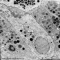 |
Parotid gland (rat) | Electronmicroscopy. Part of a serous acinus with characteristic secretion granules supranuclearly. Note different densities of the granules without any signs of fusion. | oral cavity; serous gland | Poja Histology Collection - Oral Cavity Subset |
| 148 |
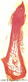 |
Permanent tooth - canine, human, adult; low magnification of labiolingual section | Stain: Hematoxylin and scarlet red. From top to bottom: Crown region with dentin but without enamel (decalcified specimens); Neck region at the attachment of the gingiva to dentin (left and right); Cementum is visible as a dark small rim from the neck region to the bottom of the tooth; Periodontal l... | oral cavity; alveolar process | Poja Histology Collection - Oral Cavity Subset |
| 149 |
 |
Premolar permanent tooth (human, adult; low magnification of labiolingual section) | Stain: Hematoxylin and eosin. From top to bottom: Crown region with enamel-dentin boundary (there is no enamel present in decalcified specimens); dentin (blue-pink) enwraps the whole pulp chamber (light) and here the lines of dentinal tubules appear S-shaped. Neck region at the attachment of the gin... | oral cavity; pulp canal | Poja Histology Collection - Oral Cavity Subset |
| 150 |
 |
Periodontal ligament with epithelial rests of Malassez - longitudinal section of root of tooth; human, adult | Stain: Hematoxylin and eosin. From left to right: connective tissue of periodontal ligament with epithelial rests of Malassez as the persistent remnants of the epithelium of Hertwig's epithelial root sheath. The three islets close to the cemental zone are slightly darker stained (note cluster of nuc... | oral cavity; cementoblasts; Sharpey's fibers; acellular cementum | Poja Histology Collection - Oral Cavity Subset |