The Health Education Assets Library (HEAL) is a collection of over 22,000 freely available digital materials for health sciences education. The collection is now housed at the University of Utah J. Willard Marriott Digital Library.
TO
| Title | Description | Subject | Collection | ||
|---|---|---|---|---|---|
| 126 |
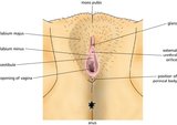 |
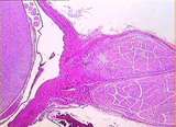
Vulva (Labeled) | External genitalia. Female. | Mons Pubis; Glans; External Urethral Orifice; Perineal Body; Opening of Vagina; Vestibule of Vagina; Labium Minus; Labium Majus | Royal College of Surgeons in Ireland Illustrations |
| 127 |
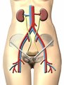 |
Urinary System | Urinary system. Kidneys. Ureter. Bladder. | Common Iliac Veins; Common Iliac Arteries | Royal College of Surgeons in Ireland Illustrations |
| 128 |
 |
Eye - Rods and Cones | Rod and cone photoreceptors have their cell bodies in the outer nuclear layer. Between these cell bodies and the retinal pigment epithelium are the photoreceptor inner segments (where proteins are synthesized) and the outer segments (the light sensitive portion). The inner segments of cones are cone... | UCLA Histology | |
| 129 |
 |
Uterus | This relatively brief phase of the endometrial cycle consists of shrinkage of the endometrium and intraendometrial hemorrhage and fragmentation. The myometrium is located to the left. A higher magnification of the ischaemic endometrium is shown in image 226-10-1. UCLA Histology Collection. | ischaemic endometrium; Uterus | UCLA Histology |
| 130 |
 |
Kidney | In this section through the renal papilla, identify the terminal collecting tubules (lined by columnar epithelium). Also note a straight segment of the loop of Henle, many thin loops of Henle, and cross-sections of the vasa recta. UCLA Histology Collection. | Kidney; renal papilla; terminal collecting tubules | UCLA Histology |
| 131 |
 |
Breast | This active, lactating breast contains a large number of lobules of tightly packed alveoli. The connective tissue of the interlobular elements is highly compressed. Note the lactating glands, the excretory duct and some fat. UCLA Histology Collection. | Breast; lactating breast | UCLA Histology |
| 132 |
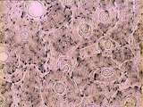 |
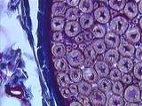
Bone - Haversian System | Haversian systems can be seen well in this image of dried compact bone. Haversian or central canals and concentrically arranged osteocytes are also visible. UCLA Histology Collection. | UCLA Histology | |
| 133 |
 |
Skin | Healing skin (rat). See lymphocyte infiltration. UCLA Histology Collection. | Skin | UCLA Histology |
| 134 |
 |
Eye - Retina | The retina consists of the retinal pigment epithelium and neurosensory retina. The neurosensory retina includes multiple layers of neurons and some glia. The outer nuclear layer contains the cell bodies of rod and cone photoreceptors. The inner nuclear layer contains the cell bodies of multiple neur... | UCLA Histology | |
| 135 |
 |
Ovary | This tissue looks like a dying corpus luteum, but that is a rare event. This is an example of a structure that we cannot identify unless we see a zona pellucida and/or a glassy membrane. If you had a section along the plane labelled A you would not be able to differentiate between a section through ... | Ovary | UCLA Histology |
| 136 |
 |
Ear | At this higher magnification, one can differentiate the hair & supporting cells, the gelatinous cupula, and the dark cells which mark the transition of the membranous labyrinth into the crista ampullaris. Bone is also evident. Movement of the endolymph against the cupula causes bending of hair cell ... | Crista Ampullaris; Ear | UCLA Histology |
| 137 |
 |
Peripheral Nervous System | This slide of myelinated nerves is stained with osmium to demonstrate the myelin sheaths. The region where two myelin sheaths covering the same axon contact one another is the Node of Ranvier. The region of myelin sheath between these nodes is known as the internode. UCLA Histology Collection. | myelin sheaths; osmium; Peripheral Nervous System | UCLA Histology |
| 138 |
 |
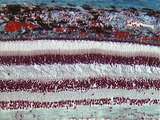
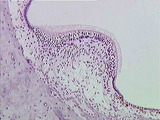
Epithelium - Cornea | The cornea of the eye is lined by simple squamous epithelium on its inner surface and stratified squamous epithelium on its outer surface. The stroma of the cornea consists of regularly arranged collagen fibers. UCLA Histology Collection. | simple squamous epithelium; stratified squamous epithelium | UCLA Histology |
| 139 |
 |
Cells / Organelles - Pituitary | Cells of the pituitary gland containing cytoplasmic secretory granules. The red-orange granules and the blue-purple granules contain different hormones. UCLA Histology Collection. | pars distalis; secretory granules | UCLA Histology |
| 140 |
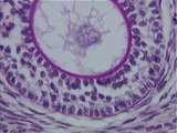 |
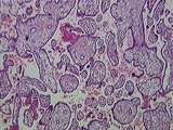
Ovary | A typical preantral follicle possessing a zona pellucida , multilayered granulosa or follicular cells and a stromal derived theca again embedded in stroma. Be sure you realize that the components within the zona pellucida are the nucleus of the ovum and its cytoplasm. UCLA Histology Collection. | Ovary; preantral follicle | UCLA Histology |
| 141 |
 |
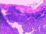
lymphoid tissue | This image shows stratified squamous epithelium overlying connective tissue. The parenchyma contains many lymphoid nodules. Crypts are present where epithelium invaginates into parenchyma. UCLA Histology Collection. | lymphoid nodule; lymphoid tissue; palatine tissue; palatine tonsil | UCLA Histology |
| 142 |
 |
Cells / Organelles - Liver | At a higher magnification, liver cell nuclei can be seen to have nucleoli. Note the heterochromatinheterochromatin lining the internal portion of the nuclear envelope. UCLA Histology Collection. | Cells and Organelles; Nucleus | UCLA Histology |
| 143 |
 |
Eye - Lens Fibers | Lens fibers elongating as they move posterior. UCLA Histology Collection. | UCLA Histology | |
| 144 |
 |
Peripheral Nervous System / Central Nervous System | The relationship between the spinal cord, the spinal roots, and the meninges is illustrated here. The surface of the spinal cord is covered by pia mater. Between the pia mater and web-like arachnoid mater is the subarachnoid space which contains CSF. The thick, collagenous dura mater is continuous w... | arachnoid mater; Central Nervous System; dura mater; meninges; Peripheral Nervous System; pia mater; spinal cord; spinal roots | UCLA Histology |
| 145 |
 |
Peripheral Nervous System | This cross sectioned peripheral nerve demonstrates the relatively thick myelin covering of axons, as well as the endoneurium which is located between the myelin sheaths. Also note the blue-stained collagen of the epineurium and a small blood vessel. UCLA Histology Collection. | axons; endoneurium; myelin; Peripheral Nervous System | UCLA Histology |
| 146 |
 |
Placenta | This image presents the basic organization of the placenta. Embedded within the maternal blood space are numerous fetal villi, some of them clearly tertiary villi , all lined by the syncytiotrophoblast . The only things missing in this image are maternal decidual cells and fetal/maternal fibrinoid. ... | Placenta | UCLA Histology |
| 147 |
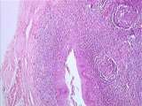 |
Epithelium - Lymphoid Tissue | Crypts of the palatine tonsils are lined by stratified squamous epithelium. The lymphoid tissue can be seen organized in nodules. A connective tissue capsule partially surrounds the tonsil. Note the skeletal muscle which is associated with the palatine tonsil. UCLA Histology Collection. | palatine tonsil | UCLA Histology |
| 148 |
 |
Kidney | The renal papilla is the site where terminal collecting ducts empty urine into the U-shaped minor calyx. The lumen of the minor calyx is lined by transitional epithelium. Strands of circular smooth muscle are found in the wall of the minor calyx. UCLA Histology Collection. | Kidney; minor calyx; renal papilla; terminal collecting ducts | UCLA Histology |
| 149 |
 |
Eye - Lens Epithelium | This enlargement of slide 236-2.5-2 reveals the lens epithelium in various phases: the anterior epithelium, area of proliferation, and area of elongation. The lens bow region consists of lens fiber nuclei in various stages of degeneration. UCLA Histology Collection. | UCLA Histology | |
| 150 |
 |
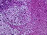
Thymus - Hassall's Corpuscle | This image shows an early and a late Hassall's corpuscle. Also visible are numerous epithelial reticular cells and even more numerous lymphocytes. UCLA Histology Collection. | epithelial reticular; Hassall's corpuscle | UCLA Histology |